Gene expression, RNA processing and protein synthesis
Gene expression converts DNA instructions into functional proteins through transcription, processing and translation.
Figure 1Gene Expression
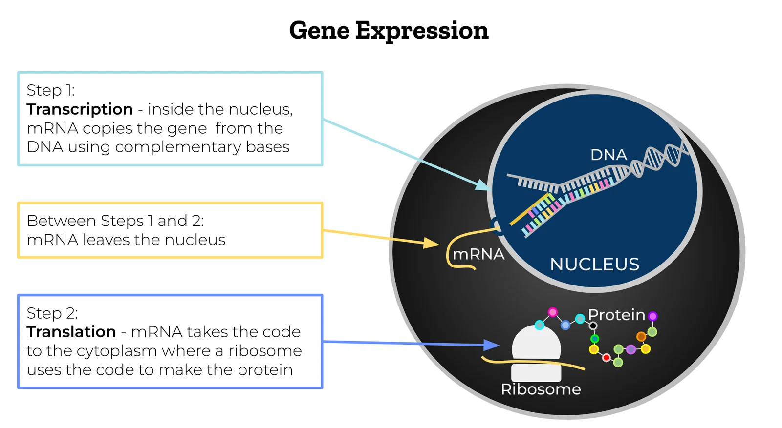
Long description
Illustration for biochemistry/gene expression rna processing and protein synthesis from Wikimedia Commons, titled 'Gene Expression': Simple steps of gene expression. The author and licence are given in the credit line.

DefinitionGene expression
Gene expression in eukaryotes is a multi‑step pathway: DNA is transcribed into a primary RNA transcript (pre‑mRNA), the transcript is modified (capping, splicing, polyadenylation), exported to the cytoplasm and translated by ribosomes. Each step is tightly regulated, allowing a single genome to generate a vast proteomic repertoire.
The lac operon of E. coli is the classic prokaryotic model for transcriptional control. It comprises three structural genes—lacZ, lacY and lacA—that encode β‑galactosidase, permease and transacetylase respectively, and a regulatory region containing the promoter, operator and the lacI repressor gene.
LacI may modulate the expression of lacZYA in the lac operon across a wide range to respond to varying growth conditions【P360】.
Key points of the lac system
- Repression: LacI binds the operator, blocking RNA polymerase.
- Induction: Allolactose (a lactose metabolite) binds LacI, reducing its affinity for the operator.
- Catabolite repression: High cAMP‑CRP levels in low glucose enhance promoter activity, integrating carbon source availability.
Promoters are DNA sequences immediately upstream of a transcription start site that recruit the core transcription machinery. In bacteria a single RNA polymerase (RNAP) with a σ factor recognises the –35 and –10 consensus motifs. In eukaryotes, multiple RNAPs exist (RNAP I, II, III) each dedicated to distinct classes of genes; RNAP II synthesises mRNA.
Enhancers are distal regulatory elements that increase transcription rates, often acting through DNA looping to contact promoters. Silencers have the opposite effect, recruiting repressor complexes that modify chromatin to a closed state. Both enhancers and silencers function primarily at the transcriptional level, consistent with the historical view that regulation occurs chiefly during transcription【P77】.
Transcription factors (TFs) bind specific DNA motifs within promoters, enhancers or silencers, influencing RNAP recruitment, initiation and promoter clearance. Co‑activators such as histone acetyltransferases remodel chromatin to a permissive configuration, whereas co‑repressors (e.g., HDACs) promote nucleosome compaction.
After RNAP II initiates transcription, the nascent pre‑mRNA undergoes three co‑transcriptional modifications that are essential for stability, nuclear export and translation.
| Modification | Enzyme complex | Functional outcome |
|---|---|---|
| 5′ Cap (7‑methylguanosine) | Capping enzyme (RNA 5′‑triphosphatase, guanylyltransferase, methyltransferase) | Protects RNA from exonucleases, promotes ribosome recruitment, assists splicing |
| Splicing (intron removal) | Spliceosome (snRNPs U1, U2, U4/U5/U6) plus auxiliary factors (e.g., U2AF65, CAPERα/β) | Generates mature mRNA; alternative splicing creates protein isoforms, exemplified by VEGF‑A isoform regulation via ASF/SF2, SRp40, SRp55 and E2F1【P107】 |
| 3′ Poly(A) tail | Poly(A) polymerase (PAP) and CPSF complex | Enhances mRNA stability, aids nuclear export, stimulates translation initiation |
Capping occurs when the 5′‑triphosphate of the nascent transcript is converted to a diphosphate, a GMP is added, and the guanine is methylated at the N7 position. This cap is recognized by the cap‑binding complex (CBC) and later by eIF4E during translation initiation.
Splicing removes introns through two transesterification reactions. The spliceosome assembles stepwise: U1 binds the 5′ splice site, U2 recognizes the branch point, and the tri‑snRNP complex (U4/U5/U6) catalyses intron excision. Alternative splicing is regulated by serine/arginine‑rich (SR) proteins and heterogeneous nuclear ribonucleoproteins (hnRNPs). For instance, m6A modification can remodel RNA secondary structure, exposing binding sites for hnRNP C and influencing splice site choice【P195】. [3]
Polyadenylation is initiated by cleavage of the pre‑mRNA at a poly(A) signal (AAUAAA). PAP then adds ~200 adenine residues, a process enhanced by poly(A) binding proteins (PABPs) that protect the tail from deadenylases.
Gene expression pathway
- DNAGene locus with promoter, enhancer, operator
- Transcription initiationRNA polymerase recruitment
- Question: Prokaryote or eukaryote?
Prokaryote
- mRNA synthesisNo processing, translation begins
Eukaryote
- Pre‑mRNACapping → Splicing → Polyadenylation
- ExportNuclear pore transport
- TranslationRibosome assembly, protein synthesis
DefinitionAmino‑acyl‑tRNA synthetase
The first step in protein synthesis is the “charging” of tRNA molecules. Each of the 20 canonical amino‑acyl‑tRNA synthetases (aaRSs) recognises a specific amino‑acid and one or more tRNA isoacceptors that carry the corresponding anticodon. The reaction proceeds in two chemically distinct stages:
- Activation of the amino‑acid – the amino‑acid reacts with ATP, producing an amino‑acyl‑adenylate intermediate (amino‑acyl‑AMP) and releasing pyrophosphate.
- Transfer to tRNA – the activated amino‑acid is transferred from AMP to the 3′‑terminal ribose of the tRNA, generating amino‑acyl‑tRNA and releasing AMP.
The overall stoichiometry can be expressed as:
Amino‑acid + tRNA + ATP → amino‑acyl‑tRNA + AMP + PPi
The high fidelity of this process is ensured by two proofreading mechanisms. First, the aaRS active site discriminates against structurally similar amino‑acids; second, an editing domain hydrolyses mis‑charged tRNAs before they leave the enzyme. Errors that escape editing are rare (≈1 error per 10⁴‑10⁵ charging events) and are usually corrected by ribosomal quality‑control pathways.
Charged tRNAs diffuse to the cytosol (or mitochondrial matrix for mitochondrial aaRSs) where they participate in translation. In mitochondria, specialised mitochondrial aaRSs (mt‑ARSs) not only supply amino‑acyl‑tRNAs for mitochondrial ribosomes but also have non‑canonical roles, for example influencing tRNA modifications such as m³C₃₂ (see flowchart)【P337】.
Translation in eukaryotes is a highly regulated, multi‑step process that converts messenger RNA (mRNA) into a polypeptide chain. The three principal phases are initiation, elongation and termination.
- Formation of the 43S pre‑initiation complex (PIC) – the 40S ribosomal subunit binds eukaryotic initiation factors (eIF1, eIF1A, eIF3, eIF5) and the ternary complex (eIF2·GTP·Met‑tRNAᵢᶠ).
- mRNA recruitment – the cap‑binding complex eIF4F (eIF4E, eIF4G, eIF4A) attaches to the 5′‑m⁷G cap, unwinds secondary structure, and bridges the mRNA to the PIC via eIF4G. Poly(A)‑binding protein (PABP) interacts with eIF4G, circularising the mRNA and enhancing re‑initiation.
- Scanning and start‑codon recognition – the PIC scans downstream until it encounters an AUG (or, less commonly, a CUG) start codon in a favourable Kozak context. Initiation factors eIF1 and eIF5 regulate the stringency of start‑codon selection. Alternative initiation at non‑AUG codons can be splice‑variant dependent, as seen for VEGF‑A isoforms where CUG initiation is splice‑independent while AUG usage varies with exon composition【P132】.
- Joining of the 60S subunit – GTP hydrolysis on eIF2 triggers release of initiation factors and recruitment of the 60S subunit, forming an 80S ribosome ready for elongation.
The elongation cycle repeats three coordinated steps for each codon:
| Step | Molecular events |
|---|---|
| Decoding | Amino‑acyl‑tRNA·eEF1A·GTP delivers the correct tRNA to the A‑site. Correct codon‑anticodon pairing stimulates GTP hydrolysis, releasing eEF1A. |
| Peptide bond formation | The peptidyl‑transferase centre of the 60S subunit catalyses peptide bond formation between the nascent chain (P‑site) and the incoming amino‑acid (A‑site). |
| Translocation | eEF2·GTP drives movement of the ribosome by one codon, shifting the deacylated tRNA to the E‑site, the peptidyl‑tRNA to the P‑site, and opening the A‑site for the next amino‑acyl‑tRNA. GTP hydrolysis resets eEF2. |
Elongation factors ensure high speed (≈5–10 amino acids per second in mammalian cells) while maintaining fidelity through kinetic proofreading.
When a stop codon (UAA, UAG, UGA) enters the A‑site, release factors eRF1 and eRF3·GTP recognise it. eRF1 mimics a tRNA and catalyses hydrolysis of the ester bond linking the nascent peptide to the tRNA, freeing the polypeptide. GTP hydrolysis by eRF3 promotes dissociation of the termination complex, after which ribosome recycling factors (ABCE1, eIF6) separate the subunits for another round of initiation.
NoteWhy PTMs matter
Post‑translational modification (PTM) is a pivotal regulatory layer that fine‑tunes protein activity, localisation, stability and interactions. Any protein can be modified either co‑translationally (while still attached to the ribosome) or post‑translationally (after release). PTMs alter charge, hydrophobicity, conformation and degradation propensity, thereby reshaping cellular physiology in a reversible manner【P3】. [1]
To date, more than 450 distinct protein modifications have been catalogued, ranging from small chemical groups (phosphate, acetyl) to complex protein tags (ubiquitin, SUMO)【P4】. The most extensively studied is phosphorylation, catalysed by over 500 kinases in mammals【P4】. Phosphorylation predominantly targets serine, threonine and tyrosine residues【P4】, modulating enzyme activity, protein‑protein interactions, subcellular localisation and degradation rates. [1]
Other major PTM classes include:
| Modification | Typical residues | Functional impact |
|---|---|---|
| Ubiquitination | Lysine | Marks proteins for proteasomal degradation or alters signalling |
| SUMOylation | Lysine (consensus ψKxE) | Influences nuclear transport, DNA repair, transcription and stress responses【P6】 |
| Acetylation | Lysine, N‑terminus | Regulates chromatin structure and enzyme activity |
| Methylation | Lysine, arginine | Affects protein–protein interactions and RNA binding |
| Glycosylation | Asparagine (N‑linked), serine/threonine (O‑linked) | Controls folding, stability and cell‑surface recognition |
- Signal transduction – Rapid phosphorylation cascades transmit extracellular cues to intracellular effectors.
- Protein turnover – Ubiquitin chains flag substrates for the 26S proteasome, while de‑ubiquitinases rescue proteins from degradation.
- Subcellular localisation – SUMOylation and phosphorylation can expose or mask nuclear localisation signals, directing proteins to the nucleus or cytoplasm.
- DNA repair and genome stability – SUMOylation is essential for maintaining genomic integrity, coordinating repair complexes and checkpoint signalling【P6】.
- Cross‑talk between PTMs – Modifications often influence one another; for example, SUMOylation can prime a substrate for subsequent phosphorylation or ubiquitination, creating combinatorial codes that fine‑tune cellular outcomes【P6】.
Most PTMs are reversible. Kinases add phosphate groups, while phosphatases remove them; ubiquitin ligases conjugate ubiquitin, and de‑ubiquitinases cleave it. This dynamic turnover enables swift adaptation to changing conditions, a principle central to immune responses, cell‑cycle progression and stress adaptation【P3】. [1]
Aberrant PTM patterns underlie many diseases. Hyper‑phosphorylation of tau protein contributes to neurofibrillary tangles in Alzheimer’s disease, while dysregulated SUMOylation is linked to cancer progression and viral infection. Understanding PTM networks therefore informs therapeutic strategies, such as kinase inhibitors or proteasome blockers.
From translation to functional protein
- tRNA chargingaaRS attaches amino‑acid to tRNA
- InitiationFormation of 43S PIC, cap binding, scanning
- ElongationDecoding → peptide bond → translocation
- TerminationStop‑codon recognition, peptide release
- Question: Is the nascent chain modified?
Yes
- Co‑translational PTMe.g., N‑terminal acetylation
- Post‑translational PTMphosphorylation, ubiquitination, SUMOylation…
No
- Mature proteinFunctional without further modification
DefinitionMolecular chaperones
During transcription and translation, a range of chaperone systems ensures that newly synthesised polypeptides attain their correct three‑dimensional conformation. In the cytoplasm, the Hsp70‑Hsp40 machinery binds emerging chains as they exit the ribosome, preventing aggregation and guiding proper folding. In the nucleus, specialised chaperones such as the SUMO‑specific ligases act as “molecular match‑makers”, attaching the small ubiquitin‑like modifier (SUMO) to target proteins and thereby influencing their localisation, stability and activity. The SUMO E3 ligases PIAS1‑4 and MMS21 belong to a class of nuclear chaperones that confer substrate specificity to the SUMO conjugation cascade, while RanBP2 (also known as NUP358) provides a cytoplasmic platform for SUMO‑dependent regulation of proteins that shuttle through the nuclear pore complex. [2] [1]
Antibiotics that target the ribosome can indirectly affect chaperone demand. Aminoglycosides, for example, cause misreading of mRNA, leading to the production of aberrant polypeptides that overload the Hsp70 system and trigger the unfolded protein response. Certain bacterial toxins, such as diphtheria toxin, ADP‑ribosylate elongation factor‑2, halting translation and consequently reducing the load on co‑translational chaperones. Conversely, some natural products act as “chemical chaperones”, stabilising protein conformations under stress; osmolytes like trehalose can mitigate the aggregation of misfolded proteins during hypoxic stress, a condition that also up‑regulates SUMO‑1 expression.
Exam trapAssuming all SUMOylation is nuclear
Students often conflate SUMOylation with ubiquitination, overlooking key mechanistic differences. SUMO attachment does not usually tag proteins for proteasomal degradation; instead, it modulates interactions, localisation and activity. A frequent error is to interpret increased expression of SUMO pathway components as uniformly oncogenic. While Ubc9 is markedly up‑regulated in early tumour development, its level falls in metastatic lesions of breast, prostate and lung cancers, reflecting a context‑dependent role. [1]
Another pitfall is neglecting the compartmentalised activity of de‑SUMOylating enzymes. Nuclear SENPs cannot de‑SUMOylate cytoplasmic proteins, and the yeast de‑SUMOylases Ulp1 and Ulp2 display distinct subcellular distributions, with Ulp1 anchored near the nucleus and Ulp2 dispersed in the cytoplasm. Overlooking this spatial separation can lead to misinterpretation of experimental data on SUMO turnover. [1]
Finally, the impact of hypoxia on the SUMO system is sometimes underestimated. Hypoxic stress elevates SUMO‑1 protein levels and induces the expression of the SUMO‑conjugation enhancer RSUME, which in turn stabilises HIF‑1α, linking oxygen sensing to transcriptional reprogramming. Ignoring this link may cause students to miss a crucial regulatory node in tumour metabolism. [1]
NoteClinical relevance of SUMO pathway modulation
The dysregulation of SUMO enzymes is a hallmark of several cancers. Overexpression of the SUMO‑specific protease SENP3 promotes ovarian cancer progression, while SENP5 enhances growth of osteosarcoma and hepatocellular carcinoma cells. Conversely, silencing of SUMO‑1 in gastric cancer cells markedly increases apoptosis, accompanied by down‑regulation of Bcl‑2, c‑Myc and p53. These observations have spurred pre‑clinical investigations of small‑molecule inhibitors of the E2 enzyme Ubc9 and of the E3 ligase RanBP2, aiming to disrupt oncogenic SUMOylation circuits. [1]
In infectious disease, the manipulation of host SUMOylation by viral proteins is a recognised virulence mechanism. Certain viral factors hijack the RanBP2‑Ubc9 complex to modify host transcription factors, thereby subverting immune responses. Understanding this interplay guides the design of antiviral agents that restore normal SUMO dynamics.
In the laboratory, researchers exploit SUMO‑tagging to improve the solubility of recombinant proteins. By fusing a SUMO moiety to a target protein, they harness the chaperone‑like properties of SUMO to promote proper folding, then remove the tag with a SUMO‑specific protease, yielding a native product.
SUMO‑mediated regulation of a transcription factor
- Signal (e.g., hypoxia) activates HIF‑1α
- RSUME expression rises
- RSUME binds Ubc9, enhancing SUMO‑1 conjugation
- Question: Is target protein a transcription factor?
Yes
- SUMO‑E3 ligase (e.g., PIAS) attaches SUMO‑1
- SUMOylated factor translocates to nucleus
- Modulates gene expression
No
- Alternative SUMO‑dependent pathway
- Cellular response (angiogenesis, metabolic shift)
- SUMO E3 ligases such as PIAS1‑4 and RanBP2 confer substrate specificity to the SUMO conjugation cascade.
- RanBP2 operates on cytoplasmic proteins linked to the nuclear pore, challenging the view that SUMOylation is solely nuclear.
- De‑SUMOylating enzymes (SENPs, Ulp1/Ulp2) are compartmentalised, dictating where SUMO removal can occur.
- Hypoxia up‑regulates SUMO‑1 and the enhancer RSUME, stabilising HIF‑1α and reshaping transcriptional programmes.
- Over‑expression of SENP3, SENP5 or SENP6 drives tumour growth in ovarian, osteosarcoma and gastric cancers respectively.
- Silencing SUMO‑1 in gastric cancer cells increases apoptosis and reduces oncogenic transcripts (Bcl‑2, c‑Myc, p53). [1]
- Antibiotics that cause mistranslation increase the burden on Hsp70 chaperones, while chemical chaperones can alleviate stress‑induced aggregation.
- Viral exploitation of the RanBP2‑Ubc9 complex illustrates the therapeutic potential of targeting SUMO pathways.
Answer each one in your head first, then open the answer.
Which SUMO E3 ligase is primarily cytoplasmic and associated with the nuclear pore complex?
Show answerHide answer
RanBP2 (NUP358)Name a de‑SUMOylating enzyme that is mainly nuclear.
Show answerHide answer
Ulp1What effect does hypoxia have on SUMO‑1 expression?
Show answerHide answer
It increases SUMO‑1 protein levels.Which SENP isoform promotes ovarian cancer progression?
Show answerHide answer
SENP3How does silencing SUMO‑1 affect gastric cancer cells?
Show answerHide answer
It markedly increases apoptosis and reduces Bcl‑2, c‑Myc and p53 expression.
- Han ZJ, Feng YH, Gu BH, et al. The post-translational modification, SUMOylation, and cancer (Review). International journal of oncology 2018;52(4):1081-1094. doi:10.3892/ijo.2018.4280. Accessed .
- Arcondéguy T, Lacazette E, Millevoi S, et al. VEGF-A mRNA processing, stability and translation: a paradigm for intricate regulation of gene expression at the post-transcriptional level. Nucleic acids research 2013;41(17):7997-8010. doi:10.1093/nar/gkt539. Accessed .
- Yue Y, Liu J, He C. RNA N6-methyladenosine methylation in post-transcriptional gene expression regulation. Genes & development 2015;29(13):1343-1355. doi:10.1101/gad.262766.115. Accessed .
Figure credits
Educational content for medical students and doctors. It is not a substitute for clinical judgement or local guidelines.