Genetic terms, population genetics and imprinting
Genetic terminology that underpins inheritance patterns and disease risk in humans.
Figure 1Genetic Variation
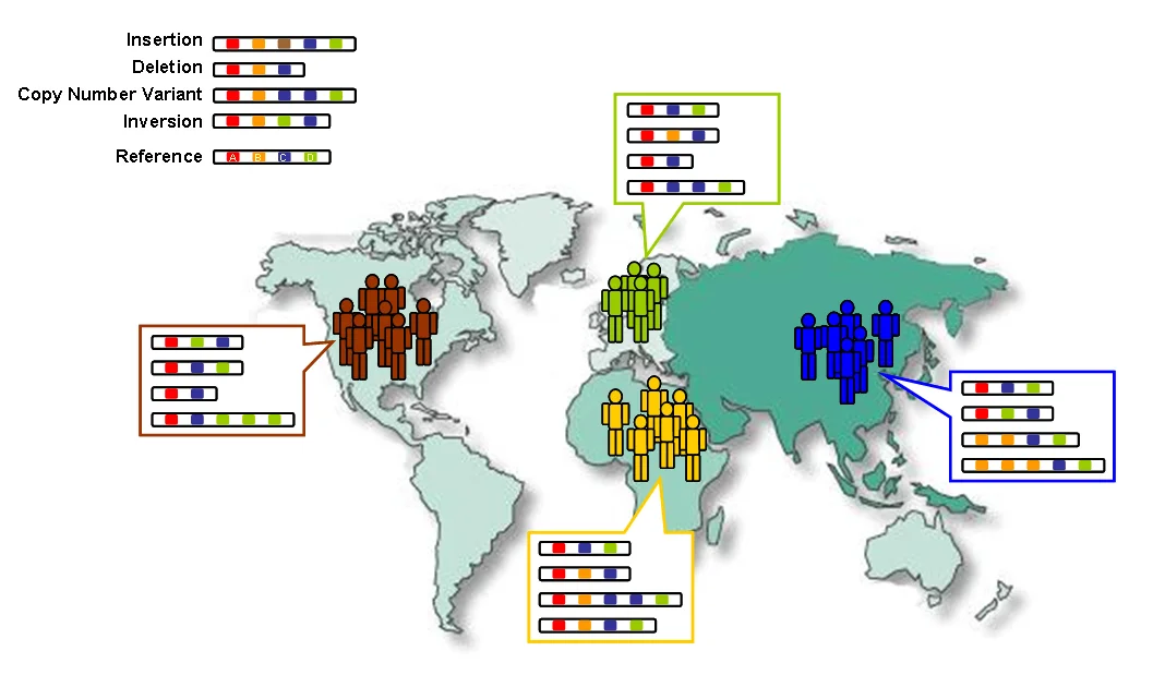
Long description
Changes in the number and order of genes (A-D) create genetic diversity within and between populations.

DefinitionGenetic term
Human genetics rests on a hierarchy of concepts that start with the DNA sequence itself and extend to how that sequence is transmitted, expressed, and sometimes modified without changing the underlying bases. The central dogma—DNA → RNA → protein—explains the flow of genetic information, but many layers of regulation intervene. Population genetics adds the dimension of allele frequencies in groups of individuals, while imprinting introduces parent‑of‑origin effects that can alter expression of otherwise identical alleles. Understanding these ideas equips medical students to interpret pedigrees, predict disease risk, and appreciate why some disorders manifest only under particular genetic or environmental contexts.
Penetrance, expressivity, pleiotropy, anticipation, epistasis, mosaicism, heteroplasmy, uniparental disomy
| Term | Meaning | Clinical illustration |
|---|---|---|
| Penetrance | Proportion of individuals with a pathogenic genotype who actually display the phenotype. | Autosomal dominant retinoblastoma shows high penetrance (>90 %). |
| Expressivity | Degree of severity or range of signs among those who are penetrant. | Neurofibromatosis type 1 may cause only café‑au‑lait spots in one person and malignant peripheral nerve sheath tumours in another. |
| Pleiotropy | One gene influences multiple, seemingly unrelated traits. | Mutations in the COL1A1 gene cause both bone fragility (osteogenesis imperfecta) and dentinogenesis imperfecta. |
| Anticipation | Successive generations present disease at an earlier age or with increased severity, often due to repeat expansion. | Huntington disease typically appears earlier in the child than in the affected parent. |
| Epistasis | Interaction where the effect of one gene is modified by one or more other genes. | The HBB sickle‑cell allele’s clinical severity is lessened when co‑inherited with α‑thalassaemia deletions. |
| Mosaicism | Presence of two or more genetically distinct cell lines derived from a single fertilised egg. | Segmental neurofibromatosis results from post‑zygotic NF1 mutation confined to a skin region. |
| Heteroplasmy | Co‑existence of normal and mutant mitochondrial DNA within a cell. | The phenotypic threshold for MELAS syndrome depends on the proportion of mutant mtDNA. |
| Uniparental disomy (UPD) | Both copies of a chromosome (or part of it) are inherited from one parent, potentially unmasking recessive mutations or disrupting imprinting. | Maternal UPD of chromosome 15 can cause Prader‑Willi syndrome, whereas paternal UPD leads to Angelman syndrome. |
Penetrance and expressivity are often confused. Penetrance is a binary concept—either the genotype produces a phenotype or it does not—whereas expressivity captures the spectrum of severity among those who are affected. A disease may be fully penetrant but highly variable in expressivity, as seen in Marfan syndrome, where some individuals develop life‑threatening aortic aneurysms while others have only mild skeletal features.
Pleiotropy explains why a single genetic test can have implications for multiple organ systems. For instance, a pathogenic BRCA1 variant raises risk for breast, ovarian, pancreatic, and prostate cancers, reflecting the gene’s role in DNA repair across diverse tissues.
Anticipation is most famously linked to trinucleotide repeat disorders. The repeat length expands during meiosis, crossing a pathogenic threshold in the next generation and thereby producing earlier onset. This phenomenon underscores the importance of measuring repeat size in families with Huntington disease, myotonic dystrophy, or fragile X syndrome.
Epistasis can mask or modify the phenotype of a primary mutation. In cystic fibrosis, certain modifier genes influence lung disease severity, illustrating that genotype‑phenotype correlations are rarely linear.
Mosaicism and heteroplasmy illustrate that not all cells carry the same genetic information. In mitochondrial diseases, the proportion of mutant mtDNA (heteroplasmy level) determines whether a patient remains asymptomatic or develops multisystem disease. Similarly, somatic mosaicism for a pathogenic variant can result in a localized skin disorder without systemic involvement.
Uniparental disomy can have two major consequences: (1) unmasking a recessive mutation when the other parental allele is absent, and (2) disrupting normal imprinting patterns. Recognising UPD is crucial when a child presents with a recessive disorder despite only one carrier parent being identified.
Linkage analysis and Hardy‑Weinberg equilibrium (HWE) are foundational tools for population genetics and for mapping disease genes.
Linkage refers to the tendency of loci that are physically close on a chromosome to be inherited together. The recombination fraction (θ) quantifies this tendency, ranging from 0 (complete linkage) to 0.5 (independent assortment). In human genetics, linkage maps are built from pedigrees using markers such as short tandem repeats (STRs) or single‑nucleotide polymorphisms (SNPs). Modern high‑density SNP arrays enable construction of linkage groups with fine resolution, analogous to the wheat linkage maps described in recent agronomic studies, where markers were organised into groups using LOD thresholds of 2–10 and average intervals of 1.66 cM/marker. Although the wheat data are plant‑specific, the principle that tighter marker spacing improves map accuracy applies equally to human linkage studies. [1]
HWE predicts genotype frequencies from allele frequencies in a large, randomly mating population with no selection, mutation, migration, or drift. The equation is:
p² + 2pq + q² = 1
where p = frequency of the dominant allele, q = frequency of the recessive allele, p² = homozygous dominant, 2pq = heterozygous, q² = homozygous recessive.
Example calculation (illustrative):
Suppose cystic fibrosis (CF) has a carrier frequency of 1 in 25 in a population. The carrier frequency equals 2pq = 1/25 = 0.04. Assuming p ≈ 1 (the disease allele is rare), q ≈ 0.02. The expected disease prevalence (q²) is 0.0004, or 1 in 2,500, matching observed CF incidence.
Deviations from HWE may signal genotyping errors, population stratification, or genuine biological forces such as selection. In clinical genetics, checking HWE for control cohorts ensures that association studies are not confounded.
When a disease locus is tightly linked to a marker (θ ≈ 0), the marker allele frequencies in affected families differ from those in the general population. This principle underlies genome‑wide association studies (GWAS), where millions of SNPs are tested for association with disease phenotypes. Significant associations often reflect linkage disequilibrium (LD) with the causal variant, a concept that parallels the high collinearity observed between genetic and physical positions of markers in wheat linkage maps. [1]
Genetic analysis pathway
- Collect pedigree or population dataFamily histories, genotype arrays, phenotype records
- Question: Is the trait monogenic?
Yes
- Perform linkage analysisCalculate recombination fractions, build LOD scores
- Identify candidate regionFine‑map with additional markers
No
- Check Hardy‑WeinbergValidate allele frequencies in controls
- Conduct GWASTest SNPs for association, interpret LD patterns
- Interpret resultsConsider penetrance, expressivity, pleiotropy, etc.
DefinitionGenomic imprinting
Imprinting disorders arise when the normal pattern of mono‑allelic expression is disturbed. The two classic neurodevelopmental syndromes that illustrate this principle are Prader‑Willi syndrome (PWS) and Angelman syndrome (AS). Both map to the same chromosomal region, 15q11‑q13, but the clinical picture differs dramatically depending on whether the paternal or maternal allele is affected.
| Mechanism | Effect in PWS | Effect in AS |
|---|---|---|
| Deletion of the critical region | Loss of paternal 15q11‑q13 | Loss of maternal 15q11‑q13 |
| Maternal uniparental disomy (UPD) | Two maternal copies, no paternal contribution | Two paternal copies, no maternal contribution |
| Imprinting centre defect | Failure to establish paternal imprint | Failure to establish maternal imprint |
| Point mutation in a single gene (e.g., UBE3A) | Rare | Often pathogenic |
- In PWS the genes that are normally active only on the paternal chromosome (e.g., SNRPN, NDN) are absent or silenced, leaving only the silent maternal copies.
- In AS the UBE3A gene, which is expressed only from the maternal allele in the brain, is lost, while the paternal copy remains epigenetically silenced.
Prader‑Willi syndrome
- Neonatal hypotonia, poor feeding → later hyperphagia and obesity
- Short stature, small hands/feet, hypogonadism
- Intellectual disability, behavioural problems
Angelman syndrome
- Severe speech impairment, frequent laughter, “happy puppet” demeanor
- Ataxia, seizures, microcephaly
- Profound intellectual disability
The contrasting phenotypes underscore how the same genomic region can give rise to opposite clinical pictures when the imprinting error involves different parental alleles.
Diagnostic pathway for 15q11‑q13 imprinting disorders
- Clinical suspicionKey features of PWS or AS
- Question: Methylation analysis?
Abnormal
- Determine patternLoss of paternal methylation → PWS; loss of maternal methylation → AS
- Question: Is deletion present?
Yes
- Array CGH / FISHDefine size of deletion
No
- UPD testingMicrosatellite analysis or SNP array
Normal
- Consider sequencingUBE3A mutation (AS) or other rare genes (PWS)
Methylation‑specific PCR or multiplex ligation‑dependent probe amplification (MLPA) is the first‑line test because it detects >99 % of cases by revealing the parental‑specific methylation pattern. If methylation is abnormal, further studies (deletion mapping, UPD analysis, sequencing) clarify the underlying mechanism.
- Both disorders are lifelong; management is multidisciplinary.
- Early nutritional support in PWS prevents failure to thrive, while later appetite control mitigates obesity.
- In AS, seizure control, speech therapy, and behavioural interventions are central.
- Growth hormone therapy improves stature and body composition in PWS; it is also used in AS for height and bone health.
Exam trapAssuming a single test is sufficient
-
Misinterpretation of methylation results – The assay distinguishes only the presence or absence of the paternal or maternal methylation imprint; it does not differentiate between a deletion and UPD. Without follow‑up testing, the precise genetic cause remains unknown, which is essential for recurrence risk counselling.
-
Overlooking mosaicism – Low‑level mosaic deletions or UPD may produce borderline methylation signals, leading to false‑negative results. Repeat testing on a different tissue (e.g., buccal cells) can improve detection.
-
Confusing phenotypic overlap – Some features (e.g., hypotonia, developmental delay) are shared with other neurogenetic conditions. A thorough clinical assessment is required before ordering imprinting studies.
-
Neglecting parental testing – Determining whether a deletion is de novo or inherited influences recurrence risk. Failure to test parents can result in inaccurate genetic counselling.
-
Assuming all cases are imprinting‑related – Approximately 5 % of Angelman cases are due to UBE3A point mutations without imprinting defects; these require sequencing.
Imprinting disorders are encountered across several clinical specialties:
- Paediatrics & genetics – First‑line assessment of infants with hypotonia, feeding difficulties, or atypical neurobehaviour.
- Endocrinology – Management of growth hormone deficiency and hypogonadism in PWS; monitoring of bone health in AS.
- Neurology – Seizure control in Angelman syndrome; evaluation of sleep disturbances common to both conditions.
- Obstetrics & prenatal diagnostics – Non‑invasive prenatal testing (NIPT) can detect 15q11‑q13 deletions; invasive testing (CVS/amniocentesis) with methylation analysis is offered when ultrasound suggests a syndrome.
- Psychiatry – Behavioural phenotypes (e.g., compulsivity in PWS, frequent laughter in AS) guide targeted behavioural therapies.
In each setting, the diagnostic algorithm begins with a clinical suspicion, proceeds to methylation testing, and then branches according to the result, as illustrated in the flowchart above.
- Genomic imprinting results in parent‑specific mono‑allelic expression.
- Prader‑Willi syndrome results from loss of paternal expression at 15q11‑q13; Angelman syndrome from loss of maternal expression.
- Methylation analysis is the first‑line diagnostic test, detecting >99 % of cases.
- Deletions, uniparental disomy, imprinting centre defects, and point mutations are the main molecular mechanisms.
- Management is multidisciplinary and lifelong, focusing on nutrition, growth, seizures, and behavioural support.
- Pitfalls include assuming a single test is definitive, overlooking mosaicism, and failing to test parents for recurrence risk.
Answer each one in your head first, then open the answer.
Which parental allele is silenced in Angelman syndrome?
Show answerHide answer
The paternal allele.What is the most common first‑line laboratory test for suspected imprinting disorders of chromosome 15?
Show answerHide answer
Methylation‑specific PCR or MLPA.Name one clinical feature that distinguishes Prader‑Willi from Angelman syndrome.
Show answerHide answer
Hyperphagia leading to obesity (PWS) versus frequent laughter and a happy demeanor (AS).Why is parental testing important after a deletion is identified?
Show answerHide answer
To determine whether the deletion is de novo or inherited, which affects recurrence risk.What rare mechanism can cause Angelman syndrome despite a normal methylation pattern?
Show answerHide answer
A point mutation in the UBE3A gene.
- Chen B, He Y, Tang Y, et al. QTL analysis in the Mianmai 902×Taichang 29 RIL population reveals the genetic basis for the high-yield of wheat cultivars Mianmai 902 in terms of spike and plant architecture. BMC plant biology 2024;24(1):1181. doi:10.1186/s12870-024-05885-w. Accessed .
Figure credits
Educational content for medical students and doctors. It is not a substitute for clinical judgement or local guidelines.