Glycogen metabolism and glycogen storage diseases
Glycogen metabolism maintains blood glucose and provides cellular energy via synthesis and breakdown pathways.
Figure 1Glycogen phosphorylase kinase control
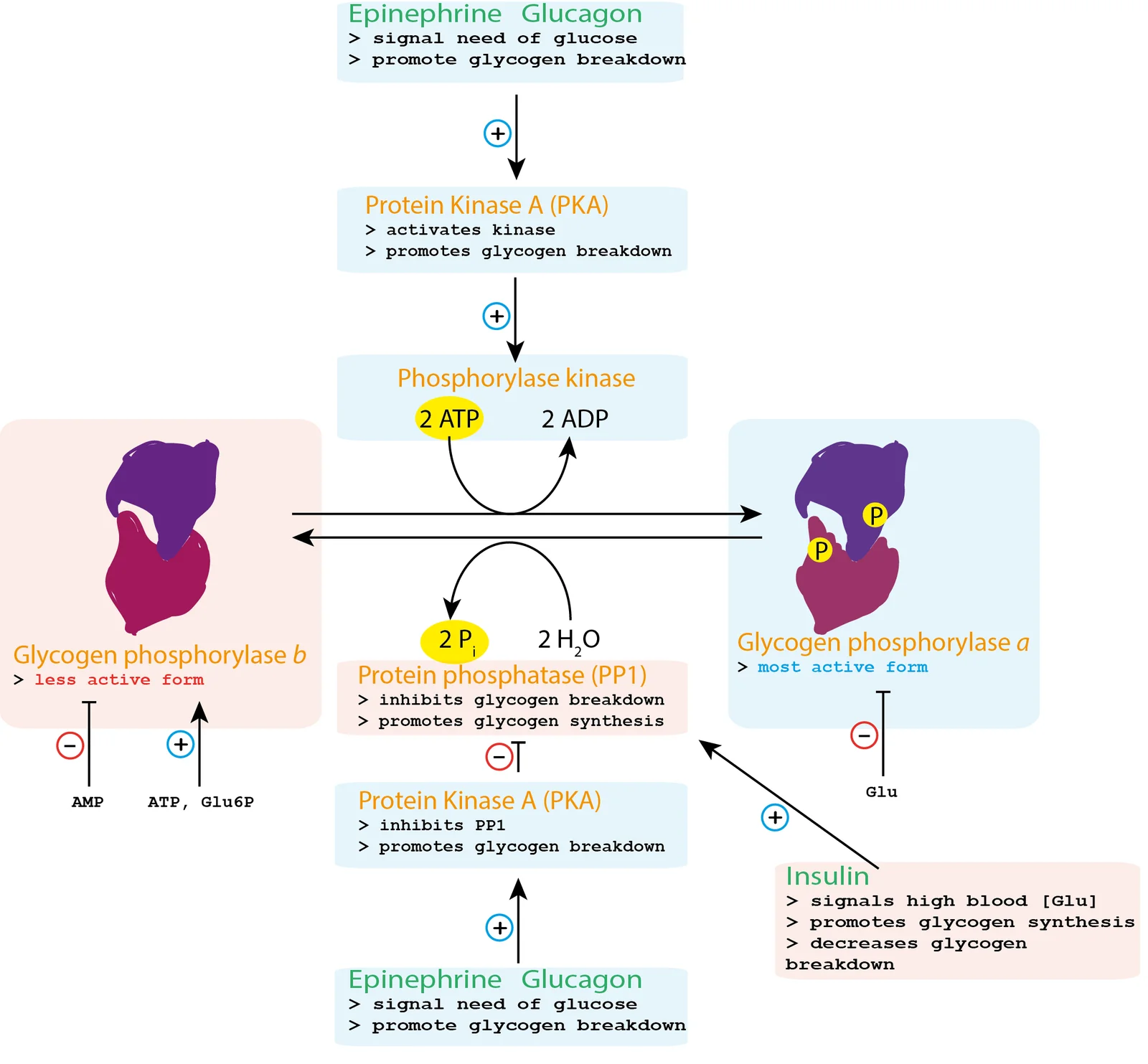
Long description
Control of glycogen metabolism involves allosteric regulation and enzyme-catalyzed modification of glycogen phosphorylase and glycogen synthase. The covalent modifications are under hormonal control.

Glycogen serves as the primary storage form of glucose within mammalian cells, holding a vital role in sustaining energy supply and maintaining systemic glucose homeostasis. The discovery of glycogen in the liver dates back to 1857, credited to Claude Bernard. While the general biochemical mechanisms governing glycogen synthesis and degradation remain uniform across all tissues, the precise regulatory controls that dictate flux through these pathways show marked tissue-specific differences. Glycogen is predominantly stored in two primary physiological depots: the liver and skeletal muscle. Smaller amounts have been identified in extrahepatic and extramuscular sites such as the brain, heart, kidney, adipose tissue, and erythrocytes, although the physiological functions of glycogen within these secondary locations remain largely undetermined. [1] [2]
The two main glycogen depots perform distinct physiological roles tailored to the demands of their surrounding tissues. Liver glycogen is mobilized primarily to supply free glucose to the bloodstream during periods of fasting, leading to a measurable decrease in hepatic glycogen content as fasting duration extends. Conversely, skeletal muscle glycogen acts as an endogenous fuel reserve, providing glucose locally to muscle fibers to sustain energy requirements during muscle contraction. Consequently, muscle glycogen content diminishes following active exercise within working muscle groups. Unlike the liver, short periods of fasting do not provoke a major decrease in muscle glycogen content. [1]
Quantitative distribution heavily favors skeletal muscle, which holds roughly 80 percent of the total bodily glycogen. This preponderance occurs because skeletal muscle accounts for 40 to 50 percent of total body weight, with a typical tissue glycogen concentration ranging from 80 to 150 mmol per kg. Structurally, glycogen is assembled into large macromolecular granules. Each individual glycogen granule contains approximately 30,000 glucose residues, arranged with numerous non-reducing ends that permit rapid enzymatic access for both glucose mobilization and polymer extension. Within skeletal muscle architecture, glycogen is not distributed uniformly; up to 90 percent of an incoming glucose load is converted into glycogen, and storage levels are consistently higher in type II fast-twitch fibers compared with type I slow-twitch fibers. Electron microscopy reveals distinct subcellular pools of muscle glycogen, categorized by their precise micro-anatomical locations: subsarcolemmal glycogen, intermyofibrillar glycogen situated close to the sarcoplasmic reticulum and mitochondria, and intramyofibrillar glycogen. [2]
Molecular disruptions in the genes encoding enzymes responsible for glycogen metabolism can precipitate inherited metabolic disorders known as glycogen storage diseases (GSDs). These conditions arise by interfering either directly with glycogen synthesis or with glycogen degradation pathways. Furthermore, specific mutations occurring in genes that code for enzymes of the glycolytic pathway are also classified under the broader umbrella of glycogen storage diseases. Overall, glycogen storage diseases are systematically classified according to their specific individual enzyme deficiencies, where each missing or dysfunctional enzyme normally regulates either the synthetic or degradative arm of glycogen metabolism. These enzymatic defects primarily impact target organ systems including the liver, skeletal muscle, the heart, the central nervous system, and the kidneys. [1] [2]
DefinitionGlycogen storage diseases
The construction and disassembly of glycogen require coordinated enzymatic machinery operating through distinct chemical steps. Glycogen synthesis relies on the formation of active sugar nucleotides and the subsequent action of glycogen synthase, an enzyme that exists in two major mammalian isoforms encoded by separate genes. Glycogen synthase 1, designated as GYS1, has a molecular mass of 84 kDa and consists of 737 amino acids, being expressed predominantly in skeletal muscle alongside other tissues. Glycogen synthase 2, designated as GYS2, has a molecular mass of 81 kDa and consists of 703 amino acids, being expressed predominantly in the liver. The hepatic isoform GYS2 shares about 70 percent amino acid sequence identity with the muscle isoform GYS1, and features several critical phosphorylation sites located near its N-terminus and C-terminus. Deficiencies in GYS1 and GYS2 result in muscle and liver glycogen storage disease type 0, respectively. Clinical manifestations linked to these specific enzyme deficiencies encompass an inability to properly form glycogen, profound muscle weakness, cardiac arrhythmia, sudden death, and recurrent hypoglycemia. [2]
Glycogen synthase cycles between two interconvertible conformational and functional states: a phosphorylated form known as glycogen synthase b, which exhibits lower activity, and a dephosphorylated form known as glycogen synthase a, which exhibits high catalytic activity. Reversible phosphorylation and dephosphorylation represent a major determinant in modulating overall enzyme activity. Phosphorylation is driven by specific protein kinases that lead to enzyme inactivation, whereas dephosphorylation is catalyzed by specialized phosphatases that result in enzyme activation. In addition to covalent modification, the catalytic activity of glycogen synthase is allosterically stimulated by the presence of glucose 6-phosphate and inhibited by increased intracellular concentrations of free glycogen. [1] [2]
Glycogen Synthase Regulation
- Glycogen Synthase bPhosphorylated, inactive or less active state.
- Question: Dephosphorylation by phosphatase?
Yes
- Glycogen Synthase aDephosphorylated, active state, stimulated by glucose 6-phosphate.
No
- Stays inactiveInhibited by high local glycogen concentration.
The degradative pathway relies heavily on glycogen phosphorylase, the key enzyme responsible for releasing glucose 1-phosphate residues directly from the linear alpha-1,4-linked chains of the glycogen polymer. The activity of glycogen phosphorylase is meticulously controlled through a dual mechanism involving allosteric effectors as well as reversible phosphorylation and dephosphorylation. Intracellular metabolites such as glucose 6-phosphate and UDP-glucose function as allosteric inhibitors of the enzyme. [1]
Cellular targeting subunits play an essential auxiliary role in organizing these pathways. Overexpression of the protein target to glycogen subunit, known as PPP1R3C, in liver or fat cells leads to a statistically significant increase in intracellular glycogen levels. The three hepatic glycogen-targeting subunits are notably smaller than the muscle-type PPP1R3A subunit, a structural divergence caused by the complete lack of a domain required for interaction with the endoplasmic reticulum in the hepatic variants. These glycogen-targeting subunits exert their glycogenic effects through three distinct mechanisms: first, they promote the dephosphorylation and consequent inactivation of glycogen phosphorylase alongside the activation of glycogen synthase; second, they enhance the physical targeting and localization of both glycogen synthase and glycogen phosphorylase to the solid glycogen particle; and third, they stabilize the glycogen synthase protein itself against premature degradation. [2]
| Enzyme / Factor | Isoform / Gene | Primary Location | Regulatory Mechanisms |
|---|---|---|---|
| Glycogen Synthase | GYS1 | Skeletal muscle | Activated by dephosphorylation and glucose 6-phosphate; inhibited by phosphorylation and high glycogen |
| Glycogen Synthase | GYS2 | Liver | Approximately 70 percent identical to GYS1; regulated by N- and C-terminal phosphorylation sites |
| Glycogen Phosphorylase | Muscle / Liver isoenzymes | Muscle and liver | Controlled by allosteric effectors (AMP, glucose 6-phosphate, UDP-glucose) and covalent phosphorylation |
| Targeting Subunits | PPP1R3A / PPP1R3C | Muscle and liver | Directs enzymes to glycogen particles, modulates phosphorylation status, and stabilizes synthase protein |
Effective glycogen metabolism is exceptionally important during physiological states characterized by high energy demand, such as periods of fasting and active muscle contraction. Because liver and skeletal muscle depots carry out disparate physiological functions, the regulatory mechanisms that modulate the activity of key enzymes like glycogen synthase differ partially between these two tissues. Glycogen synthase activity is tightly coordinated in tandem with glycogen phosphorylase activity to ensure that the cellular machinery achieves either net glycogen synthesis or net glycogen degradation in response to prevailing systemic signals. [1]
The isoenzymes of glycogen phosphorylase found in the liver versus skeletal muscle differ noticeably in their specific responsiveness to regulatory control elements. Muscle glycogen phosphorylase is strongly activated by adenosine monophosphate (AMP), signaling an acute cellular energy deficit, as well as by an increase in local glycogen concentration within the muscle fiber. Beyond hormonal and allosteric controls, muscle glycogen content itself acts as an independent physiological regulator that influences ongoing glycogen synthesis in healthy humans. Specifically, a high internal glycogen concentration exerts a strong negative feedback inhibition on the synthesis of fresh glycogen in skeletal muscle. [1]
Dietary carbohydrate intake alters these stores in a highly tissue-specific manner. In periods of muscle rest, neither routine fasting nor standard food ingestion alters glycogen stores within skeletal muscle to any large degree. However, muscle glycogen content increases slightly following the acute intake of large amounts of carbohydrates. Following an oral glucose tolerance test, approximately 15 percent of the total ingested glucose load is successfully stored as skeletal muscle glycogen in healthy human subjects. Interestingly, prolonged intake of high amounts of carbohydrates does not increase baseline glycogen content in skeletal muscle among untrained subjects. Furthermore, glycogen levels in completely inactive muscles remain entirely unmodified by high carbohydrate delivery. Skeletal muscle biopsies harvested from healthy subjects demonstrate that while glycogen concentration increases only slightly in resting or inactive muscle, previously working muscles augment their glycogen content markedly when carbohydrate-rich food is ingested following exercise. [1]
Clinical pearlExercise priming
Effective metabolic integration ensures that hormonal cascades rapidly mobilize hepatic glucose when systemic levels fall. Hepatic glycogenolysis is stimulated promptly by glucagon and epinephrine during fasting or stress, securing adequate blood glucose for the brain and erythrocytes, whereas muscular glycogenolysis is tailored directly to local work rate through calcium fluxes and AMP accumulation.
Deficit of the enzymes involved in glycogen metabolism causes various glycogenoses. These inherited metabolic conditions disrupt normal synthesis or degradation pathways, leading to abnormal tissue accumulation of glycogen or structurally atypical polysaccharides. A prominent example is glycogen storage disorder type IV (GSDIV), an autosomal recessive disorder associated with glycogen branching enzyme deficiency. This condition leads to an accumulation of insoluble polysaccharide particles and is a severe disorder with variable onset age and clinical severity. GSDIV includes a classical hepatic form in neonates and children that progresses to cirrhosis, also known as Andersen disease, as well as a neuromuscular form classified into four subtypes: perinatal, congenital, juvenile and adult onset. Additionally, late-onset adult polyglucosan body disease represents a neurological variant affecting mainly the Ashkenazi Jewish population. [1] [2]
GP defects in Hers disease patients are associated with hepatomegaly, growth retardation, ketotic hypoglycaemia and post-prandial lactic acidosis [2]
DefinitionGlycogen storage diseases
A group of inherited metabolic disorders caused by enzymatic defects in glycogen synthesis or degradation, leading to organ-specific accumulation of glycogen or aberrant polymers.
| Disorder | Subtype / Form | Enzyme Defect | Key Clinical Features |
|---|---|---|---|
| GSD III (Cori disease) | Type 3A | Debranching enzyme (liver and muscle) | Hepatomegaly, hypoglycaemia, short stature, myopathy [2] |
| GSD III | Type 3B | Debranching enzyme (liver only) | Hepatic involvement without muscle enzyme deficiency |
| GSD IV (Andersen disease) | Hepatic form | Branching enzyme | Neonatal or childhood onset, progressive cirrhosis |
| Hers disease | Hepatic | Glycogen phosphorylase | Hepatomegaly, growth retardation, ketotic hypoglycaemia |
Glycogen degradation takes place both in the cytoplasm and inside the lysosomes. In the cytosol, glycogen breakdown is accomplished by the coordinated action of two enzymes: glycogen phosphorylase, which releases glucose 1-phosphate by untangling the alpha-1,4-glycosidic linkages, and glycogen debranching enzyme, which unfastens the branch points, releasing free glucose. Glycogen phosphorylase serves as the rate-limiting enzyme of glycogenolysis. It cleaves the alpha-1,4 linkage to remove glucose residues from the chain as glucose 1-phosphate. However, the action of glycogen phosphorylase is blocked when it reaches four glucose residues from the glycogen branch point, a structure known as the limit dextrin. Complete glycogen degradation therefore requires the activity of the bifunctional glycogen debranching enzyme. Using its transferase activity, the debranching enzyme transfers three glucose residues to an adjacent branch of the glycogen chain. Subsequently, through its glucosidase activity, it cleaves the alpha-1,6 linkage to release a free glucose molecule from the branch point. Glucose 1-phosphate derived from glycogen in the cytosol may be isomerized into glucose 6-phosphate, which is then dephosphorylated to free glucose by glucose 6-phosphatase in order for glucose to leave the cell via glucose transporters. In the lysosomes, glycogen breakdown is accomplished by the lysosomal enzyme acid alpha-glucosidase, also known as acid maltase. [1] [2]
Glycogen synthesis occurs in the cytosol using extracellular glucose transported into cells via glucose transporters or via an indirect pathway utilizing lactate and amino acids. The initial step involves the autoglucosylation of the core protein glycogenin, which provides an oligosaccharide primer. Glycogen synthase then elongates the glucose chain by attaching uridine diphosphate (UDP)-glucose units through alpha-1,4 glycosidic linkages. When the elongating chain reaches around 12 glucose units, the glycogen branching enzyme transfers a chain of 6 to 8 units, or a chain of 7 units, to an adjacent chain via an alpha-1,6 linkage, thereby creating a multi-branched polymer. Glycogen synthase continues to elongate the main chain while the branching enzyme produces new branches. Enzyme regulation is tightly controlled; protein phosphatase 1 (PP1) acts as a central regulator, activated by glycogen targeting subunits of the PPP1R3 family that recruit it to glycogen via a conserved RVxF motif. PP1 reciprocally activates glycogen synthase by dephosphorylation while simultaneously inactivating glycogen phosphorylase. Similarly, insulin inhibits glycogen phosphorylase while inducing simultaneous activation of glycogen synthase to promote hepatic glycogen synthesis. [1] [2]
Clinical pearlBranching and debranching limits
Glycogen phosphorylase halts four residues away from an alpha-1,6 branch point, necessitating the transferase and glucosidase actions of the debranching enzyme for complete breakdown.
Cytosolic Glycogen Pathway Outline
- Glucose uptake / PrimingExtracellular glucose enters via transporters; glycogenin provides an oligosaccharide primer.
- Question: Synthesis or Breakdown?
Synthesis
- Glycogen SynthaseElongates chains via alpha-1,4 linkages using UDP-glucose.
- Branching EnzymeTransfers 6-8 glucose units to form alpha-1,6 branch points.
Breakdown
- Glycogen PhosphorylaseRate-limiting step releasing glucose 1-phosphate until four residues remain from the branch.
- Debranching EnzymeUses transferase and glucosidase activities to clear branch points and release free glucose.
Glycogen metabolism serves as a vital energy reservoir, balancing rapid glucose demands with systemic metabolic homeostasis. In human tissues, glycogen is a branched polymer of glucose residues linked primarily by alpha-1,4-glycosidic bonds, with alpha-1,6-glycosidic bonds forming branch points. The initiation of glycogen synthesis requires glycogenin, a self-glucosylating protein primer. In humans, glycogenin exists in two main isoforms: GYG1, which has a mass of 39 kilodaltons and comprises 350 amino acids, and is expressed predominantly in skeletal muscle; and GYG2, which has a mass of 55 kilodaltons and comprises 501 amino acids, and is expressed in the liver, heart, and pancreas. Glycogenin contains a conserved domain of approximately 250 amino acids at its amino-terminus that is required for uridine diphosphate glucose binding and catalysis, alongside a highly conserved domain of 30 to 35 amino acids at its carboxy-terminus required for glycogen synthase binding. [2]
Elongation and branching of the glucose polymer are executed by glycogen synthase and branching enzyme. The regulation of these enzymes is tightly controlled by phosphorylation cascades and targeting subunits. At least seven genes encode glycogen targeting subunits that direct protein phosphatase 1 to glycogen particles. These include PPP1R3A encoding the muscle-specific subunit (124 kilodaltons), PPP1R3B encoding the liver and muscle subunit (33 kilodaltons), and the protein target to glycogen (PTG or Gc/R5/PPP1R3C, 36 kilodaltons) which is highly expressed in skeletal and cardiac muscle and the liver. Additional subunits include GD/R6 PPP1R3D (33 kilodaltons) and GE PPP1R3E (31 kilodaltons) expressed most in skeletal muscle and heart, GF PPP1R3F (79 kilodaltons) expressed at high levels in the brain, and GG PPP1R3G (38 kilodaltons) expressed at high levels in the liver. The amino-terminal region of the glycogen targeting subunit binds protein phosphatase 1, the central region binds glycogen, and the carboxy-terminal region binds glycogen phosphorylase, glycogen synthase, and phosphorylase kinase. [1] [2]
Pathological disruption of these pathways results in glycogen storage diseases. For instance, glycogenin 1 deficiency causes a muscle glycogen storage disease known as type XV. Furthermore, mutations causing impaired interaction of glycogenin-1 with glycogen synthase or deficiencies in glycogenin-1 lead to a distinct muscle glycogen disorder characterised by the accumulation of polyglucosan bodies. Glycogen storage diseases also affect the central nervous system. Laforin disease, resulting from rare mutations in malin and laforin, increases the rate of glycogen accumulation in neurons during aging. Laforin acts as a phosphatase of glycogen synthase and binds glycogen directly through a carbohydrate-binding domain, whereas malin is an E3-ubiquitin ligase recruited to substrates via interaction with laforin. Together, laforin and malin form a complex that, alongside glycogenolytic enzymes such as glycogen debranching enzyme 1 and the brain isoform of glycogen phosphorylase, orchestrates the degradation of polyglucosan bodies. [2]
DefinitionGlycogen targeting subunits
Management strategies for hepatic glycogen storage diseases focus on maintaining euglycemia and preventing secondary metabolic complications through tailored dietary regimens. For patients with hepatic glycogen storage diseases, dietary management requires frequent feeds, typically every three to four hours during the day and night, using oral uncooked cornstarch or continuous nocturnal nasogastric or gastrostomy tube glucose infusion. A meta-analysis indicates that overnight intermittent administration of uncooked cornstarch prevents nocturnal hypoglycemia in glycogen storage disease type Ia children more effectively than continuous nocturnal feeding of dextrose. Specialized extended-release formulations, such as waxy maize extended-release cornstarch administered at bedtime to preserve overnight glucose levels, have been shown to improve the quality of life of patients with glycogen storage disease type I while maintaining adequate metabolic control. Dietary restrictions are vital in glycogen storage disease type I, requiring the strict avoidance of sucrose, fructose, and galactose to prevent untoward intracellular metabolite accumulation. [4]
Hepatic GSD Management
- Assess hepatic GSDEvaluate baseline metabolic control and fasting tolerance.
- Question: Fasting stability?
Compensated
- Prescribe frequent feeds every 3 to 4 hours with standard uncooked cornstarch.
Unstable or nocturnal drops
- Initiate continuous nocturnal nasogastric or gastrostomy tube glucose infusion or waxy maize extended-release cornstarch.
For glycogen storage disease type Ib specifically, specialized treatment addresses neutropenia and associated gastrointestinal inflammation. Granulocyte colony-stimulating factor is utilised for severe neutropenia to reduce infection risk, while sodium-glucose cotransporter-2 inhibitors, such as empagliflozin, are employed to manage neutropenia and associated inflammatory bowel disease manifestations by clearing toxic 1,5-anhydroglucitol-6-phosphate. Enzyme replacement therapy is available for Pompe disease, utilising recombinant human acid alpha-glucosidase, specifically alglucosidase alfa or avalglucosidase alfa, to supplement deficient lysosomal enzyme activity. [4]
Management of glycogen storage disease type V centers on exercise modification, requiring pre-exercise simple carbohydrate intake, moderate aerobic exercise, and the avoidance of strenuous anaerobic exertion that precipitates acute rhabdomyolysis and myoglobinuria. Organ transplantation indications include liver transplantation for recurrent hepatic adenomas or uncontrollable metabolic derangement, and cardiac transplantation for severe cardiomyopathy refractory to enzyme replacement therapy in infantile Pompe disease.
Clinical pearlDietary rule in GSD I
A major pitfall in managing disorders of glycogen metabolism is assuming uniform tissue responses to pharmacological and metabolic interventions. Glycogen kinetics and storage capacities differ profoundly across organs. The average glycogen content in the adult brain is around 3 to 12 micromoles, which is much less compared to the substantial reserves found in the liver and skeletal muscle. Despite this lower concentration, brain glycogen is indispensable for supporting synaptic activit [2]
Another clinical pitfall is overlooking the intrinsic metabolic activity of neurons. Although early paradigms assumed astrocytes exclusively supported neurons via the glycogen shunt, it is established that neurons maintain an active glycogen metabolism that contributes directly to hypoxia tolerance. Neurons continuously synthesise and degrade small amounts of this polysaccharide, using it not as a long-term energy store but as a rapid, small, and constant source of energy. Glycogen metabolism in neurons generates 3 molecules of adenosine triphosphate, whereas glycolysis from free glucose yields 2 molecules of adenosine triphosphate. Furthermore, neurotransmitters and neuromodulators stimulate the mobilisation of astrocytic glycogen stores, converting them to lactate for neuronal utilisation. [2]
Misinterpreting the systemic effects of therapeutic agents on glycogen stores can also lead to adverse outcomes. Pharmacological agents used in neuropsychiatric and oncological practice frequently perturb glycogen homeostasis. Lithium chloride inhibits glycogen synthesis and decreases glycogen content in cortical cultured astrocytes and rat liver, yet it paradoxically stimulates glycogen synthesis and glycogen synthase activity in the rat diaphragm and salivary glands. Valproate, a histone deacetylase inhibitor, increases the brain isoform of glycogen phosphorylase and decreases pathological glycogen accumulation in the skeletal muscle of patients with McArdle disease. Clinicians must recognise these divergent tissue-specific responses when managing patients with underlying metabolic myopathies or central nervous system disorders.
Clinical pearlEnergy yield
Glycogen Pathway Assessment
- Evaluate presenting symptomsExamine for muscle fatigue, exercise intolerance, or neurological manifestations.
- Question: Is tissue involvement primarily muscular?
Yes
- Assess for myophosphorylase or glycogenin-1 defectsConsider McArdle disease or glycogen storage disease type XV.
No
- Question: Are polyglucosan bodies present?
Yes
- Investigate Laforin-malin complex mutationsEvaluate for progressive myoclonus epilepsy and Lafora disease pathways.
No
- Review central nervous system and hepatic energy fluxesAssess astrocyte-neuron lactate shuttling and systemic metabolic stressors.
In clinical and investigative practice, understanding glycogen metabolism is essential for managing exercise physiology, oncology, and metabolic disorders. Glucose released from glycogen serves as a major energy source for contracting muscles, and high-intensity physical exercise depletes glycogen stores in active skeletal muscle. Skeletal muscle biopsies from healthy volunteers demonstrate that glycogen content is markedly reduced following cycling exercise in working muscles, whereas glycogen levels in inactive muscles remain unchanged. The endurance capacity of skeletal muscle is directly associated with muscle glycogen content, and fatigue develops when glycogen storage is exhausted in active muscles. Strategies to spare muscle glycogen include dietary interventions, administration of fructose—which has been reported to be comparable to or better than glucose or placebo—and maintaining elevated concentrations of free fatty acids in plasma during exercise.
Beyond exercise physiology, glycogen metabolism is increasingly targeted in oncology. Metabolic reprogramming is a hallmark of cancer cells, contributing to tumour microenvironment adaptation and resistance to anticancer therapies. Glycogen metabolism is a recognised feature of cancer cells because it is upregulated in many tumour types, indicating a critical role in cancer cell pathophysiology under stress conditions such as hypoxia, glucose deprivation, and anticancer treatment. Diagnostic evaluation involves detecting glycogen in tumours in vivo, and therapeutic strategies increasingly focus on pharmacological modulators of glycogen metabolism as components of combinational cancer therapy.
Therapeutic exploration of glycogen-modulating drugs bridges neurology and oncology. Lithium is under clinical evaluation for prostate cancer (clinicaltrials.gov identifier NCT02198859), while valproate is being evaluated in clinical trials for advanced cancers in combination with temsirolimus and bevacizumab (clinicaltrials.gov identifier NCT01552434). Recognising how these therapeutic agents interact with glycogen synthase, glycogen phosphorylase, and regulatory phosphatases allows clinicians to anticipate metabolic side effects and harness metabolic pathways for novel therapeutic gains. [2]
Exam trapExercise exhaustion
Among the numerous classifications, specific subtypes dominate clinical presentations. Glycogen storage disease type I, type II, type III, and type IX represent the most common forms encountered in clinical practice. Glycogen storage disease type Ia accounts for approximately 80 percent of patients with glycogen storage disease type I. [4]
Clinical pearlSubtype proportions
GSD Ia constitutes about 80% of all GSD I cases, making molecular diagnosis of the G6PC gene a high-yield initial test for these individuals.
Significant regional variations and ethnic predilections alter the baseline epidemiological landscape across populations. Furthermore, glycogen storage disease type III presents with different anatomical distributions: most patients ex
| Condition | Common Subtype Breakdown / Proportion | Ethnic or Regional Predilection |
|---|---|---|
| GSD I | Type Ia represents approximately 80% of patients | High prevalence of GSD Ia in Ashkenazi Jewish population |
| Significant regional variations and ethnic predilections alter the baseline epidemiological landscape across populations. Furthermore, glycogen storage disease type III presents with different anatomical distributions: most patients ex |
The underlying aetiology of glycogen storage diseases involves inherited molecular changes in genes encoding enzymes or transporter proteins that regulate glycogen synthesis, transport, or degradation. The vast majority of these conditions follow an autosomal recessive inheritance pattern, although specific subtypes diverge. [4]
DefinitionInheritance divergence
While most glycogen storage disorders are autosomal recessive, specific isoforms like GSD IXa follow an X-linked recessive mode of inheritance.
Classical clinical phenotypes map directly to specific gene loci and chromosomal locations. Pompe disease, or glycogen storage disease type II, is an autosomal recessive disorder caused by congenital deficiency of functional acid alpha-glucosidase d
Additional classical disruptions include glycogen storage disease type VI (Hers disease), an autosomal recessive disorder caused by loss of liver glycogen phosphorylase activity owing to mutations in the PYGL gene on chromosome 14q21–22. Phosphorylase kinase deficiencies involve multiple subunit genes: mutations in PHKA1 cause skeletal muscle phosphorylase kinase deficiency (GSD VIII), mutations in the PHKA2 gene that encodes the liver isoform of the α-subunit of PHK cause phosphorylase kinase
Classical clinical phenotypes map directly to specific gene loci and chromosomal locations. Glycogen storage disease type Ia arises from mutations in the G6PC gene encoding glucose 6-phosphatase-1, while glycogen storage disease type Ib is caused by mutations in the SLC37A4 (also known as G6PT) gene encoding glucose 6-phosphate translocase. Pompe disease, or glycogen storage disease type II, is an autosomal recessive disorder caused by congenital deficiency of functional acid alpha-glucosidase
Glycogen storage diseases are systematically classified into hepatic and muscle or myopathic types based on the predominant tissue affected and the clinical manifestations. The classical spectrum comprises a detailed numbering system spanning GSD 0, GSD I with subtypes Ia and Ib, GSD II or Pompe disease divided into infantile-onset and late-onset forms, GSD III with subtypes IIIa and IIIb, GSD IV, GSD V or McArdle disease, GSD VI, GSD VII or Tarui disease, GSD IX, and GSD XI.
Classical clinical phenotypes map directly to specific gene loci and chromosomal locations. Glycogen storage disease type Ia arises from mutations in the G6PC gene encoding glucose 6-phosphatase-1, while glycogen storage disease type Ib is caused by mutations in the SLC37A4 (also known as G6PT) gene encoding glucose 6-phosphate translocase. Pompe disease, or glycogen storage disease type II, is an autosomal recessive disorder caused by congenital deficiency of functional acid alpha-glucosidase d
DefinitionHepatic versus Muscle GSDs
Hepatic glycogen storage disorders primarily impair glucose homeostasis leading to fasting hypoglycemia, whereas myopathic GSDs disrupt energy provision during contraction, causing exercise intolerance, cramps, and rhabdomyolysis.
| GSD Type | Affected Enzyme / Gene | Primary Tissues | Key Clinical Hallmarks |
|---|---|---|---|
| GSD 0 (Muscle) | Glycogen synthase (GYS1) | Skeletal muscle, heart | Myopathy, hypertrophic cardiomyopathy, long QT |
| GSD I (Ia / Ib) | Glucose-6-phosphatase complex | Liver, kidneys, neutrophils | Severe fasting hypoglycemia, massive hepatomegaly, lactic acidosis |
| GSD II (Pompe) | Acid alpha-glucosidase | Skeletal muscle, heart | Floppy baby, hypertrophic cardiomyopathy, respiratory failure |
| GSD III (Cori) | Glycogen debranching enzyme (AGL) | Liver, skeletal muscle, heart | Muscle weakness, reduced exercise capacity, cardiomyopathy |
| GSD V (McArdle) | Glycogen phosphorylase (PYGM) | Skeletal muscle | Exercise-induced cramps, second-wind phenomenon, myoglobinuria |
| GSD VII (Tarui) | Phosphofructokinase | Skeletal muscle, erythrocytes | Exercise intolerance, hemolytic anemia, hypertrophic cardiomyopathy |
Clinical presentations vary widely depending on whether hepatic or muscle depots are compromised. GSD I, or von Gierke disease, presents with severe fasting hypoglycemia, doll-like facies, massive hepatomegaly, lactic acidosis, hyperlipidemia, and hyperuricemia, with neutropenia characteristically manifesting in subtype Ib.
Infantile-onset GSD II, known as Pompe disease, presents as a floppy baby characterized by profound hypotonia, hypertrophic cardiomyopathy, macroglossia, and progressive respiratory failure. Late-onset Pompe disease typically manifests as a progressive proximal limb-girdle myopathy.
Muscle-specific GSDs share overlapping acute manifestations. Exercise intolerance with cramps and episodes of rhabdomyolysis and myoglobinuria usually occur in myopathic GSDs. In McArdle disease (GSD V), diminished glycogen phosphorylase activity limits glucose availability at the initiation of muscle contraction, inducing fatigue, cramps, and a rise in heart rate out of proportion to the workload. After a few minutes, fatigue and heart rate spontaneously subside as patients experience the second wind phenomenon, driven by active muscle utilization of blood-borne glucose, lactate, free fatty acids, and branched-chain amino acids. [4]
GSD III patients show reduced exercise capacity and lower skeletal muscle glucose oxidation alongside higher fatty acid oxidation compared to healthy individuals. Deficiency of AGL in GSD III produces a cardiomyopathy that echocardiographically mimics idiopathic hypertrophic cardiomyopathy. In GSD VII (phosphofructokinase deficiency), clinical features include exercise intolerance associated with hemolytic anemia and possible hypertrophic cardiomyopathy, with muscle biopsies revealing excessive glycogen storage. [4]
Special clinical scenarios require tailored management. Patients with GSD III may require frequent meals high in carbohydrate supplemented with uncooked cornstarch, or sometimes a ketogenic diet, to avoid hypoglycemia, while fructose ingestion can improve their exercise tolerance. Similarly, a ketogenic diet has achieved clinical improvement in phosphofructokinase deficiency. Regular exercise training programs enhance the ability of muscle to oxidize fatty acids and reduce reliance on glycogen
Clinical pearlThe Second Wind Phenomenon
In McArdle disease, initial exercise-induced tachycardia and cramps subside after a few minutes because working muscles successfully switch to burning alternative systemic substrates like free fatty acids and blood-borne glucose.
Evaluation of Exercise Intolerance in Myopathic GSDs
- Presenting symptomExercise intolerance, cramps, or myoglobinuria
- Question: Is there a second-wind phenomenon?
Yes
- Consider GSD V (McArdle disease)Defective muscle glycogen phosphorylase
No
- Question: Associated features present?
Hemolytic anemia
- Consider GSD VII (Tarui disease)Phosphofructokinase deficiency
Fixed weakness or cardiomyopathy
- Consider GSD III or GSD II (Pompe)Debrancher or acid alpha-glucosidase defect
When evaluating neonates or infants presenting with hepatomegaly and hypoglycemia, clinicians must consider a broad differential diagnosis that extends beyond hepatic glycogen storage diseases. Fatty acid oxidation disorders present with hypoketotic hypoglycemia and hepatomegaly, which can closely mimic hepatic glycogen storage diseases, and certain hepatic glycogen storage conditions like glycogen debranching enzyme deficiency may clinically mimic fatty acid oxidation disorders with altered plasma concentrations of medium-chain fatty acids [P730]
In the evaluation of metabolic myopathies, conditions such as carnitine palmitoyltransferase II deficiency and mitochondrial myopathies present with exercise intolerance, muscle cramps, and rhabdomyolysis, overlapping significantly with muscle glycogen storage diseases like McArdle disease. For infants exhibiting hypertrophic cardiomyopathy and profound hypotonia, the differential diagnosis must distinguish Pompe disease from spinal muscular atrophy, which causes severe hypotonia without cardiomyopathy, and Danon disease, an X-linked glycogenosis that also presents with skeletal myopathy and marked cardiac involvement. Congenital deficiency of PGM1 should be designated glycogenosis type XIV and can present with fasting intolerance, hypoglycemia, rhabdomyolysis, hepatopathy, dilated cardiomyopathy, and cardiac arrest. Furthermore, hepatic glycogen synthase deficiency due to GYS2 mutations leads to postprandial hyperglycemia, glucosuria, and hyperlactatemia, along with fasting hypoglycemia and ketone formation, requiring differentiation from other defects in carbohydrate homeostasis. [4]
| Disorder | Primary features | Distinguishing laboratory findings |
|---|---|---|
| Fatty acid oxidation disorders | Hepatomegaly, hypoglycemia | Hypoketotic hypoglycemia, elevated C8 and C10 |
| Pompe disease | Hypotonia, hypertrophic cardiomyopathy | Massive LVH, shortened PR interval, vacuolar myopathy |
| GSD type 0 (GYS2) | Postprandial hyperglycemia, fasting hypoglycemia | Hyperlactatemia, ketosis, low hepatic glycogen |
| PGM1 deficiency (GSD XIV) | Exercise intolerance, myopathy, cardiac involvement | Basal elevated creatine kinase, normal lactate on exercise test |
Establishing the definitive diagnosis of glycogen storage diseases requires a combination of clinical evaluation, biochemical testing, and advanced molecular or enzymatic analysis. For major glycogen storage disease types, definitive molecular criteria involve identifying pathogenic mutations in the specific genes encoding the relevant metabolic enzymes or transporters using comprehensive panel testing or sequencing. Enzymatic criteria require demonstrating deficient activity of the specific enzyme in affected tissues, which traditionally involves a tissue biopsy such as liver or skeletal muscle, though leukocyte or fibroblast assays are useful for specific conditions.
DefinitionDiagnostic standard
For glycogen storage disease type I, diagnosis is confirmed by identifying mutations in the glucose-6-phosphatase catalytic subunit gene for type Ia or the glucose-6-phosphate transporter gene for type Ib, or by demonstrating impaired hepatic glucose-6-phosphatase activity on liver biopsy. In Pompe disease, also known as glycogen storage disease type II, diagnosis is established by demonstrating deficient acid alpha-glucosidase activity in lymphocytes, cultured fibroblasts, or skeletal muscle, corroborated by genetic testing of the GAA gene. For glycogen storage disease type V, or McArdle disease, diagnosis is confirmed by identifying pathogenic mutations in the muscle glycogen phosphorylase gene or by demonstrating absent phosphorylase activity on a skeletal muscle biopsy. Fanconi-Bickel disease, classified as glycogen storage disease type XI, is diagnosed by identifying mutations in the GLUT2 gene, which causes impaired glucose and galactose utilization alongside characteristic proximal tubular nephropathy features. [4]
| Disease Type | Defective Protein or Enzyme | Definitive Diagnostic Method |
|---|---|---|
| GSD I | Glucose-6-phosphatase / Transporter | Mutation analysis or liver enzyme assay |
| Pompe Disease | Acid alpha-glucosidase (GAA) | Leukocyte/fibroblast assay or GAA gene sequencing |
| GSD V | Muscle glycogen phosphorylase | PYGM gene mutation or muscle biopsy |
| GSD XI (Fanconi-Bickel) | GLUT2 transporter | GLUT2 gene mutation analysis |
Laboratory baseline testing for suspected glycogen storage diseases includes fasting glucose, lactate, uric acid, a lipid panel, creatine kinase, and an acylcarnitine profile. In GSD type Ia, caused by mutations in the G6PC1 gene encoding glucose 6-phosphatase-1, the inability to release glucose from the liver during fasting induces fasting hypoglycemia, while intracellular elevation of glucose 6-phosphate promotes alternative pathways leading to hypertriglyceridemia, lactic acidosis, and hyperuricemia. For suspected muscle glycogenoses such as GSD V, a forearm non-ischemic exercise test demonstrates a characteristic failure of lactate to rise alongside an exaggerated ammonia rise. Enzymatic assays performed in leukocytes, erythrocytes, or dried blood spots are critical for definitive diagnosis, such as the acid alpha-glucosidase assay used to confirm Pompe disease. Modern first-line confirmation increasingly relies on genetic testing utilizing targeted multi-gene next-generation sequencing panels. [4]
Tissue biopsies of the liver or muscle provide definitive histological insights. Histopathology typically reveals vacuolar degeneration and glycogen granules residing within lysosomes of muscle cells, while autophagic vacuoles are visible occasionally, and excess glycogen is also present in the capillary wall of muscle and skin. Periodic acid-Schiff staining highlights accumulated glycogen, and electron microscopy reveals ultrastructural storage patterns, though specific entities like aldolase A deficiency show negative PAS staining and no glycogen accumulation on electron microscopy. Cardiac evaluation via electrocardiogram and echocardiogram is mandatory when Pompe disease is suspected, frequently demonstrating marked PR interval shortening and massive left ventricular hypertrophy. Management adjustments such as frequent meals every four hours with uncooked cornstarch can ameliorate symptoms in conditions like GSD type 0. [4]
Diagnostic pathway for metabolic myopathies and hepatopathies
- Initial evaluationAssess fasting glucose, lactate, uric acid, lipids, and creatine kinase.
- Question: Clinical presentation?
Exercise intolerance
- Forearm non-ischemic exercise testLook for failure of lactate rise with exaggerated ammonia.
- Enzymatic and genetic panelsTargeted NGS panels for definitive molecular confirmation.
Infantile hypotonia and cardiomegaly
- Cardiac imagingCheck for PR shortening and massive LVH on ECG/echo.
- Enzyme assayPerform acid alpha-glucosidase assay in blood spots or leukocytes.
Glycogen storage diseases provoke extensive multi-organ manifestations that span hepatic, renal, metabolic, and myopathic domains. Hepatic complications are particularly prominent in hepatic glycogen storage disorders such as type I and type III disease, where chronic metabolic derangement drives the development of hepatic adenomas. These benign hepatocellular adenomas carry a substantial risk of malignant transformation to hepatocellular carcinoma, necessitating rigorous surveillance and monitoring for any suspicious radiological progression or concerning biochemical shifts. [3]
Renal complications are exceptionally characteristic of glycogen storage disease type I, manifesting through a constellation of structural and functional abnormalities. Affected individuals frequently experience glomerular hyperfiltration, microalbuminuria, and profound renal tubular dysfunction. Patients may exhibit proximal tubular defects resulting in urinary wasting phenomena, alongside nephrocalcinosis and the formation of calcium oxalate or uric acid kidney stones. Over time, these persistent insults can culminate in progressive chronic kidney disease and eventual chronic kidney failure.
Metabolic and systemic complications add further clinical complexity. Severe hypertriglyceridemia can precipitate acute pancreatitis, which represents a life-threatening surgical emergency requiring intensive management. Disturbed purine metabolism frequently leads to hyperuricemia and clinical gout or acute gouty arthritis. Furthermore, patients affected with glycogen storage disease type Ia often suffer from generalized osteoporosis or osteopenia, reflecting impaired bone mineral density regulation. [4]
Myopathic complications dominate muscle-specific glycogenoses such as glycogen storage disease type V, known as McArdle disease. In these myopathic disorders, physical exertion can trigger severe energy crises within skeletal muscle tissue, culminating in acute rhabdomyolysis. The massive breakdown of skeletal muscle releases intracellular myoglobin into the circulation, which can precipitate pigment-induced acute kidney injury if not promptly recognized and aggressively managed with intravenous hydration and supportive care.
Exam trapMalignant transformation
| System | Complication | Associated Subtypes |
|---|---|---|
| Hepatic | Hepatic adenomas and malignant transformation | GSD I and III |
| Renal | Hyperfiltration, microalbuminuria, CKD, stones | GSD I |
| Metabolic | Gout, acute pancreatitis, osteoporosis | GSD I and others |
| Myopathic | Acute rhabdomyolysis and myoglobinuria | GSD V |
The natural history, functional trajectory, and ultimate life expectancy in glycogen storage diseases vary dramatically across the different genetic subtypes, ranging from severe, life-threatening infantile phenotypes to entirely normal lifespans when optimal therapeutic management is established early. Understanding these prognostic trajectories is essential for guiding clinical counseling, setting expectations, and implementing timely life-prolonging interventions.
In infantile Pompe disease, which represents glycogen storage disease type II, the classical presentation without enzyme replacement therapy is characterized by profound generalized muscle weakness, massive hypertrophic cardiomyopathy, and rapid deterioration leading to death from cardiorespiratory failure within the first year of life. Conversely, the advent of targeted enzyme replacement therapy has fundamentally altered this grim natural history, dramatically improving survival and altering the developmental trajectory of affected infants when administered prior to irreversible organ damage.
For myopathic disorders such as well-managed McArdle disease (glycogen storage disease type V), patients typically experience a normal overall lifespan, provided they learn to recognize and respect their physical limitations and employ strategies such as the second-wind phenomenon to prevent severe exercise-induced episodes of acute rhabdomyolysis. Their primary prognostic challenge lies in avoiding catastrophic acute kidney injury resulting from severe myoglobinuria during unaccustomed exertion.
In hepatic glycogenoses, long-term outcomes are heavily dictated by the severity of metabolic control, the prevention and management of hepatic adenomas, and the stabilization of renal function. Individuals who maintain strict metabolic control can avoid many devastating systemic sequelae. However, those who develop advanced hepatic complications such as large adenomas with malignant transformation or progressive chronic kidney disease face significantly curtailed long-term survival unless definitive interventions, such as liver or kidney transplantation, are successfully undertaken.
Clinical pearlTherapeutic impact
Prognostic stratification and management pathways
- Assess GSD subtype and clinical presentationDifferentiate hepatic, myopathic, and lysosomal phenotypes.
- Question: Is enzyme replacement therapy available?
Yes (e.g. Pompe disease)
- Initiate ERT earlyPrevents rapid cardiopulmonary deterioration and alters natural history.
No (e.g. GSD I, III, V)
- Question: Is it a myopathic form like McArdle?
Yes
- Dietary and exercise counselingPrevents acute rhabdomyolysis and preserves normal lifespan.
No (Hepatic/Renal forms)
- Strict metabolic control and surveillanceMonitor for hepatic adenomas, malignant transformation, and progressive CKD.
Preventative strategies and screening protocols for glycogen storage diseases encompass a comprehensive framework combining early biochemical identification, carrier evaluation, and genetic risk assessment for high-risk families. Newborn screening protocols utilize advanced laboratory methodologies, specifically incorporating dried blood spot tandem mass spectrometry and fluorometry for specific conditions such as Pompe disease, enabling identification of affected neonates prior to the onset of irreversible organ damage. For families with a documented history of glycogen storage disorders, com
DefinitionNewborn Screening
Laboratory testing of dried blood spots using tandem mass spectrometry or fluorometry allows early detection of metabolic disorders before symptoms appear.
| Intervention | Method | Clinical Goal |
|---|---|---|
| Newborn Screening | Dried blood spot tandem mass spectrometry or fluorometry | Early detection of Pompe disease and other inherited disorders |
| Family Planning | Carrier testing, prenatal diagnosis, pre-implantation genetic diagnosis | Assessment and reduction of recurrence risks |
| Ultrasound Surveillance | Liver ultrasound every 6 months starting at age 50 years | Detection of hepatic malignancy when risk exceeds 1.5 percent |
Preventative Screening Pathway
- Newborn ScreeningPerform dried blood spot tandem mass spectrometry or fluorometry.
- Question: Positive Screening Result?
Yes
- Confirmatory TestingInitiate diagnostic workup and early multidisciplinary intervention.
No
- Routine CareStandard pediatric developmental and metabolic surveillance.
Long-term follow-up and surveillance protocols for patients with glycogen storage diseases require structured monitoring regimens and a coordinated multidisciplinary team to manage multisystem complications. Surveillance imaging is a cornerstone of hepatic management, requiring routine liver ultrasound or magnetic resonance imaging every 6 to 12 months for the detection of hepatic adenomas, which can develop starting in childhood or adolescence as seen in disorders like PHKG2 deficiency where hepatic adenomas have been occasionally reported. Monitoring protocols must also include continuous glucose monitoring to prevent dangerous metabolic troughs, regular assessment of urinary microalbumin and creatinine clearance to evaluate renal function, periodic measurement of serum alpha-fetoprotein as part of hepatic surveillance, and comprehensive fasting lipid panels to manage dyslipidemia. Managing these complex patients requires a dedicated multidisciplinary team comprising a metabolic geneticist, a specialized metabolic dietitian, a cardiologist, a nephrologist, and a physical therapist. The clinical spectrum of these disorders is broad; for example, the newborn usually presents with cardiomyopathy and profound muscular hypotonia leading to respiratory distress that requires mechanical ventilation. In early childhood, patients with glycogen branching enzyme deficiency usually develop liver dysfunction and progressive liver cirrhosis that may require liver transplantation, while some patients experience muscle weakness and dilated cardiomyopathy. Conversely, the late-onset variant of glycogen branching enzyme deficiency is known as adult polyglucosan body disease, a condition that affects predominantly the nervous system and is found mostly in individuals of Ashkenazi Jewish ancestry. Similarly, congenital deficiency of the liver isoform of the gamma-subunit of phosphorylase kinase causes an autosomal recessive disease with a variable clinical phenotype ranging from a mild disease characterized by a tendency to hypoglycemia, to liver fibrosis that may progress to liver cirrhosis during childhood. [4]
Clinical pearlMultidisciplinary Care
Long-term survival and quality of life depend on coordinated input from metabolic geneticists, dietitians, cardiologists, nephrologists, and physical therapists.
| Parameter | Surveillance Modality | Frequency |
|---|---|---|
| Hepatic Adenomas | Liver ultrasound or MRI | Every 6 to 12 months starting in childhood or adolescence |
| Metabolic Control | Continuous glucose monitoring and lipid panels | Ongoing regular intervals |
| Renal Complications | Urinary microalbumin and creatinine clearance | Periodic evaluation |
| Tumor Markers | Serum alpha-fetoprotein | Monitored alongside imaging |
Long-term Follow-up Workflow
- Multidisciplinary Review
Clinical pearlClassic signs
High-yield clinical associations define the glycogen storage diseases and allow immediate bedside recognition. Patients with GSD type I exhibit characteristic physical features including a doll-like face alongside profound metabolic derangements such as lactic acidosis, hyperuricemia, and hyperlipidemia. In contrast, infants with GSD type II present with massive cardiomegaly, profound generalized hypotonia, and early death if untreated. A vital classic sign in McArdle disease is the second-wind phenomenon, which is clinically explained by a compensatory metabolic shift to hepatic glucose mobilization and free fatty acids that bypasses the muscular glycolytic block.
Differentiating subtypes requires meticulous attention to overlapping features. The GSD Ib differentiator is that patients manifest all the classic features of GSD Ia alongside distinct neutropenia, recurrent infections, and Crohn-like enteritis. The key biochemical discriminator between major hepatic forms is that GSD I exhibits pronounced lactic acidosis, whereas GSD III features ketotic hypoglycemia without marked lactic acidosis and with normal kidneys. Furthermore, GSD III shows normal kidn
| Disease | Primary Enzyme Defect | Distinctive Metabolic Features | Associated Clinical Findings |
|---|---|---|---|
| GSD Ia | Glucose-6-phosphatase | Lactic acidosis, hyperuricemia, hyperlipidemia | Doll-like face, hepatomegaly |
| GSD Ib | Glucose-6-translocase | Lactic acidosis, neutropenia | Recurrent infections, Crohn-like enteritis |
| GSD III | Amylo-1,6-glucosidase | Ketotic hypoglycemia, normal kidneys | Absence of marked lactic acidosis |
| GSD IV | Branching enzyme | Polyglucosan accumulation | Dilated or hypertrophic cardiomyopathy |
DefinitionPathophysiology
Comprehensive coverage of GSD type I encompasses both von Gierke disease Ia and Ib, driven by defects in the glucose-6-phosphatase complex. Pathophysiologically, the inability to hydrolyse glucose-6-phosphate traps glucose within hepatocytes and renal tubular cells, leading to hepatomegaly, nephromegaly, and fasting hypoglycemia. Detailed review of GSD type II, known as Pompe disease, centers on the deficiency of lysosomal acid alpha-glucosidase (GAA), which leads to intralysosomal glycogen accumulation. Management of Pompe disease utilizes enzyme replacement therapy with human recombinant and transgenic acid alpha-glucosidase, though limitations include variable tissue response and neutralizing antibody formation.
Detailed review of GSD type V, or McArdle disease, involves mutations in the muscle glycogen phosphorylase gene (PYGM). This enzymatic defect abolishes glycogenolysis in skeletal muscle, causing the hallmark exercise intolerance, muscle cramps, and myoglobinuria that can precipitate acute renal failure. The second-wind phenomenon in McArdle disease typically occurs after ten minutes of rest or slowed activity, when hepatic glucose output and circulating free fatty acids become available to fuel working myocytes.
Management framework for hepatic and myopathic GSDs
- Assess presentationDifferentiate hepatic hypoglycemia from myopathic exercise intolerance.
- Question: Hepatic GSD suspected?
Yes
- Initiate dietary therapyUncooked cornstarch dosing and frequent complex carbohydrate feeds.
- SurveillanceRegular ultrasound and AFP for hepatic adenomas and HCC.
No
- Evaluate myopathic GSDAssess for second-wind phenomenon, CK elevation, and myoglobinuria.
- Enzyme or genetic testingConfirm PYGM or GAA deficiency where appropriate.
Treatment protocols for hepatic GSDs rely heavily on uncooked cornstarch to maintain normoglycemia during fasting. Uncooked cornstarch acts as a slow-release glucose source, typically dosed at 1.75 to 2.5 grams per kilogram body weight every 4 to 6 hours. Complications of overtreatment with cornstarch include nocturnal weight gain, excessive hyperinsulinemia, and rebound hypoglycemia if a dose is delayed. Surveillance protocols for hepatic adenomas and hepatocellular carcinoma in hepatic GSDs
- Glycogen initiation relies on glycogenin isoforms GYG1 in skeletal muscle and GYG2 in liver, heart, and pancreas.
- Muscle glycogen storage disease type XV is caused by glycogenin 1 deficiency.
- Glycogen targeting subunits direct protein phosphatase 1 activity across diverse tissues via specialised structural domains.
- Astrocytes support neurons through the glycogen shunt by producing lactate during metabolic stress.
- Neurons possess active glycogen metabolism yielding 3 adenosine triphosphate molecules, exceeding free glucose glycolysis.
- Laforin and malin mutations impair polyglucosan body degradation, leading to neurodegenerative storage disorders.
- Tumour cells upregulate glycogen metabolism to survive hypoxia, glucose deprivation, and anticancer treatments.
Answer each one in your head first, then open the answer.
Which glycogenin isoform is expressed predominantly in skeletal muscle?
Show answerHide answer
GYG1What is the primary cellular source of lactate supplied to neurons via the glycogen shunt?
Show answerHide answer
AstrocytesHow many adenosine triphosphate molecules are generated from glycogen metabolism in neurons compared to free glucose?
Show answerHide answer
3 molecules from glycogen versus 2 from free glucoseMutations in which two proteins are responsible for Lafora disease and increased neuronal glycogen accumulation?
Show answerHide answer
Laforin and malinGlycogen storage disease type XV is caused by a deficiency in which specific protein?
Show answerHide answer
Glycogenin 1
- Adeva-Andany MM, González-Lucán M, Donapetry-García C, et al. Glycogen metabolism in humans. BBA clinical 2016;5:85-100. doi:10.1016/j.bbacli.2016.02.001. Accessed .
- Zois CE, Harris AL. Glycogen metabolism has a key role in the cancer microenvironment and provides new targets for cancer therapy. Journal of molecular medicine (Berlin, Germany) 2016;94(2):137-154. doi:10.1007/s00109-015-1377-9. Accessed .
- Wang B, Bonkovsky HL, Lim JK, Balwani M. AGA Clinical Practice Update on Diagnosis and Management of Acute Hepatic Porphyrias: Expert Review. Gastroenterology 2023;164(3):484-491. doi:10.1053/j.gastro.2022.11.034. Accessed .
- Adeva-Andany MM, González-Lucán M, Donapetry-García C, et al. Glycogen metabolism in humans. BBA clinical 2016;5:85-100. doi:10.1016/j.bbacli.2016.02.001. Accessed .
Figure credits
Educational content for medical students and doctors. It is not a substitute for clinical judgement or local guidelines.