Glycolysis, pyruvate dehydrogenase and the TCA cycle
Glycolysis, pyruvate dehydrogenase and the TCA cycle are central metabolic pathways linking glucose to energy production.
Figure 1Regulation of glycolysis and TCA cycle by HIF
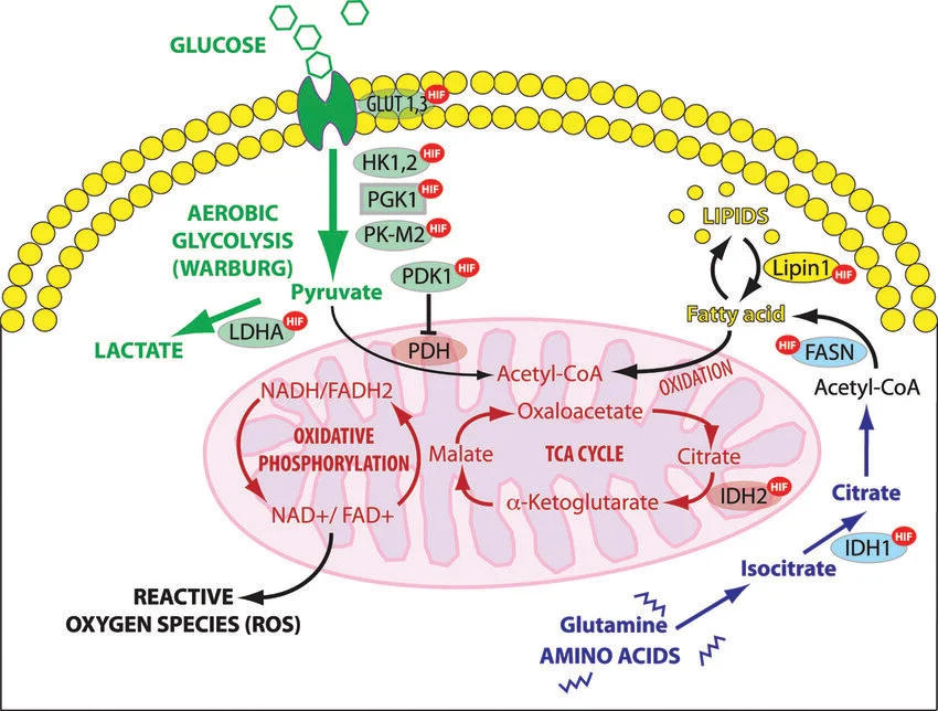
Long description
Regulation of glycolysis and TCA cycle (tricarboxylic acid cycle or citric acid cycle) by HIF. HIF-1 enhances aerobic glycolysis in the cytoplasm by which glucose is metabolized to lactate rather than converted to acetyl

Glycolysis is the cytosolic conversion of glucose to pyruvate, generating a net gain of two ATP molecules and two NADH. The pyruvate produced is either reduced to lactate by lactate dehydrogenase (LDH) or transported into mitochondria where pyruvate dehydrogenase (PDH) converts it to acetyl‑CoA, the entry substrate for the tricarboxylic acid (TCA) cycle. The TCA cycle then produces NADH and FADH₂, which feed the electron transport chain to generate the majority of ATP via oxidative phosphorylation (OXPHOS).
The activity of PDH is tightly regulated by pyruvate dehydrogenase kinase (PDK) and phosphatases. When PDK phosphorylates PDH, the enzyme is inactivated, favouring lactate production; dephosphorylation activates PDH, promoting mitochondrial oxidation of pyruvate.
The ten enzymatic steps of glycolysis are:
- Hexokinase (or glucokinase in liver) phosphorylates glucose to glucose‑6‑phosphate.
- Phosphoglucose isomerase converts glucose‑6‑phosphate to fructose‑6‑phosphate.
- Phosphofructokinase‑1 (PFK‑1) catalyses the irreversible conversion of fructose‑6‑phosphate to fructose‑1,6‑bisphosphate, the key regulatory step.
- Aldolase splits fructose‑1,6‑bisphosphate into glyceraldehyde‑3‑phosphate (G3P) and dihydroxyacetone phosphate (DHAP).
- Triose phosphate isomerase interconverts DHAP and G3P.
- Glyceraldehyde‑3‑phosphate dehydrogenase oxidises G3P to 1,3‑bisphosphoglycerate, producing NADH.
- Phosphoglycerate kinase transfers a phosphate from 1,3‑bisphosphoglycerate to ADP, forming ATP and 3‑phosphoglycerate.
- Phosphoglycerate mutase converts 3‑phosphoglycerate to 2‑phosphoglycerate.
- Enolase dehydrates 2‑phosphoglycerate to phosphoenolpyruvate (PEP).
- Pyruvate kinase converts PEP to pyruvate, generating the second ATP of the pathway.
Regulation centres on PFK‑1 and pyruvate kinase. PFK‑1 is allosterically activated by AMP and fructose‑2,6‑bisphosphate (F2,6BP) and inhibited by ATP and citrate, thereby linking glycolytic flux to cellular energy status and mitochondrial activity. Pyruvate kinase activity is modulated by isoform expression (PKM1, PKM2, PKR, PKL) and post‑translational modifications such as tyrosine phosphorylation, which can alter its catalytic efficiency and influence metabolic decisions between glycolysis and oxidative metabolism. [2]
DefinitionPyruvate kinase isoforms
Four isoforms exist: PKM1, PKM2, PKR and PKL, each with tissue‑specific expression patterns that affect glycolytic capacity and metabolic flexibility. [2]
F2,6BP is a potent allosteric activator of PFK‑1, thereby stimulating glycolysis. It is synthesised from fructose‑6‑phosphate by phosphofructokinase‑2 (PFK‑2) and degraded by fructose‑2,6‑bisphosphatase (FBPase‑2). The bifunctional enzyme PFK‑2/FBPase‑2 switches between these activities in response to hormonal signals: insulin promotes the kinase activity, increasing F2,6BP and glycolysis, whereas glucagon and epinephrine favour the phosphatase activity, reducing F2,6BP and slowing glycolysis.
The concentration of F2,6BP is a critical determinant of the balance between glycolytic flux and gluconeogenesis, especially in hepatocytes where it modulates the activity of key enzymes such as PFK‑1 and fructose‑1,6‑bisphosphatase.
| Enzyme | Key regulator | Effect |
|---|---|---|
| Hexokinase | ATP (feedback) | Inhibition |
| PFK‑1 | F2,6BP (activation), ATP (inhibition) | Major control point |
| Pyruvate kinase | PKM isoform, tyrosine phosphorylation | Modulates pyruvate production |
PDK phosphorylates PDH, inhibiting the conversion of pyruvate to acetyl‑CoA. Inhibition of PDK (e.g., by dichloroacetate, DCA) restores PDH activity, enhancing mitochondrial oxidation of pyruvate and reducing lactate production. This shift has therapeutic implications in conditions such as ischemia/reperfusion injury and cancer, where metabolic reprogramming favours glycolysis.
Clinical pearlPDK inhibition boosts oxidative metabolism
Inhibition of PDHK1 increases PDH activity, leading to higher rates of pyruvate oxidation and reduced lactate production, thereby improving energy metabolism in retinal cells. [2]
Regulation of pyruvate fate
- Glucose uptakeTransport into cell
- Question: Energy status?
High ATP
- PFK‑1 inhibitedReduced glycolysis
Low ATP
- PFK‑1 activated by F2,6BPIncreased glycolysis
- Pyruvate productionVia pyruvate kinase
- Question: PDH phosphorylation?
Phosphorylated (PDK active)
- Pyruvate → lactate (LDH)Anaerobic pathway
Dephosphorylated (PDK inhibited)
- Pyruvate → acetyl‑CoA (PDH)TCA cycle entry
Glycolysis, pyruvate dehydrogenase and the TCA cycle form an integrated network that balances energy production with biosynthetic demands. Regulation at key enzymatic steps—particularly by F2,6BP and PDK—allows cells to adapt rapidly to changes in nutrient availability and oxygen tension, ensuring metabolic homeostasis.
DefinitionPyruvate kinase deficiency (PKD)
A hereditary haemolytic anaemia caused by loss‑of‑function mutations in the PKLR gene, leading to reduced activity of the red‑cell pyruvate kinase enzyme.
Pyruvate kinase (PK) is the final catalytic step of glycolysis, converting phosphoenolpyruvate (PEP) to pyruvate and generating ATP. In erythrocytes, PK is the sole source of ATP; any reduction in its activity compromises red‑cell energy homeostasis and membrane integrity, precipitating intravascular haemolysis. PKD is inherited in an autosomal recessive manner and is characterised by a spectrum of clinical severity, from mild anaemia to life‑threatening haemolysis requiring transfusion or splenectomy.
The prevalence of PKD varies geographically, reflecting the distribution of pathogenic PKLR variants. A systematic literature review estimated a prevalence of 51 per million, with a standard error of 32.5 per million, based on an “index mutation” c.1529G > A that was assumed to have 100 % penetrance in affected individuals. In a cohort of consecutive newborns in Hong Kong, the prevalence of total PK deficiency was often observed. However, the high prevalence in this study may be influenced by qualitative diagnostic methods and potential bias towards jaundiced in [1]
Patients with PKD typically present with chronic non‑immune haemolytic anaemia, jaundice, splenomegaly, and a propensity for iron overload due to repeated transfusions. The degree of haemolysis correlates with residual PK activity; a threshold of < 50 % of normal activity is often used to define deficiency, though this criterion may miss milder cases. The clinical spectrum ranges from asymptomatic carriers to severe disease requiring splenectomy or chronic transfusion support. [1]
No specific drug therapies are currently approved for PKD. Historical attempts to use riboflavin and sulphydryl compounds were unsuccessful. A pharmacologic activator of red‑cell PK is in early phase clinical trials; in mice it enhances glycolytic flux and in healthy volunteers it increases ATP and decreases 2,3‑DPG levels, but data in PKD patients are pending. Standard care remains supportive: folic acid supplementation, transfusion for symptomatic anaemia, and splenectomy for refractory haemolysis. Emerging gene‑editing approaches are under investigation but are not yet clinically available.
DefinitionPyruvate dehydrogenase complex (PDHc)
A mitochondrial multienzyme complex that converts pyruvate into acetyl‑CoA, linking glycolysis to the tricarboxylic acid (TCA) cycle.
The PDHc is essential for aerobic metabolism. It comprises three catalytic subunits (E1, E2, E3) and multiple regulatory proteins. Dysregulation of PDHc activity, whether by genetic mutations or post‑translational modification, leads to a spectrum of metabolic disorders, most notably pyruvate dehydrogenase deficiency (PDHD), characterised by lactic acidosis, neuro‑developmental delay, and hypoglycaemia.
PDHD arises from biallelic pathogenic variants in genes encoding PDHc subunits or its regulatory kinases/phosphatases. Loss of PDHc activity forces pyruvate to be shunted to lactate, increasing lactate production and impairing the TCA cycle. The resultant energy deficit is particularly detrimental to the brain, explaining the severe neurological phenotype.
Patients with PDHD often present in infancy with failure to thrive, hypotonia, seizures, and progressive neuro‑developmental regression. Laboratory findings include elevated blood lactate, normal or low blood glucose, and a normal anion gap metabolic acidosis. Neuroimaging may reveal basal ganglia lesions or diffuse cerebral atrophy.
Current management focuses on metabolic support: a ketogenic diet to provide alternative energy substrates, thiamine supplementation to enhance residual PDH activity, and avoidance of glucose‑rich feeds that exacerbate lactate production. In some cases, dichloroacetate (DCA), a pyruvate dehydrogenase kinase (PDHK) inhibitor, has been used to reactivate PDHc by preventing its inhibitory phosphorylation. In non‑small cell lung cancer (NSCLC) models, DCA combined with EGFR tyrosine kinase inhibitors (TKIs) redirected metabolism towards pyruvate oxidation, reducing lactate production and enhancing therapeutic efficacy. While these findings are pre‑clinical, they illustrate the broader principle that PDHc modulation can influence cellular metabolism and disease outcomes.
Clinical pearlMetabolic integration
Pyruvate sits at a metabolic crossroads: it can be converted to lactate in the cytosol or to acetyl‑CoA in mitochondria for entry into the TCA cycle.
The glycolytic pathway breaks down glucose to pyruvate, generating a net gain of two ATP molecules per glucose molecule. In the presence of oxygen, pyruvate is transported into mitochondria and converted by the PDHc to acetyl‑CoA, which condenses with oxaloacetate to form citrate, the first TCA cycle intermediate. The TCA cycle then oxidises acetyl‑CoA, producing NADH and FADH₂, which feed the electron transport chain to generate the majority of cellular ATP.
PDHc activity is tightly regulated by phosphorylation: PDHKs phosphorylate the E1 subunit, inhibiting the complex, whereas pyruvate dehydrogenase phosphatases (PDPs) dephosphorylate and activate it. In cancer cells, up‑regulation of PDHK1 promotes a shift towards glycolysis (the Warburg effect) and away from oxidative phosphorylation, supporting rapid proliferation. Inhibition of PDHK1 with DCA can reverse this metabolic reprogramming, as demonstrated in NSCLC models. [2]
Defects in any component of this metabolic axis can lead to profound clinical syndromes. PKD disrupts the final step of glycolysis in red cells, while PDHD impairs the conversion of pyruvate to acetyl‑CoA, both culminating in energy deficiency. Understanding the molecular underpinnings of these disorders informs targeted therapeutic strategies, such as enzyme activators or metabolic modulators, and highlights the importance of metabolic flexibility in health and disease.
Arsenic is a well‑known inhibitor of the pyruvate dehydrogenase complex (PDC). The complex is a multienzyme assembly that links glycolysis to the tricarboxylic acid (TCA) cycle by converting pyruvate into acetyl‑CoA. Arsenic binds to the lipoic acid cofactor of the E2 subunit, preventing the transfer of the acetyl group to CoA and thereby blocking the entry of carbon into the TCA cycle. Clinically, chronic arsenic exposure can lead to a spectrum of metabolic disturbances, including impaired energy production and increased lactate accumulation.
Thiamine (vitamin B1) is an essential cofactor for the first enzyme of the TCA cycle, α‑ketoglutarate dehydrogenase, and for the pyruvate dehydrogenase complex itself. Thiamine pyrophosphate (TPP) is required for the decarboxylation of pyruvate to acetyl‑CoA and for the conversion of α‑ketoglutarate to succinyl‑CoA. A deficiency in thiamine therefore mimics the biochemical effects of arsenic poisoning: reduced flux through the TCA cycle, decreased ATP production, and a shift toward anaerobic glycolysis. In patients with thiamine deficiency, laboratory tests often reveal elevated lactate and ketone bodies, reflecting the reliance on glycolysis for energy.
Both arsenic toxicity and thiamine deficiency illustrate how disruption of the pyruvate‑to‑acetyl‑CoA step can have profound metabolic consequences. In the clinic, measurement of plasma thiamine levels and urinary arsenic excretion can aid in diagnosis, while supplementation with thiamine or chelation therapy can reverse the biochemical derangements.
The TCA cycle is a central hub of cellular metabolism, oxidising acetyl‑CoA to CO₂ while generating NADH, FADH₂ and GTP (or ATP). Each turn of the cycle produces three molecules of NADH, one of FADH₂, and one GTP, which feed the electron transport chain to generate the bulk of ATP in aerobic cells.
| Intermediate | Role in the cycle | Regulatory point |
|---|---|---|
| Citrate | First product, exits mitochondria for fatty‑acid synthesis | Citrate‑inhibits phosphofructokinase‑1 (PFK‑1) in glycolysis |
| α‑Ketoglutarate | Substrate for α‑ketoglutarate dehydrogenase | Inhibited by NADH and ATP |
| Succinyl‑CoA | Substrate for succinyl‑CoA synthetase (produces GTP) | Activated by CoA and ADP/ATP |
| Succinate | Substrate for succinate dehydrogenase (Complex II) | Inhibited by high levels of succinate (feedback) |
| Fumarate | Substrate for fumarase | Inhibited by high levels of fumarate |
| Malate | Substrate for malate dehydrogenase | Inhibited by NADH |
The TCA cycle is tightly regulated by the energy status of the cell. High concentrations of ATP, NADH, or acetyl‑CoA inhibit the first and last enzymes of the cycle (citrate synthase and isocitrate dehydrogenase), thereby preventing over‑production of reducing equivalents when the cell’s energy demand is low. Conversely, low ATP and NADH levels relieve this inhibition, allowing the cycle to accelerate.
Insulin promotes the activity of pyruvate dehydrogenase by stimulating the pyruvate dehydrogenase phosphatase, which dephosphorylates and activates the complex. Glucagon and epinephrine, through cAMP‑dependent protein kinase A, phosphorylate and inactivate pyruvate dehydrogenase, favouring gluconeogenesis and glycogenolysis in the liver.
The TCA cycle is interconnected with other pathways. For example, oxaloacetate can be diverted to aspartate synthesis, and α‑ketoglutarate can be used for glutamate production. These anaplerotic and cataplerotic reactions allow the cycle to adapt to varying biosynthetic demands.
-
Assuming the TCA cycle is always the main ATP source
In hypoxic tissues or rapidly proliferating cells, glycolysis can dominate ATP production despite the presence of oxygen (the Warburg effect). Relying solely on TCA activity to explain energy status can be misleading. -
Neglecting the role of anaplerotic flux
Many cells replenish TCA intermediates via anaplerotic reactions (e.g., pyruvate carboxylase converting pyruvate to oxaloacetate). Ignoring these pathways may underestimate the cycle’s capacity to sustain biosynthesis. -
Overlooking post‑translational regulation
Enzyme activity is modulated not only by substrate levels but also by phosphorylation, acetylation, and allosteric effectors. Failing to consider these modifications can lead to incorrect conclusions about metabolic control. -
Misinterpreting lactate accumulation
Elevated lactate can arise from increased glycolytic flux, impaired mitochondrial oxidation, or both. Assuming lactate is solely a marker of anaerobic metabolism may obscure underlying mitochondrial dysfunction. -
Ignoring tissue‑specific isoforms
Different tissues express distinct isoforms of key enzymes (e.g., PKM1 vs PKM2). Assuming a uniform enzyme profile across tissues can result in inaccurate metabolic models.
The principles of glycolysis, pyruvate dehydrogenase activity, and the TCA cycle underpin many clinical and research applications:
-
Diagnostic testing for metabolic disorders
Measurement of pyruvate dehydrogenase activity, lactate/pyruvate ratios, and TCA intermediates assists in diagnosing inborn errors of metabolism such as pyruvate dehydrogenase deficiency or mitochondrial DNA mutations. -
Cancer metabolism research
Tumour cells often exhibit upregulated glycolysis and altered TCA flux. Targeting enzymes like PKM2 or modulating pyruvate dehydrogenase activity is a strategy in developing anticancer therapies. -
Pharmacological interventions
Drugs that influence pyruvate dehydrogenase regulation (e.g., dichloroacetate) are investigated for treating lactic acidosis and certain neurodegenerative diseases. -
Nutritional supplementation
Thiamine supplementation is routinely used in patients with alcoholism or malnutrition to restore pyruvate dehydrogenase function and improve energy metabolism. -
Exercise physiology
Understanding how the TCA cycle responds to increased energy demand informs training protocols and recovery strategies for athletes.
Metabolic decision point in a hypoxic cell
- High glucose uptakeGlucose transported into the cell
- Question: Oxygen availability?
Sufficient
- Pyruvate enters mitochondriaConverted to acetyl‑CoA by PDC
- TCA cycle activeOxidative phosphorylation generates ATP
Limited
- Pyruvate converted to lactateRegenerates NAD+
- ATP from glycolysisLower yield but rapid
- Arsenic blocks pyruvate dehydrogenase by binding lipoic acid.
- Thiamine is essential for pyruvate dehydrogenase and α‑ketoglutarate dehydrogenase.
- The TCA cycle produces NADH, FADH₂ and GTP, feeding the electron transport chain.
- Allosteric inhibitors (ATP, NADH, acetyl‑CoA) regulate the cycle’s pace.
- Hormones (insulin, glucagon) modulate pyruvate dehydrogenase activity.
- Metabolic disorders are diagnosed by measuring pyruvate dehydrogenase activity and lactate/pyruvate ratios.
Answer each one in your head first, then open the answer.
Which cofactor is required for pyruvate dehydrogenase activity?
Show answerHide answer
Thiamine pyrophosphate (TPP)What is the main product of the pyruvate dehydrogenase complex?
Show answerHide answer
Acetyl‑CoAName one allosteric inhibitor of isocitrate dehydrogenase.
Show answerHide answer
ATP or NADHWhich enzyme converts pyruvate to lactate during anaerobic glycolysis?
Show answerHide answer
Lactate dehydrogenase (LDH)Why might lactate accumulation not always indicate hypoxia?
Show answerHide answer
It can also result from impaired mitochondrial oxidation or increased glycolytic flux.
- Secrest MH, Storm M, Carrington C, et al. Prevalence of pyruvate kinase deficiency: A systematic literature review. European journal of haematology 2020;105(2):173-184. doi:10.1111/ejh.13424. Accessed .
- Dyrstad SE, Lotsberg ML, Tan TZ, et al. Blocking Aerobic Glycolysis by Targeting Pyruvate Dehydrogenase Kinase in Combination with EGFR TKI and Ionizing Radiation Increases Therapeutic Effect in Non-Small Cell Lung Cancer Cells. Cancers 2021;13(5):941. doi:10.3390/cancers13050941. Accessed .
Figure credits
Educational content for medical students and doctors. It is not a substitute for clinical judgement or local guidelines.