Haem synthesis, bilirubin and iron metabolism
Physiological pathway of haem biosynthesis, iron metabolism, bilirubin handling and lead toxicity in humans.
Figure 1Heme synthesis Dutch text
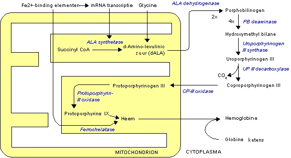
Long description
Illustration for biochemistry/haem synthesis bilirubin and iron metabolism from Wikimedia Commons, titled 'Heme synthesis Dutch text': Heme synthesis Dutch text.png. The author and licence are given in the credit line.

Haem is a vital prosthetic group composed of a cyclic porphyrin ring, specifically protoporphyrin IX, coordinated with a central ferrous iron atom. It serves as the functional prosthetic moiety for numerous hemoproteins essential for life, including haemoglobin, myoglobin, cytochromes within the mitochondrial electron transport chain, catalase, and nitric oxide synthase. Understanding haem synthesis requires appreciating how iron and bilirubin intersect with this pathway. Although human physiology lacks an active physiological excretion system for excess iron, the body maintains systemic iron homeostasis through tightly regulated intestinal absorption, storage within ferritin, and recycling of iron from senescent erythrocytes by reticuloendothelial macrophages. [2]
Dietary iron is acquired via two distinct molecular pathways in the gastrointestinal tract corresponding to nonhaem iron and haem iron. A typical Western diet contains 7 mg of iron per 1000 kcal, yet only 1 to 2 mg is absorbed daily. Nonhaem iron constitutes up to 90% of ingested dietary iron and is predominantly found in plant sources, cereals, beans, and herbs. Because nonhaem iron exists largely in the oxidized ferric (Fe3+) state, it tends to precipitate in solutions where the pH exceeds 3. Consequently, gastric acid must solubilize and chelate ferric iron before it reaches the less acidic proximal small intestine. Ferrous iron (Fe2+) remains soluble at neutral pH and is more readily transported across the apical membrane of enterocytes in the duodenum and upper jejunum. [2]
By contrast, haem iron accounts for 10% of dietary iron but contributes a disproportionately large share of total iron absorption due to its superior bioavailability. Haem iron accounts for 10 to 15% of total iron intake. Because haem iron is absorbed with an efficiency of approximately 15 to 35%, it can account for more than 40% of total intestinal iron absorption. Dietary factors, luminal chelators, and the physical state of iron as ferrous or ferric strongly influence nonhaem iron absorption, whereas haem iron absorption is less affected by dietary inhibitors. Once inside the enterocyte, iron from both haem and nonhaem sources enters a shared intracellular pool where it can be utilized, stored, or exported into the circulation via basolateral ferroportin. [2]
DefinitionHaem and Iron Balance
Haem is an iron-protoporphyrin complex essential for oxygen transport and cellular respiration, regulated by systemic iron availability and precise enzymatic steps.
The biosynthesis of haem is a universal cellular pathway that occurs partly in the mitochondria and partly in the cytosol of all mammalian cells, with the highest rates occurring in developing erythroid cells in the bone marrow and hepatocytes in the liver. The pathway consists of eight distinct enzymatic reactions.
The first step takes place within the mitochondrial matrix. Glycine and succinyl-CoA are condensed to form delta-aminolevulinic acid (ALA) in a reaction catalyzed by the enzyme ALA synthase (ALAS). This is universally recognized as the rate-limiting step of the entire pathway. Mammals express two main isoforms: ALAS1, which is found ubiquitously and regulated by intracellular free haem via feedback inhibition and repression, and ALAS2, which is erythroid-specific and regulated primarily by iron availability through an iron-responsive element in its mRNA untranslated region.
Following its synthesis in the mitochondria, ALA is transported into the cytosol, where the next several steps occur. Two molecules of ALA are condensed by porphobilinogen synthase, also known as aminolevulinic acid dehydratase (ALAD), to form the monopyrrole porphobilinogen (PBG). Next, four molecules of porphobilinogen are linked together by hydroxymethylbilane synthase (also called porphobilinogen deaminase) to yield the linear tetrapyrrole hydroxymethylbilane. This unstable intermediate is cyclized by uroporphyrinogen III synthase to form uroporphyrinogen III, an asymmetric macrocycle that serves as the precursor for all natural porphyrins.
Uroporphyrinogen decarboxylase then removes the four acetate side-chain carboxyl groups, converting uroporphyrinogen III to coproporphyrinogen III within the cytosol. Coproporphyrinogen III is subsequently transported back into the mitochondrial intermembrane space or matrix, where coproporphyrinogen III oxidase converts it to protoporphyrinogen IX. Protoporphyrinogen oxidase then oxidizes protoporphyrinogen IX to protoporphyrin IX through the removal of six hydrogens, creating a fully conjugated planar macrocycle.
The final step of haem synthesis occurs in the mitochondrial matrix, where ferrochelatase inserts a ferrous iron (Fe2+) molecule into the centre of protoporphyrin IX, yielding haem. The newly formed haem is subsequently exported to the cytoplasm to be incorporated into hemoproteins or degraded into biliverdin and subsequently bilirubin by the enzyme haem oxygenase when erythrocytes undergo physiological senescence in the reticuloendothelial system.
| Cellular Compartment | Enzyme | Reaction / Intermediate |
|---|---|---|
| Mitochondrion | ALAS | Glycine + succinyl-CoA to ALA |
| Cytosol | ALAD (PBG synthase) | ALA to porphobilinogen (PBG) |
| Cytosol | Hydroxymethylbilane synthase | PBG to hydroxymethylbilane |
| Cytosol | Uroporphyrinogen III synthase | Hydroxymethylbilane to uroporphyrinogen III |
| Cytosol | Uroporphyrinogen decarboxylase | Uroporphyrinogen III to coproporphyrinogen III |
| Mitochondrion | Coproporphyrinogen III oxidase | Coproporphyrinogen III to protoporphyrinogen IX |
| Mitochondrion | Protoporphyrinogen oxidase | Protoporphyrinogen IX to protoporphyrin IX |
| Mitochondrion | Ferrochelatase | Insertion of Fe2+ into protoporphyrin IX to form haem |
Lead poisoning is a classic environmental and occupational toxicity that profoundly disrupts haem biosynthesis, leading to characteristic haematological and neurological manifestations. Lead is a heavy metal that interferes with multiple enzyme systems by binding avidly to sulfhydryl groups and substituting for essential divalent cations such as calcium, zinc, and iron. Within the haem synthetic pathway, lead exerts its most potent inhibitory effects on two key enzymes: ALA dehydratase (ALAD) in the cytosol and ferrochelatase in the mitochondria.
When lead inhibits ALAD, the condensation of delta-aminolevulinic acid into porphobilinogen is impaired, causing a marked accumulation and elevated urinary excretion of upstream ALA. When lead inhibits ferrochelatase inside the mitochondria, the insertion of ferrous iron into protoporphyrin IX is blocked. Consequently, the mitochondrion shunts zinc instead of iron into protoporphyrin IX, resulting in the formation and accumulation of zinc protoporphyrin (ZPP) within circulating erythrocytes. The impairment of haem synthesis coupled with shortened erythrocyte survival culminates in a microcytic, hypochromic anaemia, frequently accompanied by basophilic stippling of red blood cells representing aggregated degenerate ribosomes.
Clinical identification and management require structured public health and medical interventions. Case identification and treatment for lead poisoning should first be extended to villages in which a child with a blood lead level (BLL) equal to or greater than 45 micrograms per decilitre was identified, followed by villages with childhood lead poisoning, lead contamination, or both. Children with frank lead poisoning require long-term follow-up and support. Healthcare providers in endemic or exposed regions must be trained to diagnose and treat lead poisoning, and medical treatment must be coordinated with environmental interventions because chelation therapy alone remains ineffective if children continue to reside in contaminated homes. [3]
The clinical presentation of lead toxicity can be insidious and non-specific. Symptoms of lead poisoning are generally non-specific, and blood lead testing may be unavailable during the initial phases of an outbreak. In historical or severe outbreak analyses, clinical indicators such as convulsions and deaths have been utilized. However, convulsions and death are not sensitive markers of lead poisoning because they typically occur at blood lead levels that are higher than the levels detected in most blood samples collected from affected communities, and convulsions and death are not specific for lead poisoning since they can result from other locally endemic conditions, including malaria and bacterial meningitis. Furthermore, surveillance protocols often focus on young children, omitting older populations such as children aged 5 years or older, adults, or livestock. [3]
Lead Toxicity Pathway on Haem Synthesis
- Lead exposureIngestion or inhalation of environmental lead particles.
- Question: Enzymatic inhibition?
ALAD inhibited
- ALA accumulatesIncreased urinary excretion of delta-aminolevulinic acid.
Ferrochelatase inhibited
- ZPP formationZinc substituted into protoporphyrin IX, causing microcytic anaemia.
Clinical pearlZPP Elevation
Inhibition of mitochondrial ferrochelatase by lead leads to zinc incorporation into protoporphyrin instead of iron, producing elevated zinc protoporphyrin levels.
Porphyrias represent a group of metabolic disorders caused by inherited or acquired defects in the enzymatic pathway responsible for the biosynthesis of haem. Haem is a vital prosthetic group containing a central iron atom enclosed in a porphyrin ring, essential for oxygen transport, mitochondrial respiration, and numerous enzymatic reactions across the human body. The biosynthesis of haem involves eight sequential enzymatic steps distributed between the mitochondrial matrix and the cytosol. The pathway begins in the mitochondria with the condensation of glycine and succinyl-CoA, catalysed by the enzyme aminolaevulinic acid synthase (ALAS). This initial rate-limiting reaction produces delta-aminolaevulinic acid (ALA). ALAS exists as two major isoforms: ALAS1, which is expressed ubiquitously and regulated by intracellular free haem via negative feedback, and ALAS2, which is restricted to erythroid cells and controlled by iron availability. Following its synthesis in the mitochondria, ALA is transported into the cytosol, where the subsequent four enzymatic steps occur. [1]
In the cytosol, two molecules of ALA condense to form porphobilinogen via the enzyme porphobilinogen synthase, also known as delta-aminolaevulinic acid dehydratase. Next, four porphobilinogen molecules are joined together by hydroxymethylbilane synthase, traditionally called porphobilinogen deaminase, to yield the linear tetrapyrrole hydroxymethylbilane. Hydroxymethylbilane is then cyclised and spatially rearranged by uroporphyrinogen III synthase to produce uroporphyrinogen III. This cyclic intermediate undergoes decarboxylation of its acetate side chains by uroporphyrinogen decarboxylase to form coproporphyrinogen III. Coproporphyrinogen III is subsequently transported back into the mitochondrial intermembrane space or matrix.
Inside the mitochondria, coproporphyrinogen oxidase converts coproporphyrinogen III into protoporphyrinogen IX. Protoporphyrinogen oxidase then oxidises protoporphyrinogen IX to protoporphyrin IX. The final step of the pathway takes place in the mitochondrial matrix, where ferrochelatase inserts a ferrous iron atom into the protoporphyrin IX ring, completing the synthesis of haem. Mutations affecting any of these individual enzymes disrupt the pathway, resulting in the characteristic accumulation and excretion of specific upstream pathway intermediates, such as porphyrin precursors or porphyrins, which underlie the clinical manifestations of the porphyrias.
DefinitionEnzymatic Blockade
Clinical classification of the porphyrias typically separates them into acute hepatic porphyrias and cutaneous porphyrias, determined by the primary site of metabolic overproduction and the predominant clinical presentation. Acute hepatic porphyrias, such as acute intermittent porphyria caused by a deficiency in hydroxymethylbilane synthase, primarily present with neurovisceral symptoms, including severe abdominal pain, peripheral neuropathy, and psychiatric disturbances. These acute neurovisceral attacks are frequently precipitated by factors that induce hepatic ALAS1 activity, such as certain medications, hormonal fluctuations, or caloric restriction. In contrast, cutaneous porphyrias, such as porphyria cutanea tarda resulting from hepatic uroporphyrinogen decarboxylase deficiency or erythropoietic protoporphyria caused by ferrochelatase deficiency, manifest with skin fragility, blistering, or acute photosensitivity upon exposure to sunlight. When porphyrins accumulate in the skin, they absorb light energy and react with molecular oxygen to generate reactive oxygen species, causing cellular damage and localized cutaneous inflammation.
| Porphyria type | Affected enzyme | Cellular location | Clinical hallmark |
|---|---|---|---|
| Acute intermittent porphyria | Hydroxymethylbilane synthase | Cytosol | Neurovisceral attacks |
| Porphyria cutanea tarda | Uroporphyrinogen decarboxylase | Cytosol | Cutaneous photosensitivity |
| Erythropoietic protoporphyria | Ferrochelatase | Mitochondria | Painful solar urticaria |
Porphyria Classification Pathway
- Identify clinical presentationAssess whether symptoms are primarily acute neurovisceral or cutaneous.
- Question: Are symptoms acute neurovisceral?
Yes
- Consider acute hepatic porphyriasEvaluate enzymes such as hydroxymethylbilane synthase.
No
- Consider cutaneous porphyriasEvaluate enzymes such as uroporphyrinogen decarboxylase or ferrochelatase.
The physiological breakdown of haem represents the primary route for the disposal of ageing red blood cells and other haem-containing proteins, such as myoglobin and cytochromes, occurring predominantly within the reticuloendothelial system of the spleen, liver, and bone marrow. The initial and rate-limiting step of haem catabolism is catalysed by the microsomal enzyme haem oxygenase. Haem oxygenase requires molecular oxygen and reducing equivalents from NADPH-cytochrome P450 reductase to cleave the alpha-methene bridge of the protohaem ring. This reaction consumes one molecule of oxygen and yields equimolar amounts of carbon monoxide, free ferrous iron, and biliverdin. The liberated iron is rapidly sequestered by ferritin or transported by transferrin for reuse in erythropoiesis, whereas carbon monoxide is eliminated via the pulmonary system. Biliverdin, a green-pigmented tetrapyrrole, is subsequently reduced to bilirubin, a yellow-orange pigment, by the cytosolic enzyme biliverdin reductase, utilizing NADPH as a cofactor. Because newly formed unconjugated bilirubin is hydrophobic and tightly bound to serum albumin for transport through the blood, it must undergo chemical modification in the liver to become water-soluble and capable of excretion.
Clinical pearlBilirubin Transport
Upon reaching the sinusoidal membrane of the hepatocyte, unconjugated bilirubin dissociates from albumin and enters the cell via facilitated carrier-mediated transport proteins, including organic anion transporting polypeptides. Once inside the hepatocyte cytosol, unconjugated bilirubin is bound by intracellular carrier proteins, specifically cytosolic ligandins such as glutathione S-transferases, which prevent its efflux back into the bloodstream. The insoluble bilirubin is then transported to the endoplasmic reticulum, where it undergoes enzymatic conjugation to polar glucuronide groups. This crucial solubilising reaction is catalysed by the enzyme uridine diphosphate glucuronosyltransferase 1A1. Uridine diphosphate glucuronosyltransferase 1A1 transfers glucuronic acid from uridine diphosphate glucuronic acid to one or both of the propionic acid side chains of bilirubin, forming bilirubin monoglucuronide and bilirubin diglucuronide. This conjugation transforms the hydrophobic, lipid-soluble pigment into a hydrophilic, water-soluble compound that can no longer cross cell membranes passively and is protected from reabsorption.
| Parameter | Unconjugated bilirubin | Conjugated bilirubin |
|---|---|---|
| Water solubility | Insoluble | Soluble |
| Plasma carrier | Bound to albumin | Free in solution |
| Cell membrane permeability | High | Low |
| Reaction with diazo reagent | Indirect | Direct |
Following hepatic conjugation, bilirubin diglucuronide and bilirubin monoglucuronide are actively secreted across the canalicular membrane of the hepatocyte into the bile canaliculi. This energy-dependent transport step is mediated primarily by the canalicular multidrug resistance-associated protein 2. Once secreted into bile, the conjugated bilirubin flows into the small intestine as a component of bile. In the terminal ileum and colon, bacterial beta-glucuronidases hydrolyse the glucuronide groups off the bilirubin molecules, and subsequent reduction by intestinal anaerobes converts the pigment into colourless urobilinogens. A fraction of these urobilinogens is reabsorbed through the intestinal mucosa, entering the enterohepatic urobilinogen circulation, where most is re-extracted by the liver and re-secreted into bile, while a small portion escapes into the systemic circulation and is excreted by the kidneys as urobilin. The remaining unabsorbed urobilinogens in the colonic lumen are further oxidised to stercobilin, which imparts the characteristic brown colour to human faeces.
Bilirubin Processing Pathway
- Haem degradationHaem oxygenase converts haem to biliverdin, which is then reduced to unconjugated bilirubin.
- Question: Is bilirubin conjugated?
No (Unconjugated)
- Albumin transportTransported in blood bound to albumin to hepatocytes.
Yes (Conjugated)
- Biliary excretionSecreted into bile via multidrug resistance-associated protein 2 into the intestine.
Inherited hyperbilirubinaemias represent a fascinating group of disorders caused by genetic defects in bilirubin metabolism, transport, or conjugation. To understand these conditions from first principles, one must first consider the normal physiological handling of bilirubin. Bilirubin is the end-product of haem catabolism, derived primarily from the breakdown of senescent red blood cells in the reticuloendothelial system. Haem is converted into biliverdin by the enzyme haem oxygenase, and biliverdin is subsequently reduced to unconjugated bilirubin by biliverdin reductase. Unconjugated bilirubin is lipid-soluble and tightly bound to albumin in the circulation, which prevents it from crossing cell membranes indiscriminately. Upon reaching the liver, unconjugated bilirubin dissociates from albumin, enters hepatocytes via carrier-mediated uptake transporters, and is rapidly conjugated with glucuronic acid by the enzyme uridine diphosphate glucuronosyltransferase 1A1 (UGT1A1) to form water-soluble bilirubin mono- and diglucuronides. These conjugated bilirubins are then excreted actively across the canalicular membrane into the bile via the multidrug resistance-associated protein 2 (MRP2) transporter. Disruptions at any stage of this pathway—hepatic uptake, intracellular binding, conjugation, or canalicular excretion—result in distinct inherited hyperbilirubinaemias characterised by predominant unconjugated or conjugated hyperbilirubinaemia. [1]
Unconjugated hyperbilirubinaemias are generally caused by reduced activity of UGT1A1 or impaired hepatic uptake. Gilbert syndrome is a very common, benign inherited disorder characterised by mild, fluctuating unconjugated hyperbilirubinaemia typically provoked by fasting, stress, illness, or fatigue. It arises from promoter region mutations or insertions in the UGT1A1 gene that reduce the transcriptional expression of the enzyme. In contrast, Crigler-Najjar syndrome represents a severe, life-threatening group of disorders resulting from severe mutations in the coding regions of the UGT1A1 gene. Crigler-Najjar type 1 is characterised by the complete absence of UGT1A1 activity, leading to profound neonatal unconjugated hyperbilirubinaemia and a high risk of kernicterus unless aggressively managed with phototherapy or liver transplantation. Crigler-Najjar type 2 features residual UGT1A1 activity, lower serum bilirubin levels, and a response to phenobarbital induction.
Conjugated hyperbilirubinaemias, on the other hand, arise from defects in biliary excretion subsequent to normal hepatocellular conjugation. Dubin-Johnson syndrome is caused by mutations in the ABCC2 gene, which encodes the canalicular multispecific organic anion transporter MRP2. The loss of MRP2 function impairs the excretion of conjugated bilirubin and other organic anions into the bile, leading to a mild to moderate conjugated hyperbilirubinaemia and a characteristically pigmented, dark liver due to the accumulation of epinephrine-like pigment polymers in lysosomes. Rotor syndrome is another rare, benign autosomal recessive disorder featuring conjugated hyperbilirubinaemia, but it differs from Dubin-Johnson syndrome in that the liver is not pigmented and the gallbladder is typically visualised on oral cholecystography. Rotor syndrome is caused by the simultaneous deficiency of two organic anion transporting polypeptides, OATP1B1 and OATP1B3, which impairs both hepatic storage and the re-uptake of conjugated bilirubin back into hepatocytes. Understanding these distinct enzymatic and transporter defects allows clinicians to differentiate between unconjugated and conjugated presentations through simple laboratory testing, avoiding unnecessary invasive investigations for benign familial conditions. [1]
DefinitionGilbert syndrome
| Disorder | Primary Defect | Bilirubin Type | Key Feature |
|---|---|---|---|
| Gilbert syndrome | UGT1A1 promoter mutation | Unconjugated | Mild, stress-induced |
| Crigler-Najjar Type 1 | UGT1A1 absent | Unconjugated | High kernicterus risk |
| Dubin-Johnson syndrome | MRP2 / ABCC2 deficiency | Conjugated | Pigmented liver |
| Rotor syndrome | OATP1B1 / OATP1B3 loss | Conjugated | Non-pigmented liver |
Approach to Inherited Hyperbilirubinaemias
- Measure serum bilirubin fractionsDetermine whether the hyperbilirubinaemia is predominantly unconjugated or conjugated.
- Question: Predominantly unconjugated?
Yes
- Assess for Gilbert syndromeCheck for mild elevation provoked by fasting or illness with normal liver enzymes.
- Consider Crigler-NajjarEvaluate for severe neonatal jaundice if bilirubin is markedly elevated.
No
- Assess for conjugated hyperbilirubinaemiaCheck liver architecture and canalicular excretion.
- Question: Liver pigmented?
Yes
- Dubin-Johnson syndromeConfirmed by impaired biliary excretion via MRP2 deficiency.
No
- Rotor syndromeCaused by combined OATP1B1 and OATP1B3 transporter defects.
Iron absorption is a tightly regulated physiological process occurring primarily in the duodenum and upper jejunum, designed to maintain systemic iron balance in the absence of an active excretory pathway for iron. Dietary iron exists in two main forms: heme iron, derived from animal haemoglobin and myoglobin, and nonheme iron, found in plant-based foods and fortified products. Heme iron is absorbed intact across the apical membrane of enterocytes via heme carrier protein 1 (HCP1), after which intracellular heme oxygenase releases free ferrous iron. Nonheme iron is predominantly in the ferric state and must be reduced to the ferrous state by apical ferrireductases, such as duodenal cytochrome b (DCYTB), before being transported across the brush border membrane by divalent metal transporter 1 (DMT1). Once inside the enterocyte, iron can be stored within ferritin or exported across the basolateral membrane into the bloodstream via ferroportin, assisted by oxidising enzymes like hephaestin or ceruloplasmin, enabling transport bound to transferrin. Because systemic iron overload is toxic, intestinal absorption is modulated by numerous dietary enhancers and inhibitors, alongside systemic regulatory feedback mediated by the peptide hormone hepcidin. [2]
Dietary factors exert a profound influence on the efficiency of nonheme iron absorption. Ascorbic acid (vitamin C) is a potent enhancer that acts in a dose-dependent manner to reduce ferric iron to more soluble ferrous iron, thereby increasing absorption when both nutrients are consumed together. Specifically, iron absorption gradually increases from 0.8% to 7.1% when increasing amounts of ascorbic acid, ranging from 25 to 1000 mg, are added to a liquid formula meal containing 4.1 mg of nonheme [2]
Conversely, various dietary components act as potent inhibitors of iron absorption. Polyphenols—found abundantly in vegetables, cereals, spices, tea, coffee, red wine, and cocoa—function similarly to phytates by binding iron to form insoluble complexes, thereby inhibiting its bioavailability. However, the inhibitory impact depends on concentration; for instance, 20 mg of bean polyphenols introduced to noninhibitory bread meals where phytic acid had been destroyed did not affect iron absorption. Other dietary factors such as phytates, specific proteins, and calcium decrease iron absorption. Calcium is unique among these inhibitory nutrients because it decreases the absorption of both nonheme and heme iron, although the exact molecular mechanism for this dual inhibition has not yet been fully elucidated. Similarly, clinical studies examining fortified products reveal complex physiological interactions; consumption of an iron-fortified milk product supplying 100% of the required daily iron intake failed to improve iron status in iron-deficient women over four months in a randomised controlled trial, with the authors concluding that the presence of calcium and casein in the product inhibited adequate iron utilisation. In contrast, innovative strategies such as fortifying yogurt with iron and supplementing it with long-chain inulin have demonstrated promising impacts in treating iron deficiency by enhancing iron absorption, likely because prebiotic fermentation by beneficial microorganisms in the colon produces short-chain fatty acids that lower luminal pH and maintain iron solubility. Condiments and seasonings fortified with iron alone may also slightly reduce iron deficiency and improve iron status compared with unfortified alternatives, offering practical public health interventions to combat global iron deficiency anaemia. [2] [1]
Clinical pearlAscorbic acid timing
| Dietary Factor | Effect on Nonheme Iron | Mechanism / Detail |
|---|---|---|
| Ascorbic acid | Enhances | Reduces ferric to ferrous iron dose-dependently |
| Animal tissues | Enhances | Increases absorption by 150% via luminal transport |
| Polyphenols | Inhibits | Forms insoluble complexes with iron |
| Calcium | Inhibits | Decreases absorption of both heme and nonheme iron |
| Prebiotic inulin | Enhances | Promotes short-chain fatty acid production in the colon |
Dietary Modulation of Iron Absorption
- Ingestion of dietary ironHeme and nonheme iron enter the gastrointestinal lumen.
- Question: Enhancers present?
Ascorbic acid / Meat
- Enhanced reduction and uptakeFerric iron reduced to ferrous state; luminal transporters facilitate mucosal transfer.
Inhibitors present?
- Question: Polyphenols or Calcium?
Yes
- Reduced bioavailabilityInsoluble complexes formed, or direct inhibition of both heme and nonheme transport.
No
- Standard absorptionIron processed normally via DMT1 and ferroportin.
Iron is an essential heavy metal for human nutrition and plays critical roles in oxygen and electron transport, cell division, differentiation, and regulation of gene expression. It also takes part in energy production, DNA, RNA, and protein synthesis due to its electron exchange features, and is involved in the structure and function of many enzymes. In the human body, 70% of the iron binds to haemoglobin, the red blood cell pigment, while the remaining proportion binds to proteins such as myog [2]
Dietary iron deficiency leads to iron deficiency anaemia, which is estimated to cause about 66.1% of anaemia cases among males and 56.8% among females. Individuals with iron deficiency anaemia may experience inadequate iron intake, impaired absorption, or defective transport within the body, alongside losses caused by disease or specific life stages. Other contributing factors to anaemia include infectious diseases such as malaria, tuberculosis, HIV, and parasitic infections, as well as inherited disorders of haemoglobin structure and deficiencies in folate, vitamin B12, and vitamin A. Populations with limited access to meat and fish products, or those avoiding them by personal preference, face a higher risk of iron deficiency and anaemia. [1]
To satisfy physiological demands, recommended dietary allowances vary widely across demographics. For adults over 19 years of age, the recommended daily allowance value is 8 mg per day for males and 18 mg per day for females. Lactating women younger than 18 years require 10 mg per day, whereas those over 18 require 9 mg per day. Vegetarian diets demand adjusted targets: a vegetarian man requires 14 mg per day, while vegetarian females aged 14 to 18 years, 19 to 50 years, and over 51 years require 26 mg per day, 14 mg per day, and 33 mg per day, respectively. When iron requirements are unmet, iron deficiency develops, potentially precipitating clinical complications. [2]
Dietary fortification strategies must navigate chemical inhibitors such as phytic acid, phenolic compounds, and calcium present in food vehicles or diets. Selection of appropriate fortifying compounds is essential to balance bioavailability and prevent adverse sensory reactions in food products. Commonly utilised iron fortification salts include ferrous sulphate, ferrous fumarate, and chelated compounds like sodium iron EDTA, also referred to as NaFeEDTA or sodium iron ethylene diamine tetra acetate. Vehicles deployed across trials include salt, fish sauce, soy sauce, curry powder, and seasoning powder. Modern delivery systems utilise encapsulation, such as liposomal iron prepared via microencapsulation with micronized iron inside a liposomal layer, offering high absorption and lower side effects. Emulsions, including water in oil and oil in water types as well as double emulsions, provide controlled release, chemical stability, and enhanced bioaccessibility. [1] [2]
| Population group | Iron requirement or vehicle |
|---|---|
| Adult males over 19 | 8 mg/day |
| Adult females over 19 | 18 mg/day |
| Vegetarian males | 14 mg/day |
| Vegetarian females (19 to 50 years) | 14 mg/day |
| Vegetarian females (over 51 years) | 33 mg/day |
| Common fortification vehicles | Salt, fish sauce, soy sauce, curry powder |
Diagnosing anaemia and iron deficiency requires laboratory investigation rather than clinical observation alone. Anaemia is primarily assessed by measuring haemoglobin concentration, which must be interpreted in the context of age, sex, pregnancy status, altitude, and smoking status since these variables alter physiological iron needs. Measuring haemoglobin concentration remains a relatively inexpensive and feasible test, even in resource-poor settings. Conversely, assessing iron status requires multiple biochemical markers, including serum ferritin, serum iron, transferrin saturation, soluble transferrin receptor, and erythrocyte protoporphyrin. [1]
A common pitfall in clinical evaluation is relying solely on serum ferritin without accounting for confounding conditions. Ferritin is the most widely used indicator for assessing iron status and body iron depletion, reflecting a close relationship between total stored iron and serum concentration in normal individuals. However, serum ferritin is an acute-phase reactant and is significantly affected by inflammation, which can falsely elevate values and mask true iron deficiency. Misinterpreting elevated ferritin in the presence of chronic disease, infection, or inflammatory bowel disease leads to missed diagnoses of concomitant iron deficiency. [1]
Laboratory evaluation framework
- Measure haemoglobinAssess for anaemia, adjusting for age, sex, altitude, and smoking status.
- Question: Is haemoglobin low?
Yes
- Evaluate iron statusCheck serum ferritin, transferrin saturation, and soluble transferrin receptor.
- Question: Is ferritin high or normal despite deficiency risk?
Yes
- Account for inflammationRecognise that inflammation elevates serum ferritin and obscures depletion.
No
- Confirm iron deficiencyProceed with targeted nutritional or therapeutic intervention.
No
- Consider other causesInvestigate folate, vitamin B12, vitamin A, or chronic disease.
Another frequent pitfall involves overlooking dietary inhibitors when planning iron repletion or fortification. Dietary factors such as phytic acid, phenolic compounds, and calcium strongly inhibit iron absorption and can render oral supplementation or fortified foodstuffs ineffective unless properly managed. Furthermore, failing to select the correct iron compound leads to poor patient compliance due to gastrointestinal side effects or unpalatable sensory changes in food vehicles. Healthcare providers must also recognise that anaemia is multifactorial; assuming all anaemia is caused by iron deficiency without ruling out folate, vitamin B12, vitamin A deficiencies, or chronic infections like tuberculosis, HIV, and malaria will result in therapeutic failure.
Exam trapInflammatory interference
In clinical and public health practice, managing iron metabolism disorders centres on diagnosing and correcting iron deficiency anaemia across diverse patient groups. Public health programmes utilise food fortification to combat high population-level burdens of iron deficiency anaemia, which accounts for 66.1% of cases in males and 56.8% in females. When designing fortification programmes, public health nutritionists select suitable food vehicles, such as salt, fish sauce, soy sauce, curry powder, or seasoning powder. They must also incorporate protective iron compounds like sodium iron EDTA, ferrous sulphate, or ferrous fumarate to overcome dietary inhibitors like phytic acid and phenolic compounds.
In therapeutic practice, novel iron formulations are increasingly deployed to improve tolerance and efficacy. For instance, microencapsulated liposomal iron pyrophosphate has been studied in nonpregnant women of reproductive age suffering from iron deficiency anaemia. In a trial involving 558 women treated for 12 weeks, haemoglobin levels rose significantly from 8.71 plus or minus 2.24 to 10.47 plus or minus 1.69 g/dL. Similar encapsulation technologies are used for patients with complex medical backgrounds, such as individuals with inactive or mildly active inflammatory bowel disease complicated by iron deficiency anaemia, who receive oral liposomal iron formulations over an 8-week course to bypass gastrointestinal irritation. [2]
Clinical pearlLiposomal iron efficacy
Clinical management also requires careful dietary counselling, particularly for vulnerable groups such as strict vegetarians whose recommended daily allowances reach up to 33 mg per day for older females, contrasting sharply with standard adult requirements. Clinicians must correlate laboratory indices—such as serum ferritin, transferrin saturation, and soluble transferrin receptor—with clinical histories that include dietary habits, socioeconomic access to meat and fish products, and potential blood losses from disease. By integrating biochemical testing and understanding iron transport mechanisms, clinicians effectively target iron repletion while avoiding the pitfalls of unrecognised inflammatory states.
- Dietary iron deficiency anaemia accounts for approximately 66.1% of male and 56.8% of female anaemia cases.
- Serum ferritin is the most widely used indicator of iron status but is elevated by inflammation.
- Anaemia diagnosis relies on measuring haemoglobin concentration adjusted for age, sex, altitude, and smoking.
- Dietary inhibitors such as phytic acid and phenolic compounds reduce iron absorption and require strategic vehicle selection.
- Adult recommended daily allowances are 8 mg/day for males and 18 mg/day for non-vegetarian females over 19.
- Vegetarian females over 51 years require a higher recommended daily allowance of 33 mg/day.
- Microencapsulated liposomal iron significantly improves haemoglobin levels in iron deficiency anaemia.
Answer each one in your head first, then open the answer.
What percentage of anaemia cases in males is estimated to be caused by dietary iron deficiency?
Show answerHide answer
66.1%Which biochemical marker is most commonly used to assess iron status and depletion?
Show answerHide answer
Serum ferritinWhat is the adult male recommended daily allowance for iron for individuals over 19?
Show answerHide answer
8 mg/dayWhich dietary components act as inhibitors to iron absorption during fortification?
Show answerHide answer
Phytic acid, phenolic compounds and calciumWhat compound is also known as sodium iron ethylene diamine tetra acetate?
Show answerHide answer
Sodium iron EDTA
- Jalal CS, De-Regil LM, Pike V, Mithra P. Fortification of condiments and seasonings with iron for preventing anaemia and improving health. The Cochrane database of systematic reviews 2023;9:CD009604. doi:10.1002/14651858.cd009604.pub2. Accessed .
- Piskin E, Cianciosi D, Gulec S, et al. Iron Absorption: Factors, Limitations, and Improvement Methods. ACS omega 2022;7(24):20441-20456. doi:10.1021/acsomega.2c01833. Accessed .
- Lo YC, Dooyema CA, Neri A, et al. Childhood lead poisoning associated with gold ore processing: a village-level investigation-Zamfara State, Nigeria, October-November 2010. Environmental health perspectives 2012;120(10):1450-1455. doi:10.1289/ehp.1104793. Accessed .
Figure credits
Educational content for medical students and doctors. It is not a substitute for clinical judgement or local guidelines.