Nucleotide synthesis, salvage and the genetic code
Synthesis of purine and pyrimidine nucleotides and their recycling sustain DNA, RNA and energy metabolism.
Figure 1Nucleotide synthesis
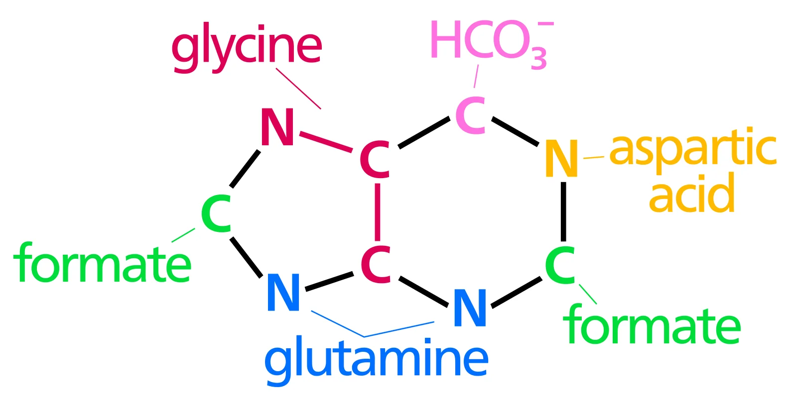
Long description
The biosynthetic origins of purine ring atoms

DefinitionNucleotide
The cell obtains nucleotides by two complementary strategies:
- De novo synthesis – construction of the purine ring on a ribose‑5‑phosphate backbone and assembly of the pyrimidine ring from simple precursors.
- Salvage pathways – recycling of free bases and nucleosides that arise from nucleic‑acid turnover or dietary intake.
Both routes converge on the formation of the activated nucleotides IMP, UMP, GMP and CMP, which are then phosphorylated to the triphosphate forms used for nucleic‑acid polymerisation, signalling and co‑factor synthesis.
The purine pathway builds the heterocyclic ring on 5‑phosphoribosyl‑1‑pyrophosphate (PRPP). The first committed step is catalysed by PRPP amidotransferase, which converts PRPP and glutamine to 5‑phosphoribosyl‑amine (PRA) while releasing glutamate. Subsequent reactions add carbon and nitrogen atoms from glycine, formyl‑tetrahydrofolate, CO₂ and aspartate, ultimately yielding inosine monophosphate (IMP). IMP is the branch point for the synthesis of adenosine monophosphate (AMP) and guanosine monophosphate (GMP) via distinct enzymatic routes. [1]
Key regulatory features:
- Feedback inhibition – AMP and GMP inhibit PRPP amidotransferase, preventing excess purine production.
- PRPP availability – high PRPP levels stimulate the pathway; low PRPP limits flux.
Pyrimidine synthesis begins with the formation of carbamoyl phosphate from glutamine, CO₂ and ATP, catalysed by carbamoyl phosphate synthetase II (CPSII). Carbamoyl phosphate condenses with aspartate to form carbamoyl‑aspartate, which is cyclised to dihydroorotate, oxidised to orotate, and finally attached to PRPP to give orotidine‑5′‑monophosphate (OMP). Decarboxylation of OMP yields uridine‑5′‑monophosphate (UMP), the precursor for all pyrimidine nucleotides.
Regulation mirrors the purine pathway:
- Feedback inhibition – UTP inhibits CPSII, while PRPP stimulates it.
- Cross‑pathway control – high levels of purine nucleotides can suppress pyrimidine synthesis, balancing the overall nucleotide pool.
Several anticancer and antimicrobial agents target these pathways:
| Drug class | Primary target | Clinical use |
|---|---|---|
| Antimetabolites (e.g., methotrexate) | Dihydrofolate reductase → ↓ 10‑formyl‑THF for purine synthesis | Oncology, rheumatoid arthritis |
| 6‑Mercaptopurine, azathioprine | Thiopurine nucleotides incorporated into DNA; also inhibit IMP dehydrogenase | Leukemia, immunosuppression |
| Fluorouracil | Thymidylate synthase (pyrimidine pathway) → ↓ dTMP | Colorectal and breast cancers |
| Pemetrexed | Multiple folate‑dependent enzymes (including GAR transformylase) | Mesothelioma, non‑small cell lung cancer |
| Mycophenolic acid | IMP dehydrogenase → ↓ GMP synthesis | Transplant immunosuppression |
These agents exploit the reliance of rapidly dividing cells on abundant nucleotide pools; inhibition leads to replication stress and cell death.
DefinitionSalvage pathway
The key enzyme hypoxanthine‑guanine phosphoribosyltransferase (HPRT) converts hypoxanthine to IMP and guanine to GMP, using PRPP as the ribose donor. In HPRT deficiency, the substrates accumulate and are oxidised to uric acid by xanthine oxidase, while excess PRPP fuels de novo synthesis, amplifying uric‑acid production. The combined effect of reduced recycling and increased synthesis explains the marked hyperuricaemia seen in this disorder. [1]
Other purine salvage enzymes include:
- Adenine phosphoribosyltransferase (APRT) – adenine → AMP.
- Purine nucleoside phosphorylase (PNP) – inosine/guanosine → hypoxanthine/guanine, which then re‑enter the HPRT route.
Pyrimidine bases are salvaged by phosphoribosyltransferases:
- Uracil phosphoribosyltransferase (UPRT) – uracil → UMP.
- Thymidine kinase (TK) – thymidine → TMP (phosphorylated directly to dTMP).
- Cytidine kinase – cytidine → CMP.
These reactions are especially important in tissues with high turnover (e.g., bone marrow, intestinal epithelium) and in organisms that lack a complete de novo pathway.
Defects in salvage enzymes produce characteristic metabolic phenotypes:
- Lesch‑Nyhan syndrome – HPRT deficiency, leading to hyperuricaemia, neurological dysfunction and self‑injurious behaviour. [1]
- APRT deficiency – accumulation of 2,8‑dihydroxyadenine crystals causing renal stones.
Therapeutic exploitation includes:
- Allopurinol – xanthine oxidase inhibitor reduces uric‑acid formation downstream of excess hypoxanthine/guanine.
- Purine analogues (e.g., azathioprine) rely on salvage enzymes for activation within target cells, providing selective cytotoxicity.
Nucleotide synthesis overview
- Start with glucose → ribose‑5‑phosphatePentose‑phosphate pathway supplies PRPP
- Question: Is PRPP used for de novo or salvage?
De novo
- Purine de novoPRPP amidotransferase → IMP → AMP/GMP
- Pyrimidine de novoCPSII → UMP → UTP/CMP
Salvage
- Purine salvageHPRT converts hypoxanthine/guanine → IMP/GMP
- Pyrimidine salvageTK, UPRT, cytidine kinase → TMP/UMP
DefinitionLesch‑Nyhan syndrome
Lesch‑Nyhan syndrome (LNS) sits at the severe end of a spectrum of HPRT deficiency. The disease was first described in 1964 by Lesch and Nyhan and linked to a virtually complete loss of HPRT enzymatic activity in 1967[1]. Because the HPRT gene resides on Xq26, inheritance is X‑linked recessive; consequently, affected individuals are almost exclusively male, although isolated cases in females have been reported when the X chromosome carries two pathogenic alterations[1].
HPRT catalyses the salvage of hypoxanthine and guanine to inosine monophosphate (IMP) and guanosine monophosphate (GMP). When the enzyme is absent, purine bases are shunted toward de‑novo synthesis, generating excess uric acid. The resulting hyperuricaemia explains the early‑onset gout and renal calculi that often precede neurological signs. In addition, the lack of salvage deprives cells of purine nucleotides needed for DNA/RNA synthesis, contributing to neurodevelopmental delay and movement disorders.
The classic phenotype (Group 4 in the authors’ four‑group classification) includes:
- Self‑injurious behaviour (biting lips, fingers, or head‑banging)
- Hyperkinetic movement disorders – choreoathetosis, ballismus, spasticity
- Severe motor impairment – inability to stand or walk, dependence for daily activities
Some patients may lack overt self‑destruction or overt mental retardation, illustrating phenotypic variability[1]. Partial HPRT deficiency (Kelly‑Seegmiller syndrome) presents with hyperuricaemia and gout but little or no neurological involvement[1]. The term “Lesch‑Nyhan variants” has been introduced for patients who have gout‑related hyperuricaemia together with mild neurological signs, falling between the two extremes[1].
A diagnosis rests on a combination of clinical, biochemical, enzymatic and molecular data[1]. Key steps are:
- Biochemical screening – markedly elevated urinary uric acid and serum urate, often accompanied by increased orotic acid.
- Enzyme assay – measurement of HPRT activity in erythrocytes or fibroblasts; values <5 % of normal are diagnostic of classic LNS, whereas residual activity of 5–30 % suggests a partial form.
- Molecular confirmation – sequencing of the HPRT gene (nine exons, 654 bp coding region) to identify pathogenic variants. Over 300 disease‑associated mutations have been described, with point mutations predominating in partial deficiency and larger deletions or rearrangements in classic LNS[1].
Because complete HPRT deficiency can mimic cerebral palsy, careful neuro‑developmental assessment is essential[1].
Therapeutic goals are threefold: control hyperuricaemia, mitigate neurological complications, and support functional independence.
- Urate‑lowering therapy – allopurinol or febuxostat reduces stone formation and gout attacks.
- Allopurinol‑responsive neuroprotection – there is no robust evidence that urate reduction improves neurological outcomes; treatment is therefore symptomatic.
- Behavioural strategies – protective helmets, dental guards and constant supervision are required to prevent self‑injury.
- Rehabilitation – physiotherapy, occupational therapy and speech therapy aim to maximise motor function and communication.
- Genetic counselling – carrier testing for female relatives and prenatal diagnosis are offered given the X‑linked inheritance.
DefinitionADA deficiency
Adenosine deaminase (ADA) occupies a central node of the purine salvage pathway, converting adenosine and deoxy‑adenosine to inosine and deoxy‑inosine, respectively. Mutations in the ADA gene abolish this activity, producing a form of SCID that accounts for roughly 10–15 % of SCID cases in outbred populations. The disease incidence in Europe is estimated between 1:375 000 and 1:660 000 live births. [2]
In the absence of ADA, intracellular deoxy‑adenosine is phosphorylated to deoxy‑adenosine triphosphate (dATP). Elevated dATP competitively inhibits ribonucleotide reductase, halting DNA synthesis and triggering apoptosis of developing lymphocytes. Consequently, patients exhibit profound lymphocytopaenia affecting T‑cells, B‑cells and NK cells. Extracellular adenosine also rises, overstimulating G‑protein‑coupled adenosine receptors, especially the A₂A subtype, which exerts immunosuppressive signalling and may further dampen immune responses. [3]
- Immunological phenotype – recurrent, severe infections (bacterial, viral, fungal) within the first months of life; failure to thrive; absent thymic shadow on chest radiograph.
- Non‑immune manifestations – neurodevelopmental delay, sensorineural deafness, skeletal abnormalities (e.g., rib flaring, metaphyseal dysplasia).
- Laboratory findings – lymphocyte count <1 × 10⁹/L, absent T‑cell receptor excision circles (TRECs), markedly reduced ADA activity in red cells or fibroblasts.
- Enzyme assay – ADA activity <5 % of normal in erythrocytes confirms the biochemical defect.
- Molecular testing – sequencing of the ADA gene identifies pathogenic variants; over 200 mutations have been reported.
- Immunophenotyping – flow cytometry demonstrates paucity of CD3⁺ T‑cells, CD19⁺ B‑cells and CD56⁺ NK cells.
- Functional assays – accumulation of deoxy‑adenosine and dATP can be measured by high‑performance liquid chromatography.
Because ADA deficiency is life‑threatening, early definitive therapy is mandatory.
| Modality | Principle | Typical timing |
|---|---|---|
| Hematopoietic stem‑cell transplantation (HSCT) | Replaces the defective immune system with donor‑derived progenitors; curative in >80 % of matched sibling transplants. | First year of life, ideally before severe infections. |
| Enzyme replacement therapy (ERT) – PEG‑ADA | Polyethylene glycol‑conjugated ADA provides systemic enzymatic activity, lowering toxic metabolites. | Bridge to HSCT or as definitive therapy when transplant is unavailable. |
| Gene therapy | Autologous CD34⁺ cells are transduced with a functional ADA cassette and reinfused; long‑term immune reconstitution observed in clinical trials. | Emerging first‑line option in centres with expertise. |
PEG‑ADA treatment markedly increases extracellular adenosine catabolism, but chronic exposure can up‑regulate CD73, a surface ectoenzyme that converts ATP/ADP to adenosine. While this initially compensates for the metabolic block, prolonged PEG‑ADA may paradoxically impair regulatory T‑cell (Treg) function by excessive adenosine turnover. [3]
Patients receiving successful HSCT or gene therapy achieve normal lymphocyte counts and vaccine responses, with a markedly reduced risk of infection. Those managed with long‑term PEG‑ADA require regular monitoring of serum adenosine, liver function, and immunoglobulin levels. Neurodevelopmental outcomes depend on the age at treatment; earlier intervention correlates with better cognitive scores.
Diagnostic and therapeutic pathway for ADA‑deficient SCID
- Clinical suspicion (recurrent infections, failure to thrive)
- Question: Lymphocyte count <1 × 10⁹/L?
Yes
- Measure ADA activity
- Question: ADA activity <5 %?
Yes
- Molecular confirmation (ADA gene sequencing)
- Initiate definitive therapy (HSCT, PEG‑ADA, gene therapy)
No
- Investigate other SCID forms
No
- Consider alternative diagnoses
DefinitionOrotic aciduria
Orotic aciduria results from a deficiency of the enzyme uridine‑monophosphate synthase (UMPS), which catalyses the final two steps of de‑novo pyrimidine synthesis: the conversion of orotic acid to orotidine‑5′‑monophosphate and then to uridine‑monophosphate (UMP). When UMPS activity is absent or severely reduced, orotic acid accumulates in the cytosol and is transported into the urine, giving the eponymous biochemical hallmark.
- Block in de‑novo synthesis – The lack of UMP forces cells to rely on salvage pathways that recycle pre‑formed bases and nucleosides.
- Impaired DNA synthesis – Insufficient UMP limits the pool of thymidine nucleotides, producing a megaloblastic picture despite normal folate status.
- Feedback dysregulation – Accumulated orotic acid can inhibit carbamoyl‑phosphate synthetase II, further reducing pyrimidine synthesis.
- Hematology – Macrocytic anaemia with hypersegmented neutrophils; platelets and white‑cell counts are usually normal.
- Growth – Infants may present with failure to thrive, developmental delay, or recurrent infections due to ineffective haematopoiesis.
- Renal – Persistent orotic aciduria is detectable on routine urine organic‑acid analysis.
- Urine organic‑acid chromatography shows a markedly elevated orotic acid peak.
- Serum folate and B12 are normal, helping to distinguish from nutritional megaloblastic anaemia.
- Enzyme assay (if available) demonstrates reduced UMPS activity in cultured fibroblasts or lymphocytes.
- Molecular testing can identify pathogenic variants in the UMPS gene, confirming the diagnosis and enabling carrier testing.
- Uridine supplementation bypasses the metabolic block, providing the missing pyrimidine nucleoside and correcting the haematological abnormality.
- Dietary measures are not curative but avoiding excessive protein that may increase orotic acid production can be helpful.
- Monitoring includes regular complete blood counts, growth parameters, and urine orotic acid levels to gauge treatment efficacy.
Clinical pearlUniversality and redundancy
| Feature | Description |
|---|---|
| Triplet nature | Each codon consists of three nucleotides, giving 4³ = 64 possible codons. |
| Start codon | AUG serves as the canonical initiation signal and also codes for methionine. |
| Stop codons | UAA, UAG and UGA do not specify amino acids; they terminate translation. |
| Degeneracy | 61 codons encode 20 standard amino acids; redundancy reduces the impact of point mutations. |
| Wobble | The third base of a codon can tolerate non‑standard pairing, allowing a single tRNA to recognise multiple codons. |
| Non‑standard codes | Mitochondrial genomes and a few nuclear genomes use variant assignments (e.g., AUA for methionine in mitochondria). |
- Base‑pairing rules: A↔U and G↔C are strict; G can also pair with U at the wobble position.
- tRNA identity: Each tRNA carries a specific aminoacyl‑tRNA synthetase that charges it with the correct amino acid, ensuring fidelity.
- The code likely arose from a co‑evolution of amino‑acyl‑tRNA synthetases and early metabolic pathways, favouring assignments that minimise the deleterious effects of translational errors.
- Codon bias reflects differential usage of synonymous codons, influencing translation efficiency and protein folding.
Exam trapMisinterpreting orotic aciduria
- Confusing with hereditary orotic aciduria – Some patients with urea‑cycle disorders (e.g., ornithine transcarbamylase deficiency) also excrete orotic acid; clinical context and ammonia levels differentiate them.
- Overlooking the need for uridine – Treating the anaemia with folate or B12 alone will not correct the metabolic block; uridine is the definitive therapy.
- Neglecting carrier testing – Because the condition is autosomal recessive, failure to counsel families about recurrence risk can lead to unexpected affected siblings.
In the context of the genetic code:
- Assuming absolute universality – Mitochondrial genomes and some protozoa use alternative codon assignments; prescribing antibiotics that target bacterial ribosomes must consider these differences.
- Ignoring wobble flexibility – Designing synthetic genes without accounting for codon‑anticodon pairing can result in poor expression in heterologous systems.
NoteClinical genetics and metabolic medicine
- Newborn screening – Tandem mass spectrometry can detect elevated orotic acid, prompting early uridine therapy and preventing irreversible neurodevelopmental damage.
- Pharmacogenomics – Understanding codon usage bias guides the optimisation of recombinant protein production for biologics, ensuring high yield and correct folding.
- Gene therapy – Vector design must respect the host’s codon preferences and avoid rare codons that could stall translation.
- Antimicrobial development – Exploiting differences in mitochondrial versus bacterial codon usage helps to design agents with selective toxicity.
Diagnostic approach to macrocytic anaemia with orotic aciduria
- Identify macrocytosisCBC shows MCV > 100 fL
- Question: Is folate/B12 low?
Yes
- Treat deficiencySupplement folate or B12
No
- Measure urine orotic acidOrganic‑acid chromatography
- Question: Is orotic acid markedly elevated?
Yes
- Start uridine therapyOral uridine 100 mg/kg/day
No
- Investigate other causesUrea‑cycle disorders, drug effects
- Orotic aciduria is caused by UMPS deficiency, leading to excess urinary orotic acid and a folate‑unresponsive megaloblastic anaemia.
- Uridine supplementation bypasses the metabolic block and corrects the haematological defect.
- The genetic code is triplet‑based, nearly universal, and degenerate, with three dedicated stop codons.
- Wobble pairing at the third codon position allows a single tRNA to read multiple synonymous codons.
- Mitochondrial genomes use variant codon assignments, a key consideration in pharmacogenomics.
- Misdiagnosing orotic aciduria as a simple folate deficiency delays effective treatment.
- Codon bias influences recombinant protein expression and must be accounted for in gene‑therapy vector design.
- Newborn screening can detect orotic aciduria early, enabling prompt uridine therapy.
Answer each one in your head first, then open the answer.
What enzyme deficiency underlies orotic aciduria?
Show answerHide answer
Uridine‑monophosphate synthase (UMPS)Which three codons act as translation stop signals?
Show answerHide answer
UAA, UAG and UGAWhat is the primary treatment that corrects the haematological abnormality in orotic aciduria?
Show answerHide answer
Oral uridine supplementationWhich amino acid is uniquely encoded by the start codon AUG?
Show answerHide answer
MethionineWhy must codon bias be considered when designing a recombinant gene for human expression?
Show answerHide answer
Because synonymous codons are used at different frequencies, affecting translation efficiency and protein yield
- Torres RJ, Puig JG. Hypoxanthine-guanine phosophoribosyltransferase (HPRT) deficiency: Lesch-Nyhan syndrome. Orphanet journal of rare diseases 2007;2:48. doi:10.1186/1750-1172-2-48. Accessed .
- Flinn AM, Gennery AR. Adenosine deaminase deficiency: a review. Orphanet journal of rare diseases 2018;13(1):65. doi:10.1186/s13023-018-0807-5. Accessed .
- Sauer AV, Brigida I, Carriglio N, Aiuti A. Autoimmune dysregulation and purine metabolism in adenosine deaminase deficiency. Frontiers in immunology 2012;3:265. doi:10.3389/fimmu.2012.00265. Accessed .
Figure credits
Educational content for medical students and doctors. It is not a substitute for clinical judgement or local guidelines.