Patterns of inheritance and single-gene disorders
Patterns of inheritance explain how single‑gene disorders are transmitted across generations.
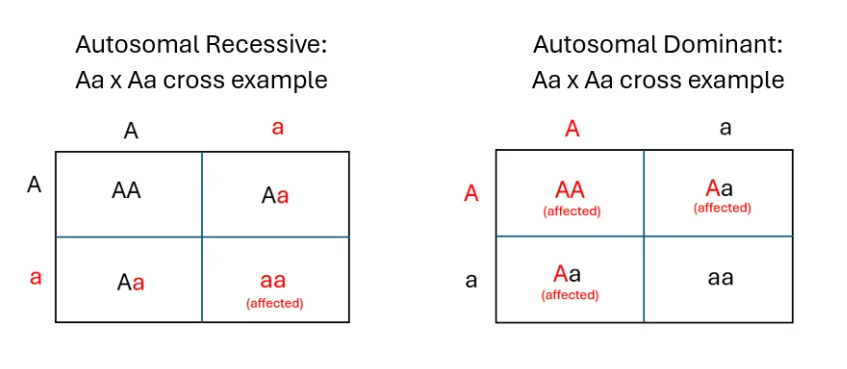
Figure 1Autosomal recessive and dominant inheritance patterns 2025
Long description
Autosomal recessive and dominant inheritance patterns demonstrated through punnett square examples

DefinitionInheritance pattern
The predictable way a pathogenic variant is passed from parent to offspring, determined by the gene’s chromosomal location and the nature of the mutation.
Single‑gene (monogenic) disorders arise when a pathogenic variant in one gene is sufficient to cause disease. The pattern of inheritance—autosomal dominant, autosomal recessive, X‑linked, or mitochondrial—governs the risk to relatives, the clinical presentation, and often the molecular testing strategy.
Autosomal genes reside on the 22 non‑sex chromosomes and are present in two copies per cell.
- Autosomal dominant (AD) – a single pathogenic allele can produce disease. Affected individuals usually have one mutant and one normal allele; each child inherits a 50 % chance of receiving the mutant allele.
- Autosomal recessive (AR) – disease manifests only when both alleles carry pathogenic variants. Carriers (heterozygotes) are typically asymptomatic; each child of two carriers has a 25 % chance of being affected, a 50 % chance of being a carrier, and a 25 % chance of being unaffected.
The same gene can display both AD and AR inheritance, depending on the mutation’s effect on the protein. For example, mutations in SLC4A1 (AE1) cause distal renal tubular acidosis (dRTA) by either AD or AR mechanisms. Dominant mutants such as R589H, R901X and S613F are retained intracellularly, producing a dominant‑negative effect, whereas recessive mutants are rescued to the cell surface by the wild‑type protein, showing a dominant‑positive effect【P582】【P591】. [4]
Mitochondrial disorders illustrate how nuclear‑encoded genes can follow either AD or AR inheritance. Multiple mtDNA deletions or depletion secondary to nuclear DNA (nDNA) mutations may be inherited in an autosomal recessive or dominant fashion; genes such as POLG, POLG2, SLC25A4, C10orf2, SPG7, DNA2, RNASEH1, TOP3A, TK2, DGUOK, RRM2B, GMPR, LIG3, and RRM1 are implicated【P254】. Early‑onset, often fatal, presentations are usually linked to AR depletion, while adult‑onset phenotypes like progressive external ophthalmoplegia (PEO) are often seen. [2]
Similarly, POLG‑related disease shows a clear genotype‑phenotype correlation: biallelic pathogenic variants cause early‑onset, autosomal recessive disease, whereas a single heterozygous variant can produce late‑onset, autosomal dominant PEO【P296】. [2]
| Gene | Inheritance pattern(s) | Typical phenotype |
|---|---|---|
| SLC4A1 | AD (dominant‑negative) / AR (dominant‑positive) | Distal renal tubular acidosis |
| POLG | AR (biallelic) – early‑onset; AD (heterozygous) – late‑onset PEO | Mitochondrial myopathy, PEO |
| POLG2, SLC25A4, C10orf2, SPG7, DNA2, RNASEH1, TOP3A, TK2, DGUOK, RRM2B, GMPR, LIG3, RRM1 | AD or AR (depends on specific mutation) | Multiple mtDNA deletions or depletion, variable neuromuscular disease |
X‑linked genes reside on the X chromosome. Because males have one X chromosome, a pathogenic variant is fully expressed in hemizygous males, while females, with two X chromosomes, are usually carriers and may show milder or variable disease due to X‑inactivation.
- X‑linked recessive – most common pattern; affected males transmit the mutant allele to all daughters (who become carriers) and none of their sons. Carrier females have a 50 % chance of passing the mutant allele to each child, producing affected sons half the time. Classic examples include Duchenne muscular dystrophy (DMD) and hemophilia A.
- X‑linked dominant – rarer; both males and females can be affected. Affected males transmit the mutation to all daughters and no sons; affected females transmit to 50 % of offspring of each sex. Lethal X‑linked dominant disorders often result in male miscarriage.
The distinction between dominant and recessive X‑linked inheritance is crucial for counselling families and for interpreting genetic test results, especially when a female carrier presents with symptoms due to skewed X‑inactivation.
Mitochondria possess their own circular DNA (mtDNA), inherited almost exclusively from the mother because the oocyte contributes the bulk of cytoplasm to the embryo. Consequently, mtDNA mutations follow a maternal (non‑Mendelian) inheritance pattern.
Pathogenic mtDNA variants can be:
- Point mutations – single nucleotide changes (e.g., NADH dehydrogenase subunit 4 mutation in Leber hereditary optic neuropathy)【P159】. [2]
- Large‑scale deletions – up to 7 kb deletions identified in mitochondrial myopathies【P159】. [2]
- Depletion – reduced copy number of mtDNA, often associated with early‑onset, severe disease【P254】.
Because each cell contains many mitochondria, heteroplasmy (a mixture of mutant and wild‑type mtDNA) determines disease severity. The proportion of mutant mtDNA can shift between tissues and across generations, leading to variable penetrance.
Mitochondrial diseases may also arise from nuclear genes that encode proteins required for mtDNA replication, transcription, translation, or OXPHOS assembly. These nDNA‑encoded mitochondrial disorders follow classic Mendelian inheritance (AD or AR) but ultimately affect mitochondrial function. The discovery of large‑scale mtDNA deletions and subsequent identification of nuclear contributors marked the start of the molecular era of mitochondrial genetics【P159】. [2]
- Maternal transmission: children of an affected mother are at risk; children of an affected father are not. [2]
- Heteroplasmy threshold: disease manifests when mutant mtDNA exceeds a tissue‑specific threshold, often around 60‑80 % for severe phenotypes.
- Antenatal testing: pre‑implantation genetic diagnosis can select embryos with low heteroplasmy levels, reducing disease risk.
Understanding the underlying inheritance guides:
- Family history assessment – pedigree analysis reveals dominant, recessive, X‑linked, or maternal patterns.
- Genetic testing strategy – targeted gene panels for AD/AR nuclear genes, mtDNA sequencing for maternal cases, and X‑chromosome analysis for suspected X‑linked disease.
- Counselling – risk estimates differ markedly: 50 % for AD, 25 % for AR, 50 % for carrier females in X‑linked recessive, and near‑100 % maternal transmission for mtDNA mutations.
Diagnostic pathway for single‑gene disorders
- Take detailed three‑generation pedigreeIdentify pattern of affected individuals
- Question: Is inheritance autosomal?
Yes
- Question: Dominant or recessive?
Dominant
- Test single gene panel (AD)Consider de novo variants
Recessive
- Test gene panel (AR) or exomeLook for biallelic variants
No
- Question: Is X‑linked?
Yes
- Test X‑chromosome genesConsider sex of proband
No
- Question: Maternal transmission?
Yes
- Sequence mtDNAAssess heteroplasmy
No
- Consider de novo or complex inheritance
DefinitionPedigree
A pedigree is a diagrammatic representation of family relationships that records the presence or absence of a trait in each individual, allowing patterns of inheritance to be visualised. [1]
Pedigrees are the first step in recognising whether a disorder follows an autosomal recessive, autosomal dominant, X‑linked, or mitochondrial pattern. In an autosomal recessive pedigree the disease appears only when a child inherits two pathogenic alleles, one from each parent. The parents are typically unaffected carriers, shown as half‑filled symbols, and the disease may skip generations. When a disease is X‑linked recessive, affected males are shown as filled squares, while carrier females are half‑filled circles; the trait often appears only in males and is transmitted through carrier mothers.
Cystic fibrosis (CF) is the classic example of an autosomal recessive single‑gene disorder. The CFTR gene on chromosome 7q31.2 encodes a chloride channel that regulates fluid transport across epithelial surfaces. Pathogenic variants reduce or abolish channel function, leading to thickened secretions in the lung, pancreas, and other organs. Because the disease requires two defective alleles, the probability that two carrier parents will have an affected child is 25 % for each pregnancy, while each child has a 50 % chance of being a carrier and a 25 % chance of being completely unaffected.
The most common pathogenic variant in populations of northern European descent is the ΔF508 deletion, which removes phenylalanine at position 508. However, more than 2 000 different CFTR mutations have been described, and the spectrum varies with ethnicity. Genetic testing for CF therefore includes a panel of the most frequent mutations plus sequencing of the entire coding region when the panel is negative but clinical suspicion remains.
When interpreting a pedigree for CF, the key features are:
- Both sexes are equally affected.
- No vertical transmission (the disease does not appear in parents).
- Consanguinity increases the likelihood of homozygosity for rare mutations.
- Carrier testing of parents and at‑risk relatives can be offered once the familial mutations are known.
Understanding these principles enables clinicians to counsel families about recurrence risk, carrier screening, and prenatal diagnosis.
DefinitionMuscular dystrophy
Muscular dystrophies are a heterogeneous group of inherited disorders characterised by progressive muscle fibre degeneration and replacement by fibrous or fatty tissue.
The muscular dystrophy spectrum ranges from congenital forms that present at birth to adult‑onset limb‑girdle phenotypes. Two broad categories are relevant for medical students:
CMDs are defined as early‑onset muscle disorders with a dystrophic pattern on biopsy, even if the classic dystrophic changes are not fully developed. Overlap exists with congenital myopathies; for example, mutations in RYR1 and SEPN1 can produce both core myopathies and CMD‑like presentations. [1]
Key genetic subtypes include:
| Gene | Typical phenotype | Notable features |
|---|---|---|
| COL6A1‑3 (COL6‑RD) | Severe contractures, joint hypermobility, cardiac involvement is not a feature | Differentiated from LMNA‑RD and Emery‑Dreifuss muscular dystrophy which do involve the heart [1] |
| LAMA2 | Partial laminin‑α2 deficiency, prominent joint contractures | Often considered in milder COL6‑RD phenotypes |
| LMNA | Early‑onset CMD (LMNA‑CMD) and Emery‑Dreifuss muscular dystrophy | Scapuloperoneal weakness, contractures, cardiomyopathy |
| FKRP | Fukuyama‑type CMD, secondary laminin‑α2 deficiency | Associated with brain malformations in the Japanese population |
| FKBP14 | Kyphoscoliotic Ehlers–Danlos overlap syndrome | Severe kyphoscoliosis with myopathy |
These genes encode proteins that maintain the extracellular matrix, nuclear envelope, or intracellular calcium handling, illustrating how diverse pathways converge on a dystrophic muscle phenotype.
LGMDs are characterised by symmetric weakness of the pelvic and shoulder girdle, usually with a later onset than CMDs. The nomenclature reflects the underlying gene (e.g., LGMD2I caused by FKRP mutations). Other “titinopathic” phenotypes, such as tibial muscular dystrophy (TMD, Udd myopathy), arise from mutations in the giant sarcomeric protein titin. [3]
- Clinical assessment – pattern of weakness, contractures, cardiac involvement, and extra‑muscular features.
- Serum creatine kinase – often markedly elevated in dystrophic processes.
- Muscle biopsy – dystrophic changes (fiber size variation, necrosis, fibrosis) support a CMD or LGMD diagnosis.
- Genetic testing – targeted panels or whole‑exome sequencing to identify pathogenic variants in the relevant genes.
Early genetic confirmation guides prognosis, informs cardiac surveillance (especially in LMNA‑related disease), and enables family counselling.
DefinitionTrinucleotide repeat expansion
A trinucleotide repeat disease results from the abnormal expansion of a three‑nucleotide sequence within a gene, leading to toxic gain‑of‑function or loss‑of‑function effects.
These disorders illustrate how a single‑gene mutation can produce a spectrum of severity that correlates with repeat length. The repeat can be located in coding, untranslated, or intronic regions, and pathogenic mechanisms include:
- Protein aggregation – expanded polyglutamine tracts (e.g., CAG repeats) form intracellular inclusions that disrupt cellular homeostasis.
- RNA toxicity – long repeats in non‑coding regions sequester RNA‑binding proteins, impairing splicing and translation.
- Loss of protein function – large expansions may silence transcription or trigger nonsense‑mediated decay.
| Disease | Gene | Repeat | Typical presentation |
|---|---|---|---|
| Huntington disease | HTT | CAG (polyglutamine) | Adult‑onset chorea, psychiatric changes |
| Myotonic dystrophy type 1 | DMPK | CTG (non‑coding) | Myotonia, cataracts, cardiac conduction defects |
| Friedreich ataxia | FXN | GAA (intron) | Progressive ataxia, cardiomyopathy |
| Spinocerebellar ataxias (multiple) | Various | CAG | Cerebellar degeneration, variable age of onset |
A hallmark of these conditions is anticipation: successive generations inherit longer repeats, leading to earlier onset and more severe disease. This phenomenon is especially evident in paternal transmission of Huntington disease, where spermatogenesis is prone to repeat expansion.
- Molecular testing – PCR or Southern blotting quantifies repeat length; values above a disease‑specific threshold confirm the diagnosis.
- Genotype‑phenotype correlation – larger repeats generally predict earlier onset, but modifiers (e.g., DNA repair capacity) can influence the threshold.
- Counselling – because repeat length can change in germ cells, predictive testing for at‑risk relatives must address uncertainty about future expansion.
Understanding trinucleotide repeat diseases reinforces the principle that a single‑gene defect can generate a spectrum of clinical phenotypes through dynamic mutation mechanisms, distinct from the static point mutations seen in many other single‑gene disorders.
DefinitionWhat is Fragile X?
Fragile X syndrome is the most common inherited cause of intellectual disability and autism, resulting from a trinucleotide (CGG) repeat expansion in the 5′‑untranslated region of the FMR1 gene on the X chromosome.
The expanded repeat leads to hyper‑methylation of the FMR1 promoter, silencing transcription and causing loss of the fragile‑mental‑retardation‑protein (FMRP). FMRP is an RNA‑binding protein that regulates synaptic plasticity by controlling the translation of many neuronal mRNAs. Without sufficient FMRP, dendritic spine morphology is abnormal and long‑term potentiation is impaired, underpinning the cognitive and behavioural phenotype.
Inheritance follows an X‑linked pattern. A mother who carries a premutation (55–200 CGG repeats) can expand to a full mutation (>200 repeats) in her oocytes, giving a 50 % chance of transmitting the disorder to sons (who will be affected) and a 50 % chance to daughters (who become carriers). Males are usually more severely affected because they have only one X chromosome.
Clinically, affected individuals display moderate to severe intellectual disability, characteristic facial features (long face, prominent ears, high‑arched palate), macroorchidism after puberty, and a high prevalence of anxiety, attention‑deficit hyperactivity disorder and autistic traits. Early intervention with speech, occupational and behavioural therapy improves outcomes, and targeted pharmacological approaches (e.g., mGluR5 antagonists) are under investigation.
DefinitionWhat is Rett syndrome?
Rett syndrome is a neurodevelopmental disorder almost exclusively affecting females, caused by pathogenic variants in the MECP2 gene on the X chromosome.
MECP2 encodes methyl‑CpG‑binding protein 2, a transcriptional regulator that modulates chromatin structure and gene expression in mature neurons. Loss‑of‑function mutations disrupt neuronal maturation and synaptic connectivity, leading to the classic regression phase of the disease.
The disorder follows an X‑linked dominant inheritance with male lethality in most cases; therefore, affected individuals are typically heterozygous females. De novo mutations account for the majority of cases, but familial transmission can occur when a mother is a carrier.
After a period of apparently normal development, children with Rett syndrome experience a rapid loss of purposeful hand skills, spoken language and gait, accompanied by the emergence of stereotypic hand‑wringing movements, breathing irregularities and seizures. Cognitive abilities are severely limited, yet many retain the capacity for social engagement.
Management is multidisciplinary: physiotherapy to maintain mobility, communication aids, seizure control, and treatment of autonomic disturbances. Recent research into gene‑replacement strategies and modulation of downstream pathways (e.g., BDNF signalling) offers hope for disease‑modifying therapies.
DefinitionMitochondrial disease overview
Mitochondrial diseases are a heterogeneous group of disorders caused by defects in oxidative phosphorylation, arising from mutations in either mitochondrial DNA (mtDNA) or nuclear DNA (nDNA) that encode mitochondrial proteins.
- mtDNA mutations are maternally inherited because the oocyte contributes the bulk of mitochondria to the embryo. Pathogenic mtDNA mutations can affect women of reproductive age. [3] [2]
- In adults, approximately 80 % of mitochondrial diseases are linked to mtDNA mutations, whereas in children the majority are due to nDNA mutations, with only 20–25 % stemming from mtDNA mutations. [2]
Large‑scale mtDNA deletions produce a spectrum of phenotypes. Pearson syndrome, Kearns‑Sayre syndrome and progressive external ophthalmoplegia (PEO) all share a common deletion, but the timing of the deletion during development influences the clinical picture: late‑stage deletions tend to cause PEO, while earlier deletions may manifest as Kearns‑Sayre or Pearson syndrome. The proportion of mutant mtDNA also matters; patients with Pearson syndrome often harbour a higher load of deleted mtDNA than those with Kearns‑Sayre or PEO. [2]
Oxidative stress is a unifying pathogenic factor. Reduced mitochondrial membrane potential (Δψm) impairs ATP synthesis, calcium handling and mitochondrial quality control (MQC). A collapse of Δψm initiates the mitochondrial integrated stress response (mt‑ISR) and mitophagy, but chronic activation can accelerate tissue damage. [2]
MQC processes such as inner‑membrane fusion, mitophagy and newly described mechanisms (migrasome‑mediated exocytosis, mitolysosome exocytosis, mitopherogenesis) aim to preserve mitochondrial integrity, yet persistent stress leads to maladaptive activation that worsens disease.
Specific disease examples illustrate these mechanisms:
- Kearns‑Sayre syndrome shows altered tau protein in cerebrospinal fluid, linking tau‑mediated ROS generation to mitochondrial dynamics and mPTP opening; cardiomyocytes display enlarged mitochondria, and arrhythmias arise from ATP‑deficient ion channel dysfunction. [2]
- Leigh syndrome models demonstrate that pharmacological activation of PPAR with bezafibrate up‑regulates mitochondrial biogenesis, improves antioxidant capacity and rescues neuronal morphology, extending survival in mouse models. [2]
The first nuclear gene causing a mitochondrial respiratory chain defect (succinate dehydrogenase complex II deficiency) was identified in 1995, paving the way for molecular diagnostics. The creation of the MITOMAP database in 1996 facilitated systematic cataloguing of mtDNA variants. The advent of next‑generation sequencing (NGS) in the 2000s allowed rapid, comprehensive screening of both mtDNA and nDNA genes, while multi‑omics approaches (transcriptomics, proteomics) now provide functional validation and phenotype correlation.
Mitochondrial disease diagnostic pathway
- Patient presents with multisystemic signse.g., neurodevelopmental delay, myopathy, cardiomyopathy
- Question: Is there a maternal inheritance pattern?
Yes
- Screen mtDNA by NGSAssess heteroplasmy level
- Question: Heteroplasmy > 60 %?
Yes
- Confirm pathogenicityReference MITOMAP
No
- Consider nuclear genes
No
- Perform nuclear gene panelInclude OXPHOS and MQC genes
- Integrate functional studiesRespiratory chain enzyme assays, fibroblast stress tests
- Provide genetic counsellingDiscuss inheritance, reproductive options
- Supportive care – optimise nutrition, manage seizures, cardiac monitoring, physiotherapy.
- Targeted therapies – agents that enhance mitochondrial biogenesis (e.g., bezafibrate) or modulate oxidative stress are under investigation.
- Future directions – gene‑editing of mtDNA, allotopic expression of nuclear‑encoded mitochondrial proteins, and modulation of MQC pathways hold promise.
Exam trapAssuming blood always reflects mtDNA status
Blood is often the first tissue sampled, but many mitochondrial deletions are confined to muscle and may be missed if only blood is examined. [2]
- Tissue bias – Large‑scale mt‑DNA deletions such as those causing progressive external ophthalmoplegia are frequently undetectable in blood, leading to false‑negative results if a muscle biopsy is not performed.
- Heteroplasmy misinterpretation – Low‑level heteroplasmy (as low as 1 % – 10 %) can be present in normal tissue; without quantitative methods the clinical relevance may be over‑ or underestimated.
- Over‑reliance on a single gene test – Single‑gene panels miss atypical presentations (e.g., POLG‑related Alpers‑Huttenlocher syndrome) that may require broader sequencing strategies.
- Neglecting inheritance mode – Autosomal recessive LGMD subtypes (LGMD2B, LGMD2L, LGMD2M) each have distinct carrier frequencies and penetrance patterns; treating them as a homogeneous group obscures counselling nuances.
- Ignoring population genetics – The SLC4A1 Southeast Asian ovalocytosis allele is lethal in the homozygous state, yet its high heterozygous frequency can be misread as a benign polymorphism.
Understanding inheritance patterns guides diagnostic algorithms, informs genetic counselling, and determines therapeutic choices.
- Clinical suspicion of a mitochondrial disorder – If the phenotype suggests a post‑mitotic tissue involvement (e.g., ophthalmoplegia, myopathy), clinicians often prioritize muscle mt‑DNA sequencing before whole‑exome or whole‑genome approaches.
- Sample selection – Blood remains the most common source for mtDNA testing, but urine and affected tissue are added to capture tissue‑specific mutations.
- Quantifying heteroplasmy – Modern NGS pipelines, often preceded by long‑range PCR enrichment, can report heteroplasmy down to 1 % – 10 %.
| Subtype | Gene | Inheritance | Frequency in studied populations | Typical CK elevation |
|---|---|---|---|---|
| LGMD2B | DYSF | Autosomal recessive | 15 % – 25 % of LGMD2 cases | 5 – 20 × normal [3] |
| LGMD2L | ANO5 | Autosomal recessive | 10 % – 20 % in Northern Europe | 5 – 20 × normal [3] |
| LGMD2M | FKTN | Autosomal recessive (hypomorphic allele) | Rare, but notable in specific cohorts | 10 – 50 × normal [3] |
These figures help clinicians estimate carrier risk and discuss prognosis with families.
- Valproate avoidance – In POLG‑related Alpers‑Huttenlocher syndrome, valproate can precipitate severe hepatic failure, a fact that must be communicated during medication selection. [2]
The SLC4A1 ΔAla400‑ΔAla408 deletion (SAO) illustrates how a heterozygous advantage (malaria protection) can drive high allele frequencies (up to 35 % in north‑coast PNG) while homozygosity is lethal. Such dynamics are relevant when interpreting carrier screening results in endemic regions. [4]
Diagnostic pathway for suspected single‑gene neuromuscular disorder
- Clinical assessmentIdentify phenotype (e.g., ophthalmoplegia, proximal weakness)
- Question: Is muscle involvement predominant?
Yes
- Obtain muscle biopsyGold‑standard for mtDNA deletions
- Sequence mtDNA from musclePrior to WES/WGS in adults
No
- Collect blood & urineStandard mtDNA source
- Perform NGS with long‑range PCR enrichmentDetect heteroplasmy ≥1 %
- Question: Heteroplasmy detected?
Yes
- Quantify levelCorrelate with severity
- Counsell familyDiscuss maternal inheritance
No
- Consider nuclear gene panele.g., POLG, DYSF, ANO5, FKTN
- Proceed to WES/WGS if needed
- Interpret results in context of inheritanceAutosomal recessive vs maternal
- Provide management planAvoid contraindicated drugs, monitor CK, refer to physiotherapy
- Mitochondrial deletions are often tissue‑specific; muscle biopsy remains the diagnostic gold standard for certain phenotypes.
- Modern NGS methods can detect heteroplasmy as low as 1 % – 10 % after long‑range PCR enrichment. [2]
- Autosomal recessive LGMD subtypes have distinct population frequencies and CK profiles (LGMD2B 15 % – 25 % with CK 5‑20×; LGMD2L 10 % – 20 % with CK 5‑20×; LGMD2M 10‑50×). [3]
- POLG‑related Alpers‑Huttenlocher syndrome requires avoidance of valproate due to risk of hepatic decompensation.
- The SAO allele illustrates how heterozygote advantage can inflate carrier frequencies while homozygosity is lethal.
Answer each one in your head first, then open the answer.
Why might a muscle biopsy be preferred over blood for mtDNA testing in an adult with suspected PEO?
Show answerHide answer
Because large‑scale deletions are often confined to post‑mitotic muscle and may be undetectable in blood.What heteroplasmy detection limit is achievable with long‑range PCR‑NGS approaches?
Show answerHide answer
1 % – 10 % heteroplasmy.Which LGMD subtype is most common in Northern Europe and what is its typical CK elevation?
Show answerHide answer
LGMD2L; CK 5‑20 × normal.What medication should be avoided in POLG‑related Alpers‑Huttenlocher syndrome?
Show answerHide answer
Valproate.What is the inheritance pattern of most LGMD2 forms discussed?
Show answerHide answer
Autosomal recessive.
- Bönnemann CG, Wang CH, Quijano-Roy S, et al. Diagnostic approach to the congenital muscular dystrophies. Neuromuscular disorders : NMD 2014;24(4):289-311. doi:10.1016/j.nmd.2013.12.011. Accessed .
- Wen H, Deng H, Li B, et al. Mitochondrial diseases: from molecular mechanisms to therapeutic advances. Signal transduction and targeted therapy 2025;10(1):9. doi:10.1038/s41392-024-02044-3. Accessed .
- Nigro V, Savarese M. Genetic basis of limb-girdle muscular dystrophies: the 2014 update. Acta myologica : myopathies and cardiomyopathies : official journal of the Mediterranean Society of Myology 2014;33(1):1-12. Accessed .
- Yenchitsomanus PT, Kittanakom S, Rungroj N, et al. Molecular mechanisms of autosomal dominant and recessive distal renal tubular acidosis caused by SLC4A1 (AE1) mutations. Journal of molecular and genetic medicine : an international journal of biomedical research 2005;1(2):49-62. doi:10.4172/1747-0862.1000013. Accessed .
Figure credits
Educational content for medical students and doctors. It is not a substitute for clinical judgement or local guidelines.