Pentose phosphate pathway and G6PD deficiency
The pentose phosphate pathway supplies NADPH and ribose‑5‑phosphate for biosynthesis and antioxidant defence.
Figure 1Pentose phosphate pathway
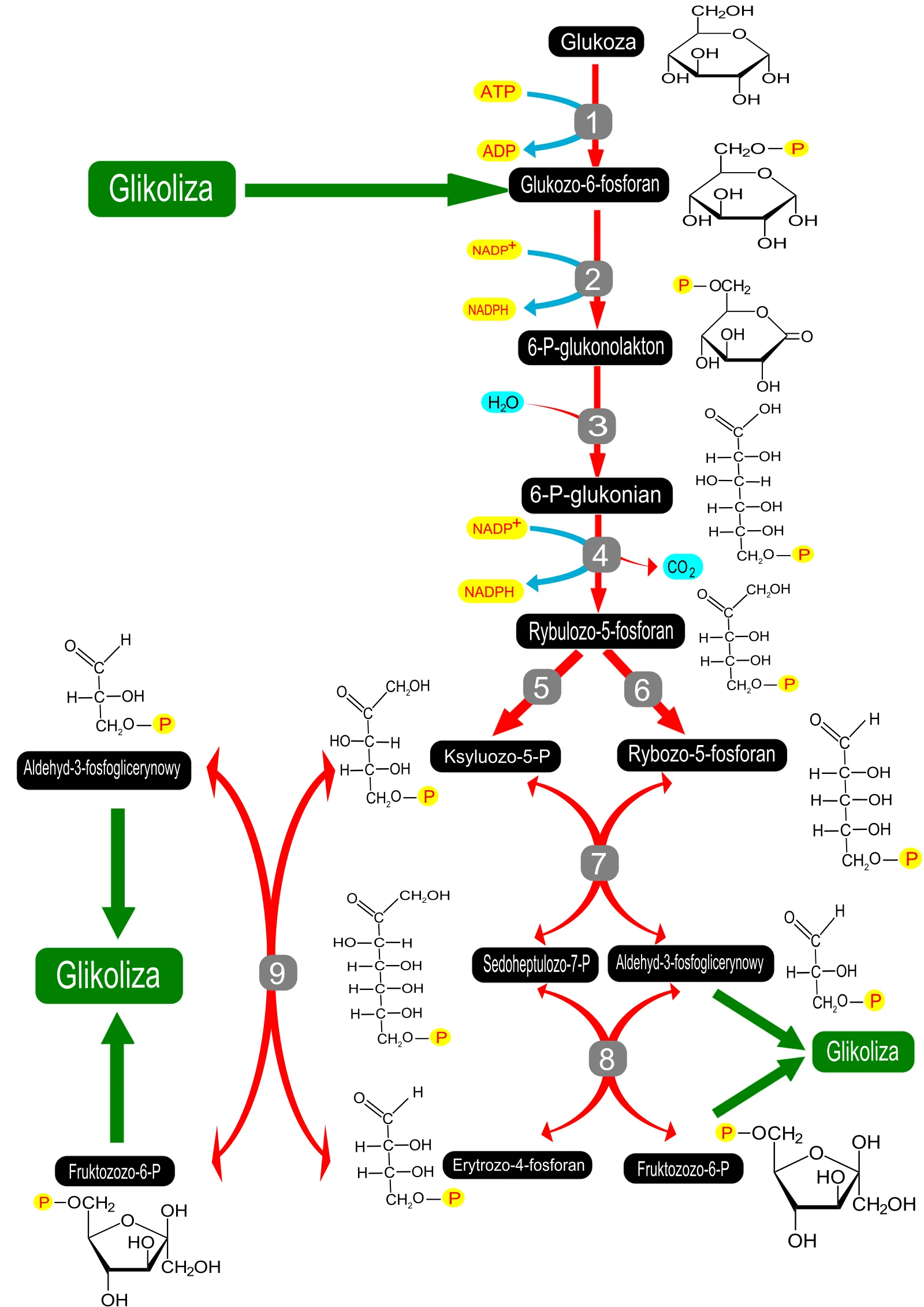
Long description
Illustration for biochemistry/pentose phosphate pathway and g6pd deficiency from Wikimedia Commons, titled 'Pentose phosphate pathway': Pentose phosphate pathway. The author and licence are given in the credit line.

DefinitionPentose phosphate pathway (PPP)
A metabolic route that oxidises glucose‑6‑phosphate to produce NADPH and ribose‑5‑phosphate, essential for reductive biosynthesis and antioxidant defence.
The PPP is divided into two interlinked branches: an oxidative phase that generates NADPH and a non‑oxidative phase that interconverts sugar phosphates for nucleotide synthesis. [2] G6PD (glucose‑6‑phosphate dehydrogenase) is the first and rate‑limiting enzyme of the oxidative phase and acts as a gatekeeper of the pathway. [2]
The oxidative phase begins with G6PD converting glucose‑6‑phosphate (G6P) to 6‑phosphoglucono‑δ‑lactone, which is hydrolysed to 6‑phosphogluconate. 6‑Phosphogluconate dehydrogenase (6PGD) then oxidatively decarboxylates it to ribulose‑5‑phosphate (Ru5P), producing two molecules of NADPH per molecule of glucose oxidised.
During this phase, NADP⁺ is the electron acceptor in both reactions, so the oxidative phase is the sole source of NADPH in the PPP.
The non‑oxidative phase is catalysed by transketolase (TKT) and transaldolase (TAL), which rearrange sugar phosphates to generate intermediates that can be fed into glycolysis or used for ribose‑5‑phosphate synthesis.
In the parasite Entamoeba histolytica, which lacks G6PDH, 6PGDH and TAL, an alternative hexose‑pentose interconversion pathway involving TKL, FBA and a PPi‑dependent phosphofructokinase supplies pentose phosphates. When exposed to oxidative stress, metabolites of the non‑oxidative branch such as glycerol and chitin biosynthesis are increased, reflecting a shift of carbon flux away from glycolysis and towards the PPP. [1]
NADPH produced by the PPP is a critical reducing equivalent for several cellular processes:
- Glutathione regeneration – NADPH donates electrons to glutathione reductase, maintaining reduced glutathione (GSH) that detoxifies reactive oxygen species (ROS). G6PD‑deficient cells are sensitive to oxidative stress because they cannot generate sufficient NADPH for GSH recycling. [2]
- Respiratory burst in phagocytes – NADPH oxidase uses NADPH to generate superoxide during the oxidative burst, a key antimicrobial mechanism.
- Drug metabolism – NADPH is required by cytochrome P450 enzymes for the oxidation of xenobiotics. Inhibition of the PPP reduces NADPH availability, impairing drug detoxification and increasing susceptibility to haemolytic drugs in G6PD‑deficient individuals.
The balance of NADP⁺/NADPH is tightly regulated. Elevated ROS or inhibition of NADPH‑generating pathways raises the NADP⁺/NADPH ratio, which in turn activates G6PD and stimulates the PPP to restore redox balance. [2]
Clinical pearlG6PD regulation
G6PD activity is controlled by a monomer‑dimer equilibrium; the dimer is active, while the monomer is inactive. p53 can bind G6PD and prevent dimerisation, whereas NADP⁺ stabilises the dimer by blocking this interaction.
| Enzyme | Reaction | Cofactor | Product |
|---|---|---|---|
| G6PD | G6P → 6‑phosphoglucono‑δ‑lactone | NADP⁺ → NADPH | 6‑phosphogluconate |
| 6PGD | 6‑phosphogluconate → ribulose‑5‑phosphate + CO₂ | NADP⁺ → NADPH | Ru5P |
| TKT | Transfers two‑carbon units between sugars | — | Various sugar phosphates |
| TAL | Transfers three‑carbon units | — | Various sugar phosphates |
G6PD deficiency is an X‑linked inherited disorder characterised by markedly reduced G6PD activity in red cells, leading to haemolytic anaemia when exposed to oxidative stressors such as certain drugs or infections. The deficiency must be considered in the differential diagnosis of haemolytic anaemia and when prescribing potentially haemolytic drugs. In areas with high prevalence, G6PD testing is essential before administering drugs like primaquine or dapsone. [3]
DefinitionG6PD deficiency
A hereditary enzymopathy characterised by reduced activity of glucose‑6‑phosphate dehydrogenase (G6PD), the first enzyme of the pentose phosphate pathway (PPP). It is inherited in an X‑linked recessive manner and is common in malaria‑endemic regions.
The clinical spectrum ranges from asymptomatic mild anaemia to severe, life‑threatening acute haemolytic anaemia (AHA) when red cells are exposed to oxidative stress. The severity of haemolysis is influenced by the specific G6PD variant and the proportion of deficient red cells. For example, the G6PD Coimbra and G6PD Mediterranean variants tend to produce more frequent and severe episodes of AHA than variants with higher mean activity such as G6PD Seattle and G6PD Kalyan‑Kerala. In a heterozygous female, the G6PD Seattle variant can cause [1]
The X‑linked inheritance pattern means that males have a single allele; a pathogenic mutation results in all red cells being deficient. Females may be homozygous, heterozygous or mosaic due to X‑chromosome inactivation. Heterozygous females therefore harbour a mixed population of normal and deficient red cells, with a variable ratio that can range widely around an average of 50:50. The number of known mutations exceeds 187, and at least 35 are common polymorphisms worldwide. None of these mutations causes complete loss of G6PD, which would be lethal; instead, they reduce enzyme stability or catalytic efficiency, accelerating the normal decline of activity as red cells age.
The residual activity of the enzyme is a key determinant of clinical risk. For instance, G6PD Mediterranean retains only about 5 % of normal activity, whereas G6PD A‑ retains about 13 %. However, the relationship between residual activity and haemolytic severity is not linear, as qualitative differences in the enzyme also play a role. [3]
DefinitionHeinz bodies
Intracellular inclusions of denatured haemoglobin that form when red cells are exposed to oxidative stress. They are visible on a peripheral smear after staining with supravital dyes and are a hallmark of G6PD‑related haemolysis.
Oxidative triggers include certain drugs, infections, and foods. Primaquine and other antimalarials are classic examples; they can precipitate AHA in G6PD‑deficient individuals. The risk of drug‑induced haemolysis persists because many clinicians still prescribe these agents without prior G6PD screening, especially in high‑prevalence populations. Other drugs such as rasburicase and dapsone have also been implicated. In a study of dapsone, 10 % of patients developed AHA, but all affected individuals were G6PD deficient, indicating that the risk in G6PD‑normal patients is essentially zero while it is near 100 % in deficient patients. [3]
Infections can generate reactive oxygen species that overwhelm the compromised PPP, leading to oxidative damage of haemoglobin. The resulting Heinz bodies are phagocytosed by splenic macrophages, contributing to extravascular haemolysis. The presence of Heinz bodies on a smear is therefore a diagnostic clue to G6PD‑related oxidative haemolysis.
The pathophysiology of drug‑induced haemolysis involves inhibition of G6PD activity or generation of oxidants that exceed the capacity of the PPP to produce NADPH, the reducing equivalent required for glutathione regeneration. Without sufficient NADPH, red cells cannot detoxify peroxides, leading to oxidative damage, Heinz body formation, and ultimately haemolysis.
DefinitionChronic granulomatous disease (CGD)
A genetic disorder characterised by defective NADPH oxidase activity in phagocytes, leading to impaired production of reactive oxygen species and recurrent infections.
Chronic granulomatous disease is caused by mutations in genes encoding subunits of the NADPH oxidase complex. The enzyme complex normally transfers electrons from NADPH to molecular oxygen, generating superoxide anion, a key microbicidal oxidant. In CGD, this electron transfer is defective, so phagocytes cannot produce sufficient reactive oxygen species to kill ingested pathogens. Consequently, patients develop granulomatous inflammation and are prone to infections with catalase‑positive organisms such as Staphylococcus aureus, Streptococcus spp., Nocardia, Listeria and Candida.
The clinical presentation is highly variable. Early childhood is the most common period for diagnosis, with recurrent bacterial and fungal infections, especially in the lungs, skin, lymph nodes and liver. Granulomas may form in almost any organ, leading to complications such as obstructive airway disease, intestinal strictures or hepatic fibrosis. Diagnosis is confirmed by functional assays of the respiratory burst (e.g., nitroblue tetrazolium test or dihydrorhodamine flow cytometry) and genetic testing for mutations in the CYBB, CYBA, NCF1, NCF2 or NCF4 genes.
Management of CGD involves prophylactic antibiotics (trimethoprim‑sulfamethoxazole and fluconazole) and antifungals (voriconazole or posaconazole), interferon‑γ to boost innate immunity, and aggressive treatment of infections. Hematopoietic stem cell transplantation offers a potential cure, particularly in patients with severe disease or refractory infections. Gene therapy is an emerging approach, with early trials demonstrating durable correction of the NADPH oxidase defect.
Exam trapMisinterpretation of G6PD activity during acute haemolysis
During acute haemolytic anaemia, reticulocytes with higher G6PD activity can mask an underlying deficiency, leading to a false‑normal result. A repeat test after 2 months is recommended. [3]
Clinical pearlRapid G6PD testing in emergencies
A point‑of‑care G6PD test can be performed in 20 minutes, with results available within an hour if performed on‑site, which is critical when haemolytic drugs are considered.
NoteDNA‑based testing limitations
DNA testing is useful when a known mutation is present, but if the mutation is not detected, phenotypic testing remains essential because unexpected variants may exist.
DefinitionPentose phosphate pathway (PPP)
A metabolic route parallel to glycolysis that generates NADPH and ribose‑5‑phosphate for reductive biosynthesis and nucleotide synthesis. The oxidative branch begins with G6PD‑mediated conversion of glucose‑6‑phosphate to 6‑phosphoglucono‑δ‑lactone.
The PPP is vital for cells that require large amounts of NADPH, such as erythrocytes, hepatocytes and rapidly proliferating tumour cells. In erythrocytes, NADPH maintains glutathione in its reduced form, protecting the cell from oxidative damage. In tumour biology, increased PPP flux supports anabolic growth and is often driven by oncogenic mutations; G6PD is overexpressed in many cancers, including diffuse large B‑cell lymphoma.
Clinical relevance of G6PD deficiency
-
Drug safety
Certain drugs (e.g., rasburicase, primaquine, dapsone) can precipitate haemolysis in G6PD‑deficient individuals. A G6PD test is recommended before prescribing these agents, especially in regions where the deficiency is common. In the context of tumour lysis syndrome, rasburicase is contraindicated in patients with a history of G6PD deficiency, but the recommendation should be to test before administration rather than rely on history alone. -
Malaria treatment
Primaquine is essential for radical cure of Plasmodium vivax and for gametocytocidal activity against P. falciparum. WHO recommends G6PD testing before primaquine use, yet this is rarely performed in endemic areas. The lack of testing often leads to withholding primaquine, which hampers malaria elimination efforts. [3] -
Diagnostic strategy
In acute haemolytic anaemia, a normal G6PD result may be misleading; a repeat test after the acute episode has resolved is advised to avoid misclassification. DNA‑based testing is suitable when a known mutation is identified; otherwise, phenotypic assays are mandatory. [3]
Decision tree for prescribing primaquine
- Patient requires primaquineFor radical cure of vivax malaria or gametocytocidal effect.
- Question: Is G6PD testing available?
Yes
- Perform G6PD testRapid test or laboratory assay.
- Question: Result normal?
Yes
- Administer primaquineStandard dosing.
No
- Avoid primaquineConsider alternative therapy.
No
- Assess riskHigh prevalence area?
- Question: High risk?
Yes
- Do not prescribe primaquineRisk outweighs benefit.
No
- Proceed with cautionMonitor for haemolysis.
- The PPP supplies NADPH for antioxidant defence and ribose‑5‑phosphate for nucleotide synthesis.
- G6PD is the rate‑limiting enzyme of the oxidative PPP; its deficiency impairs red cell protection against oxidative stress.
- Acute haemolysis can mask G6PD deficiency; repeat testing after 2 months is advised.
- Rapid G6PD testing is feasible within 20 minutes, crucial when haemolytic drugs are considered.
- Primaquine use for malaria requires G6PD screening, yet this is often neglected in endemic regions.
- DNA testing is useful only when known mutations are present; otherwise, phenotypic assays are essential.
Answer each one in your head first, then open the answer.
What is the main function of the pentose phosphate pathway?
Show answerHide answer
To generate NADPH and ribose‑5‑phosphate for reductive biosynthesis and nucleotide synthesis.Why can a G6PD test be falsely normal during acute haemolysis?
Show answerHide answer
Because reticulocytes with higher G6PD activity are released, masking the deficiency.How long can a rapid G6PD test be performed?
Show answerHide answer
Approximately 20 minutes, with results available within an hour if performed on‑site.Which drug is contraindicated in patients with a history of G6PD deficiency?
Show answerHide answer
Rasburicase.What is the WHO recommendation regarding primaquine and G6PD testing?
Show answerHide answer
Testing for G6PD deficiency is recommended before administering primaquine.
- Stincone A, Prigione A, Cramer T, et al. The return of metabolism: biochemistry and physiology of the pentose phosphate pathway. Biological reviews of the Cambridge Philosophical Society 2015;90(3):927-963. doi:10.1111/brv.12140. Accessed .
- Jiang P, Du W, Wu M. Regulation of the pentose phosphate pathway in cancer. Protein & cell 2014;5(8):592-602. doi:10.1007/s13238-014-0082-8. Accessed .
- Luzzatto L, Seneca E. G6PD deficiency: a classic example of pharmacogenetics with on-going clinical implications. British journal of haematology 2014;164(4):469-480. doi:10.1111/bjh.12665. Accessed .
Figure credits
Educational content for medical students and doctors. It is not a substitute for clinical judgement or local guidelines.