Acute pericarditis
How to recognise, risk-stratify and treat inflammation of the pericardium, and tell it apart from a heart attack.
- Acute pericarditis is inflammation of the two-layer sac around the heart. It hurts, it raises inflammatory markers, and it usually settles.
- The diagnosis is clinical: at least two of four findings (pericarditic pain, a friction rub, typical ECG changes, a new effusion) are enough. No single test rules it in, and no single test rules it out.
- The ECG can imitate a ST-elevation myocardial infarction (STEMI). Widespread concave ST elevation with PR depression, no territorial pattern and reciprocal changes confined to aVR favour pericarditis.
- Most cases are viral or idiopathic and are treated with a high-dose non-steroidal anti-inflammatory drug (NSAID) plus colchicine, with a slow taper. Corticosteroids are second-line because they breed recurrences.
- A few features (high fever, a subacute course, a large effusion, tamponade, failure to settle within a week) mean admission and a hunt for a specific cause such as tuberculosis, malignancy or a systemic disease.
By the end of this page you should be able to:
- Define acute, incessant, recurrent and chronic pericarditis, and describe an effusion by size.
- Explain how the common causes produce the symptoms, and name the causes that must not be missed in your setting.
- Elicit and interpret the three cardinal clinical signs, and explain why a friction rub can disappear.
- Read the ECG, stage its evolution, and separate pericarditis from STEMI using morphology, distribution and reciprocal change.
- Apply the ESC 2015 diagnostic criteria and the risk features that decide admission.
- Write a treatment plan with drug, dose, duration, taper and gastroprotection, and say when steroids and interleukin-1 blockers belong.
- Recognise myopericarditis, tamponade and constrictive pericarditis, and outline tuberculous pericarditis treatment.
DefinitionAcute pericarditis
Acute pericarditis is an inflammatory syndrome of the pericardium, with or without a pericardial effusion. It is diagnosed clinically, not by any single laboratory test: two of four characteristic findings are enough.[1]
The pericardium is a fibrous sac with an inner serous layer that reflects over the heart as the epicardium. Between the layers sits a film of fluid, normally 10 to 50 ml, that lubricates movement.[1] When the layers become inflamed, they produce exudate, rub against each other, and irritate the adjacent epicardium and pleura. Those three events explain the pain, the rub and the ECG changes.
The label "acute" is about time, not severity. The ESC 2015 guideline defines four patterns:[1]
| Pattern | Definition | Typical course |
|---|---|---|
| Acute | New-onset pericarditis | Symptoms for days to a few weeks |
| Incessant | Symptoms persist without a clear remission after the acute episode | Beyond 4 to 6 weeks |
| Recurrent | A documented first episode, a symptom-free interval of 4 to 6 weeks or more, then a new episode | Any time after the first |
| Chronic | Inflammation, or more often an effusion, that lasts | More than 3 months |
An effusive episode simply means that fluid has collected. Size is graded by the widest echo-free space on echocardiography: mild is under 10 mm, moderate 10 to 20 mm, and large over 20 mm.[1] Size matters twice over: large effusions are more often due to a specific cause, and they carry a higher risk of tamponade.[1]
Pericarditis is the commonest disease of the pericardium seen in practice, yet it is uncommon in absolute terms. An Italian urban study found 27.7 cases per 100,000 people per year.[1] It accounts for about 0.1% of hospital admissions and 5% of emergency department admissions for chest pain.[1] A Finnish national registry that counted only admitted patients found 3.32 hospitalisations per 100,000 person-years, so community incidence is certainly higher than that.[1]
- Age and sex. Men between 16 and 65 years were admitted about twice as often as women of the same age (relative risk 2.02).[1]
- Outcome in hospital. In-hospital mortality was 1.1%, higher with age and with severe co-infection such as pneumonia or sepsis.[1]
- Recurrence. About 30% of patients have a further episode within 18 months of the first.[1]
- Geography. In wealthy countries, viruses and immune-mediated processes dominate. In much of Africa and Asia, tuberculosis is the leading cause, and it often travels with HIV.[1][2] A systematic review of African studies found pericardial disease in up to 46.5% of HIV-infected patients who had cardiac symptoms.[2]
The cause decides the treatment and the prognosis, so the list is worth knowing cold. In developed countries most cases are viral or presumed viral, or immune-mediated with no agent found; the two are often grouped as "idiopathic".[1] In endemic areas, and in anyone with HIV, tuberculosis moves to the top of the list.[1][8]
| Group | Examples | What points to it |
|---|---|---|
| Viral / idiopathic | Enteroviruses, herpesviruses including Epstein-Barr virus and human herpesvirus 6, parvovirus B19, influenza; "presumed viral" when no agent is found | Prodrome of respiratory or gut illness; self-limiting course; no other cause after basic testing[1] |
| Tuberculous | Mycobacterium tuberculosis | Endemic setting, HIV, subacute course, large effusion, high unstimulated interferon-gamma or adenosine deaminase in fluid[1][8] |
| Post-cardiac injury | Post-myocardial infarction pericarditis (Dressler syndrome), post-pericardiotomy syndrome, trauma, after ablation or pacing | Latent period of a few weeks after the injury; fever, pleuritic pain, rub, effusion or pleural effusion with raised CRP[1] |
| Uraemic | Advanced kidney disease, before or shortly after starting dialysis, or in a stable dialysis patient | Often little pain (up to 30% asymptomatic), ECG frequently normal, bloody effusion[1] |
| Autoimmune / autoinflammatory | Systemic lupus erythematosus, Sjögren syndrome, rheumatoid arthritis, scleroderma, vasculitis, Behçet disease, sarcoidosis, familial Mediterranean fever | Features of the systemic disease; 5 to 15% of acute or recurrent pericarditis has an underlying autoimmune disease[1] |
| Malignant | Lung and breast cancer, lymphoma, mesothelioma, metastasis | Cancer is found in 10 to 25% of moderate to large effusions; often a large, bloody effusion[1] |
| Radiation | Chest radiotherapy, historically for Hodgkin lymphoma | Up to 20% develop pericardial disease within 2 years, with latency up to 15 to 20 years[1] |
| Drugs | Immune checkpoint inhibitors and other drug reactions | Temporal relationship; reactions are otherwise rare, and stopping the drug is the treatment[1][7] |
Tuberculosis deserves its own paragraph. It is the commonest cause of pericardial disease worldwide. In endemic countries it causes a clinically significant effusion in over 90% of HIV-infected patients and in 50 to 70% of those without HIV.[1] About 1 to 2% of people with pulmonary tuberculosis develop pericarditis, and 17 to 40% of those affected die within six months.[8] In developed countries it accounts for 4% or less of pericardial disease, which is why the pre-test probability, not the textbook list, should drive the workup.[1]
Post-cardiac injury syndromes are a family: post-infarction (Dressler), post-pericardiotomy and post-traumatic. All are thought to be immune-mediated, which fits the latent period of a few weeks and the response to anti-inflammatory drugs.[1] Dressler syndrome has become uncommon since prompt reperfusion of infarcts, but it still occurs, particularly after minor bleeding into the pericardium.[1]
Drug-induced pericarditis is rare. Immune checkpoint inhibitors have made it more visible: in a large real-world cohort, pericarditis after these drugs carried higher one- and five-year mortality than in matched patients not on them (44.3% versus 37.3% at one year).[7] That figure reflects the cancer population as much as the pericarditis, but it is a reminder to take cardiac inflammation in an oncology patient seriously.
East Africa
In East Africa, assume tuberculosis until it is excluded. HIV testing, a chest radiograph, sputum or fluid for Xpert MTB/RIF, and pericardial fluid adenosine deaminase or unstimulated interferon-gamma are the practical first steps. The 2015 ESC criteria still apply, but the pre-test probability of a poor-prognosis cause is far higher than in Europe, so a lower threshold for admission and for pericardiocentesis is reasonable.[1][8]
Four processes, in sequence or together, explain the whole clinical picture.
1. Inflammation of the pericardial layers. Cardiotropic viruses injure mesothelial and epicardial cells directly, and the immune response that follows does much of the damage. In herpesvirus and parvovirus infection, viral nucleic acid can persist without replication while the immune system keeps attacking cardiac proteins through molecular mimicry.[1] The final common pathway is activation of the NLRP3 inflammasome and release of interleukin-1 alpha and beta, which is why an IL-1 blocker can switch off a recurrence that steroids only suppress.[2][3]
2. Exudation. Inflamed layers leak protein-rich fluid. How fast it accumulates matters more than how much: a rapid bleed of even a small volume can raise intrapericardial pressure within minutes, whereas a slow leak allows a large collection to build over days to weeks before pressure rises.[1]
3. Epicardial injury. The parietal pericardium is electrically inert; the ST and PR changes come from inflammation of the epicardium beneath it.[1][5] This single fact explains several exam questions: ECG changes imply myocardial or epicardial involvement, the changes are widespread rather than territorial, and they evolve over days to weeks rather than minutes to hours.[5] A study of 351 patients found ST elevation in 34.5%, and ST elevation predicted myocardial involvement (odds ratio 1.82).[6]
4. Fibrosis. Repeated or chronic inflammation, especially tuberculous or purulent, can lay down fibrous tissue and thicken the pericardium, producing constriction months later.[1] Radiation and cardiac surgery are other routes to the same end.[1]
Pain is the leading symptom, present in more than 85 to 90% of cases.[1] It is typically sharp and pleuritic, worse on inspiration and coughing, and eased when the patient sits up and leans forward, because those positions take the inflamed surfaces apart and reduce the stretch on the irritated pericardium.[1] It may radiate to the neck, shoulders and back, and it can be mistaken for musculoskeletal pain or for ischaemia.[2]
Fever and a preceding respiratory or gastrointestinal illness suggest an infective or post-infective cause. Fever above 38°C, or a subacute course over several days without a clear onset, is also a marker of worse prognosis and a reason to look harder for a specific cause.[1]
Breathlessness has two mechanisms. A large effusion compresses the heart and lungs; tamponade physiology raises filling pressures. Both are late features, not the usual presentation of an uncomplicated case.
The pericardial friction rub is the physical sign to hunt for: a superficial, scratchy or squeaking sound, best heard with the diaphragm over the left sternal border, in systole and diastole.[1] It is present in no more than about a third of cases, so its absence means little.[1] It can also vanish when an effusion separates the layers, which is a trap: a disappearing rub in a patient who is deteriorating means more fluid, not recovery.
Clinical pearlListen in several positions
A rub is often intermittent. Listen at the apex, along the left sternal edge, with the patient supine, sitting forward and in held expiration. A rub that comes and goes during one examination is still a rub, and it is more specific than the pain.
Look for the cause as well as the sign: a malar rash or arthritis (lupus), a dialysis fistula or uraemic fetor, a recent infarct or cardiac operation, radiotherapy portals on the chest, new immunotherapy, and lymphadenopathy or weight loss.
NoteA vignette
A 29-year-old man has two days of sharp central chest pain that is worse on inspiration and easier when he sits forward. He has had a sore throat and a low-grade fever. Temperature 37.8°C, pulse 96, blood pressure 118/70, no murmur, and a scratchy sound at the left sternal edge that is there one minute and gone the next. Troponin is normal, CRP is 84 mg/L. The ECG shows concave ST elevation in I, II, aVL, aVF and V3 to V6, with PR depression in II and V5. This is acute pericarditis, probably viral, and he can be managed as an outpatient with an NSAID and colchicine if no risk feature is present.
The two questions are always the same: is this the heart, and is it dangerous? The dangerous mimic is an acute coronary occlusion. Pulmonary embolism, aortic dissection, pneumonia with pleurisy, oesophageal spasm, musculoskeletal pain and gastro-oesophageal reflux fill out the list.[2]
The ECG does most of the separating, and it is worth learning as a pattern rather than a list of criteria.[5]
| Feature | Favours pericarditis | Favours STEMI |
|---|---|---|
| ST morphology | Concave upward | Convex or obliquely straight[5] |
| Distribution | Widespread: I, II, III, aVL, aVF, V2 to V6, sparing aVR and V1 | Two contiguous leads in one territory (anterior, inferior, lateral)[5] |
| Reciprocal change | ST depression only in aVR, occasionally V1 | ST depression in leads outside the infarct territory, except V1 and aVR[5] |
| PR segment | PR depression in several leads, with PR elevation in aVR | Usually absent[5] |
| ST height | Usually under 5 mm, often 2 to 4 mm | Can be much larger; hyperacute T waves early[5] |
| Lead III versus II | Elevation in III not greater than in II | Elevation in III greater than in II suggests inferior STEMI[5] |
| Evolution | Changes over days to weeks; T inversion later | Dynamic over minutes to hours[5] |
| Troponin | Normal in pure pericarditis; raised with myocardial involvement | Raised and rising[1] |
Two practical rules follow. First, a concave ST elevation is not proof of pericarditis, because early STEMI can also be concave; it is the combination of widespread concave elevation, PR depression, absent territorial reciprocal change and a non-ischaemic clinical picture that persuades.[5] Second, if the history fits coronary disease, the troponin is rising, or the ECG shows a territorial pattern, treat as a possible acute coronary syndrome until angiography or serial testing says otherwise. The ESC guideline recommends coronary angiography when associated myocarditis is suspected, and the same logic applies to any diagnostic doubt.[1]
Figure 1PERICADITIS ECG Changes
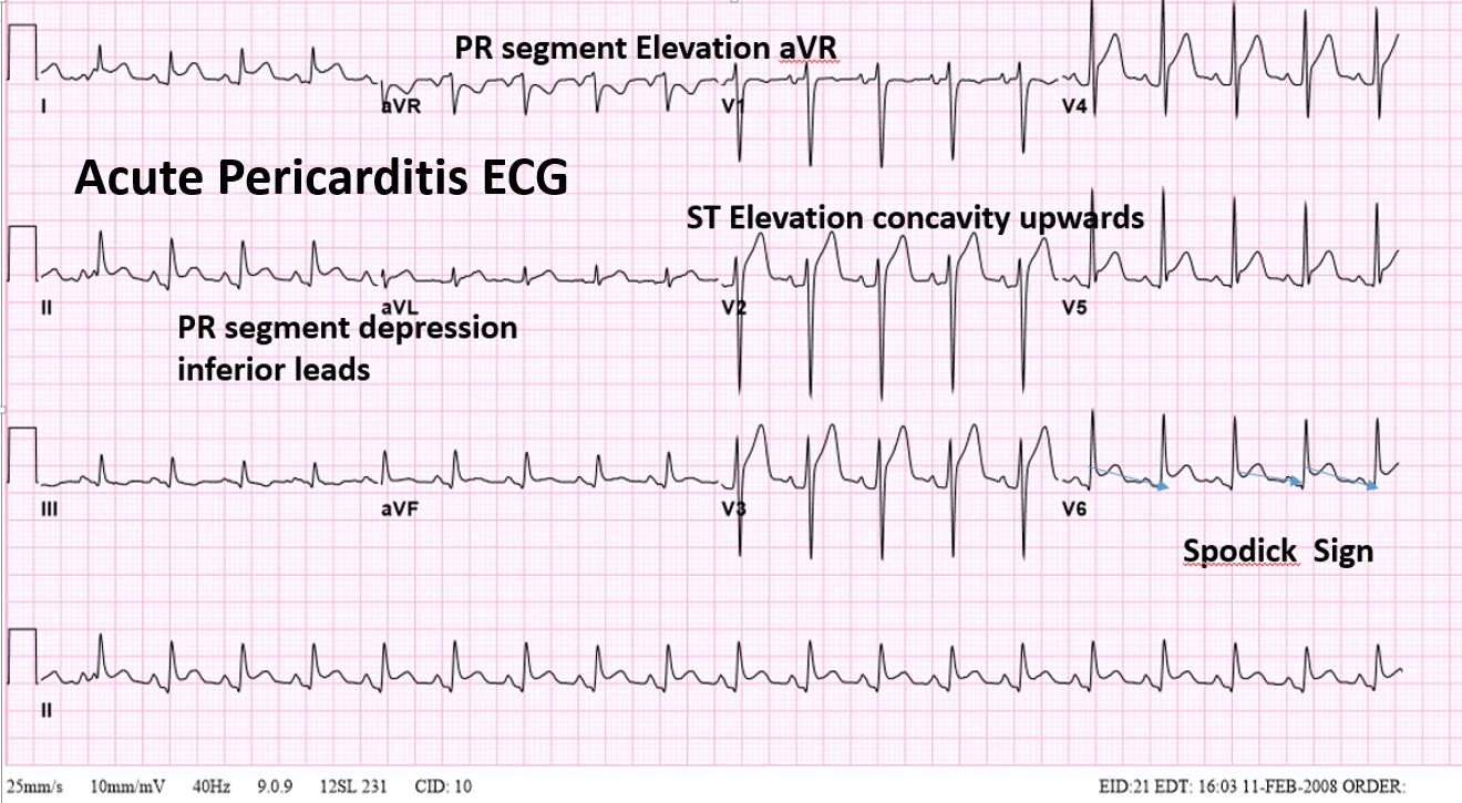
Long description
A self-published diagram captioned 'acute pericarditis: ECG changes'.

Typical ECG changes are present in up to 60% of cases, and the classic change is widespread ST elevation with PR depression.[1] The traditional four stages describe the usual evolution, though treatment and the individual patient blur the boundaries.[2]
| Stage | Timing | Findings |
|---|---|---|
| 1 | First 2 weeks | Diffuse ST elevation and PR depression, with reciprocal changes in aVR[2] |
| 2 | 1 to 3 weeks | ST segments return to baseline; T waves flatten[2] |
| 3 | 3 weeks to several weeks | T waves invert[2] |
| 4 | Several weeks onward | The ECG returns to normal[2] |
Exam trapPR depression is the quiet clue
ST elevation shouts; PR depression whispers. Look for PR depression in the lateral and inferior leads and PR elevation in aVR. It is described as almost diagnostic of an inflammatory myopericardial process, and it is the finding most often missed when a pericarditis is labelled as a STEMI.[5]
Transthoracic echocardiography is the first imaging test for every patient. It answers four questions: is there fluid, how much, is there tamponade physiology, and is left ventricular function normal?[2] It also gives a baseline for comparison if the patient returns.
Effusion size is graded as mild (under 10 mm), moderate (10 to 20 mm) or large (over 20 mm) by the widest diastolic echo-free space.[1] A large effusion should prompt a search for tuberculosis, malignancy or a purulent cause.[1] Echocardiography is normal in many patients with uncomplicated pericarditis, so a normal scan does not exclude the diagnosis.
C-reactive protein, erythrocyte sedimentation rate and white cell count are commonly raised, and they are used to support the diagnosis and to monitor activity and response.[1] CRP is the workhorse: it should be measured at diagnosis, before each taper, and at follow-up, because a normal CRP with no symptoms is the signal that it is safe to reduce treatment.[1][3]
Troponin and CK are raised when the myocardium is involved, which is the definition of myopericarditis.[1] A modest troponin rise with a normal ECG territory and preserved ventricular function is common and does not change management much.
Figure 2Perikarderguss 78M - CT axial KM arteriell und coronar pv - 001
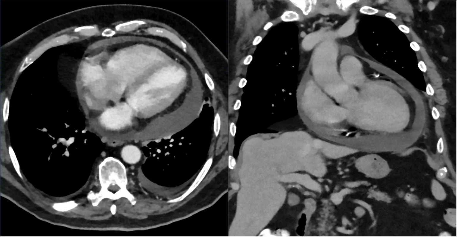
Long description
An axial contrast-enhanced CT scan showing a pericardial effusion (described as pericardial effusion on CT, axial, with arterial/coronary-phase contrast).

Cardiac magnetic resonance (CMR) is the best test for tissue characterisation. Late gadolinium enhancement detects pericardial inflammation with a sensitivity of 94% to 100%, and T2-weighted imaging shows oedema.[2][4] It is useful when the diagnosis is doubtful, when the response to treatment is poor, when recurrence is being assessed, and when a myopericardial overlap needs sorting out.[1][2]
Computed tomography shows pericardial thickness, calcium and loculation, and helps plan drainage or surgery.[1] 18F-fluorodeoxyglucose positron emission tomography with CT is used mainly for malignancy and for suspected tuberculosis, which takes up more tracer than idiopathic inflammation.[1]
NoteWhat a normal chest radiograph means
The cardiac silhouette is normal in uncomplicated pericarditis. Enlargement on the film needs an effusion of more than about 300 ml, so a normal radiograph is reassuring but not exclusionary.[1]
In an endemic setting, send pericardial fluid for acid-fast bacilli microscopy, culture, Xpert MTB/RIF and adenosine deaminase or unstimulated interferon-gamma. Unstimulated interferon-gamma is more accurate than adenosine deaminase or Xpert for microbiologically confirmed disease.[1] A definite diagnosis needs tubercle bacilli in fluid or in pericardial histology; a probable diagnosis rests on tuberculosis elsewhere plus a compatible lymphocytic exudate and response to treatment.[1]
The ESC 2015 criteria are the ones to know for exams and for the bedside. Acute pericarditis is diagnosed when at least two of four findings are present.[1]
- Pericarditic chest pain — sharp, pleuritic, improved by sitting up and leaning forward (over 85 to 90% of cases).
- Pericardial friction rub (up to about 33%).
- New widespread ST elevation or PR depression on the ECG (up to 60%).
- New or worsening pericardial effusion (up to 60%, usually mild).
Raised CRP, ESR or white cell count supports the diagnosis but is not one of the four criteria in the 2015 document.[1]
NoteThe criteria have moved on
The 2015 ESC criteria remain the classic framework, but they are no longer the only ones. A 2025 American College of Cardiology concise guidance document replaces "two of four" with pericarditic chest pain plus at least one of: friction rub, typical ECG change, new or worsening effusion, raised inflammatory markers, or CMR/CT evidence of pericardial inflammation. The 2025 ESC guideline on myocarditis and pericarditis introduced a tiered definite/possible/unlikely approach and allows atypical presentations without classic chest pain. Expect exam questions built on the 2015 version, and modern practice to use the wider criteria.[4]
Most patients with acute pericarditis can be managed at home. A minority cannot, and the decision is made on a short list of features that predict tamponade, recurrence or constriction.[1]
Major risk features[1]
- Fever above 38°C.
- A subacute course: symptoms over several days without a clear acute onset.
- A large effusion, defined as a diastolic echo-free space over 20 mm.
- Cardiac tamponade.
- Failure to respond to NSAIDs within 7 days.
Minor risk features[1]
- Myopericarditis.
- Immunodepression.
- Trauma.
- Oral anticoagulant therapy.
Any major or minor feature, or any suspicion of a specific cause such as tuberculosis or a systemic inflammatory disease, warrants admission and an aetiological search.[1] Patients with none of these features can be treated as outpatients and reviewed after one week to check the response.[1]
Triage of suspected acute pericarditis
- Diagnosis madeAt least two of: pericarditic pain, rub, typical ECG change, new effusion
- Question: Any high-risk feature?
Yes: fever >38°C, subacute course, effusion >20 mm, tamponade, NSAID failure at 7 days, myopericarditis, immunodepression, trauma, anticoagulation
- AdmitECG monitoring, echo, CRP, troponin, blood cultures if febrile
- Search for the causeTB testing in endemic areas; autoimmune screen; malignancy imaging as indicated
- Treat and reassessNSAID plus colchicine; drain if tamponade or large symptomatic effusion
No
- Outpatient managementNSAID plus colchicine, gastroprotection, written review plan
- Review at 1 weekSymptoms, CRP, ECG; escalate if not settling
Aspirin or another NSAID is the mainstay, and colchicine is added from the first episode to improve the response and prevent recurrence.[1] The drugs suppress inflammation; they do not shorten the underlying illness. The taper is part of the treatment, not an afterthought, because stopping abruptly is a common cause of recurrence.[3]
| Drug | Dose | Duration and taper |
|---|---|---|
| Aspirin | 750 to 1000 mg every 8 hours | 1 to 2 weeks, then reduce by 250 to 500 mg every 1 to 2 weeks[3] |
| Ibuprofen | 600 to 800 mg every 8 hours | 1 to 2 weeks, then reduce by 200 to 400 mg every 1 to 2 weeks[3] |
| Indomethacin | 25 to 50 mg every 8 hours | 1 to 2 weeks, then reduce by 25 mg every 1 to 2 weeks; start low to limit headache[3] |
| Colchicine | 0.5 mg twice daily if 70 kg or more; 0.5 mg once daily if under 70 kg | 3 months for a first episode; 6 months or more for recurrence[1][2] |
| Prednisone | 0.2 to 0.5 mg/kg/day | Second-line; keep until symptoms resolve and CRP normalises, then taper very slowly[1] |
NSAIDs. Choose on the basis of tolerance, other disease and what the patient already takes: aspirin suits a patient who needs antiplatelet treatment anyway.[1] Duration is guided by symptoms and CRP, but is generally one to two weeks in an uncomplicated case, followed by a taper.[1] The 2020 ACC/AHA guideline continues for two to four weeks before tapering, so the duration is a genuine point of disagreement rather than an error in either document.[2] Taper only when the patient is asymptomatic and the CRP has fallen or normalised.[3]
Colchicine. The dose is weight-adjusted because the therapeutic window is narrow. The 2015 guideline uses 0.5 mg twice daily for patients of 70 kg or more and 0.5 mg once daily below that.[1] Continue for three months after a first episode and at least six months after a recurrence.[2] A dose of 0.5 to 0.6 mg twice daily is enough; reduce to once daily in people over 70 years or under 70 kg, and reduce further in chronic kidney disease.[3] Diarrhoea is the usual reason for stopping. Tapering is optional but reasonable.[1]
United States
Colchicine is supplied as 0.5 mg and 0.6 mg tablets, so the same regimen may be written as 0.6 mg twice daily. The 0.5/0.6 mg equivalence is accepted in current reviews; what matters is the total daily dose and the adjustment for age, weight and kidney function.[3] US guidance from 2025 goes further than Europe in placing IL-1 blockers second-line for recurrent disease, rather than after corticosteroids.[4]
Gastric protection. Give a proton pump inhibitor with the NSAID, taking other risk factors into account.[3] This is not optional in older patients, in those with previous ulcer or bleeding, and in anyone on an anticoagulant or antiplatelet drug.
Exercise restriction. Restrict activity beyond ordinary sedentary life until symptoms have resolved and CRP has normalised. Athletes should not return to competition until symptoms, CRP, ECG and echocardiogram have all normalised, and a minimum of three months from onset is conventional.[1] With myocardial involvement the restriction is longer: at least six months.[1]
Corticosteroids. They relieve symptoms quickly and cause recurrences later. Use prednisone 0.2 to 0.5 mg/kg/day only when NSAIDs and colchicine are contraindicated, not tolerated or have failed, and give it with colchicine rather than instead of it.[1] Avoid high doses (1.0 mg/kg/day or equivalent), which are associated with more chronic disease and dependence.[1]
Red flagWhen not to reach for steroids
Do not start corticosteroids when a bacterial or tuberculous cause cannot be excluded, and do not use them in viral pericarditis, where they can reactivate infection and prolong inflammation.[1] If steroids are already on board and the patient relapses as the dose falls, resist the reflex to increase it: below about 10 to 15 mg/day of prednisone, reduce in steps of 1.0 to 2.5 mg every 2 to 6 weeks whenever the patient is asymptomatic and CRP is normal.[1]
Interleukin-1 blockers. For recurrent pericarditis that will not come off steroids, or that recurs despite colchicine, blockade of IL-1 works where conventional drugs fail. Anakinra, a recombinant IL-1 receptor antagonist, is given subcutaneously at 2 mg/kg/day up to 100 mg in the trial that established it, or 100 mg daily in practice; rilonacept is given as 320 mg on day 1 then 160 mg weekly.[3][4] In the RHAPSODY trial, recurrence during the randomised withdrawal period was 7% with rilonacept versus 74% with placebo; in AIRTRIP, 18% versus 90% after anakinra withdrawal.[4] They are expensive and need supervision, and stopping them abruptly invites relapse, so taper.[3]
Tuberculous pericarditis. Treat with rifampicin, isoniazid, pyrazinamide and ethambutol for two months, then rifampicin and isoniazid to complete six months; longer courses give no benefit and worsen adherence.[1] Add prednisolone for six weeks in patients without HIV: in the IMPI trial this reduced the incidence of constrictive pericarditis by 46%, with a neutral effect on the combined outcome of death, tamponade requiring drainage or constriction.[1] Avoid adjunctive steroids in HIV infection, where they were associated with an increased risk of HIV-associated malignancy.[1] Constriction develops within about six months in those who are going to develop it, and rifampicin-based treatment reduces its incidence to 17 to 40% from up to 50% in the pre-chemotherapy era.[1]
Uraemic pericarditis. It is thought to arise from retained toxic metabolites, either before dialysis or within eight weeks of starting it. The effusion is often bloody, so anticoagulation needs care.[1] Pain is often mild and the ECG often normal, so a high index of suspicion is needed in a dialysis patient with unexplained hypotension or a large effusion.[1]
Post-infarction and post-pericardiotomy. Use the same anti-inflammatory scheme, including colchicine for post-pericardiotomy syndromes with systemic inflammation.[1] Do not treat an asymptomatic post-operative effusion with NSAIDs or colchicine; the drugs cause more harm than the fluid.[1]
Purulent pericarditis. This is a different disease in tempo and danger. Suspect it in a septic patient with a pericardial effusion, perform urgent pericardiocentesis, send fluid for microscopy and culture, and give intravenous antibiotics with adequate drainage.[1]
Recurrent pericarditis. Confirm that it really is a recurrence, look for an untreated cause, and restart NSAID plus colchicine. Add steroids only as triple therapy at low dose, and consider an IL-1 blocker or, occasionally, azathioprine or intravenous immunoglobulin when steroid dependence persists.[1][3]
A treatment ladder for acute and recurrent pericarditis
- Acute episodeNSAID or aspirin at full dose + colchicine + proton pump inhibitor
- Taper after responseSymptoms gone and CRP normal; reduce stepwise over weeks, not days
- Question: Recurrence or steroid dependence?
First recurrence
- Restart NSAID + colchicineExclude a missed cause; colchicine for 6 months or more
Frequent recurrences or steroid-dependent
- Low-dose steroid as triple therapyOnly if NSAID and colchicine fail; taper very slowly
- IL-1 blockerAnakinra or rilonacept; taper, do not stop abruptly
- Specialist optionsAzathioprine, IVIG, pericardiectomy in expert centres
Cardiac tamponade. Fluid raises intrapericardial pressure until the ventricles cannot fill. It is rare in idiopathic pericarditis and more common with malignancy, tuberculosis or purulent pericarditis.[1] The speed of accumulation matters more than the volume, which is why a small bleed after a procedure can tamponade while a slow accumulation of a litre may not.[1] It requires urgent pericardiocentesis.[1]
Constrictive pericarditis. Fibrosis prevents diastolic filling. The risk depends on cause: under 1% after idiopathic or viral pericarditis, 2 to 5% after autoimmune, immune-mediated or neoplastic disease, and 20 to 30% after bacterial causes, especially tuberculosis and purulent pericarditis.[1] It usually appears months later, so any patient with persistent congestion and a normal ejection fraction after pericarditis needs re-imaging.[1]
Recurrence. This is the commonest complication. Without colchicine, 15 to 30% of patients with idiopathic acute pericarditis develop recurrent or incessant disease, and the rate can reach 50% after a first recurrence; colchicine roughly halves it.[1]
Death. Uncommon overall. In-hospital mortality was 1.1%, concentrated in older patients and those with severe infection.[1] Tuberculous pericarditis is the exception: 17 to 40% die within six months.[1][8]
When pericarditis is accompanied by myocardial injury, the term is myopericarditis; when myocardial involvement dominates, it is perimyocarditis.[1] The clinical definition is a patient who meets the criteria for acute pericarditis, has a raised troponin I or T (or CK-MB), and has no new focal or diffuse left ventricular impairment.[1] If ventricular function is newly reduced, the balance has tipped towards myocarditis.[1]
Myocardial involvement is not rare. In a 351-patient cohort it was present in 19.9%, and it has been reported in about 30% of acute idiopathic pericarditis.[6] ST elevation on the ECG was associated with it (odds ratio 1.82) but did not predict it well enough to replace troponin and imaging.[6]
Management follows pericarditis, with four differences.[1]
- Admit for monitoring and to exclude an acute coronary syndrome.[1]
- Use the lowest effective NSAID dose, because animal models of myocarditis suggest NSAIDs can worsen inflammation, although the human evidence is weak.[1]
- The 2015 guideline found insufficient evidence to recommend colchicine in myopericarditis.[1]
- Exercise is contraindicated for at least six months.[1]
The prognosis is good: observational series show no progression to heart failure or death in myopericarditis with preserved function.[1] Athletes still deserve caution, because sudden death has been reported after strenuous exertion during myocardial inflammation.[1]
Most patients with viral or idiopathic pericarditis recover fully.[1] The illness is self-limiting in the majority, and the main long-term problem is recurrence rather than death. Recurrences are troublesome and can wreck quality of life and work, but they rarely cause tamponade, and constriction has not been reported in idiopathic recurrent pericarditis.[1]
The prognosis belongs to the cause, not to the number of relapses: tuberculous and purulent pericarditis carry the mortality and the constriction risk.[1]
Pregnancy. Pericarditis in pregnancy is managed with the same anti-inflammatory principles, but the drug choices need obstetric input. The ESC guideline lists pregnancy among the specific indications for corticosteroids when NSAIDs are problematic, and any decision is shared between cardiology and obstetrics.[1]
Older adults. Colchicine toxicity rises with age and with reduced kidney function. Reduce to 0.5 to 0.6 mg daily above 70 years, and adjust for estimated glomerular filtration rate: 0.5 to 0.6 mg daily at 35 to 49 ml/min/1.73 m², and 0.5 to 0.6 mg every two to three days at 10 to 34.[3] Avoid chronic colchicine below 10 ml/min/1.73 m². NSAIDs need similar caution because of renal and gastrointestinal risk.
People with HIV. Tuberculosis is the dominant cause, and the presentation is often subacute with a large effusion. Adjunctive steroids should be avoided because of the malignancy signal.[1] Drug interactions need checking, because rifampicin changes the handling of many cardiac drugs.
Patients on anticoagulants. Anticoagulation is a minor risk factor for a worse course, and in iatrogenic effusions full anticoagulation may precipitate tamponade.[1] Do not stop a necessary anticoagulant reflexively; involve the team who prescribed it, and image early.
Athletes. Return to sport only after symptoms, CRP, ECG and echocardiogram have normalised, with a minimum of three months after isolated pericarditis and six months after myopericarditis.[1]
- Echocardiography is the test that changes management. It confirms the effusion, shows tamponade physiology, and separates pericarditis from ventricular dysfunction.[4] A portable scan at the bedside is enough.
- Assume tuberculosis in endemic areas. Send fluid for Xpert MTB/RIF, adenosine deaminase or unstimulated interferon-gamma, and start empirical antituberculous treatment when the picture fits and testing is unavailable.[1] The six-month rifampicin-based regimen is effective.[1]
- Do not chase a viral diagnosis. Serology is unhelpful and PCR on fluid or biopsy is rarely available; "presumed viral" is an honest label.[1][1]
- Colchicine and an NSAID are cheap. The combination is the highest-value intervention in the whole topic because it halves recurrence.[1]
- Drain when tamponade is present. Pericardiocentesis is life-saving; purulent or loculated fluid may need surgical drainage in addition to antibiotics.[1]
- Refer for constriction. Pericardiectomy needs cardiac surgery and experience; refer before the patient is cachectic.[1]
- Acute pericarditis is an inflammatory pericardial syndrome diagnosed by two of four findings: pericarditic pain, friction rub, typical ECG change, new effusion.
- The four temporal patterns are acute, incessant (over 4 to 6 weeks), recurrent (after a 4 to 6 week symptom-free interval) and chronic (over 3 months).
- Effusions are graded by echo-free space: mild under 10 mm, moderate 10 to 20 mm, large over 20 mm.
- Viruses and immune-mediated processes dominate in wealthy countries; tuberculosis dominates in endemic areas and in HIV, causing over 90% of significant effusions in HIV-infected patients there.
- Pain is pleuritic and relieved by sitting forward; the rub is scratchy, best at the left sternal edge, present in only about a third, and can disappear when fluid accumulates.
- The ECG shows widespread concave ST elevation with PR depression, sparing aVR and V1, with reciprocal changes confined to aVR and occasionally V1; ST elevation is usually under 5 mm and evolves over days to weeks.
- STEMI is more likely with convex or obliquely straight elevation, territorial distribution, reciprocal ST depression outside V1/aVR, elevation in III greater than II, and change over minutes to hours.
- ESC 2015 criteria use two of four findings; the 2025 ACC guidance uses pain plus one supportive finding, and the 2025 ESC guideline adds a tiered definite/possible/unlikely approach.
- Admit for fever over 38°C, a subacute course, effusion over 20 mm, tamponade, failure to respond to NSAIDs within 7 days, myopericarditis, immunodepression, trauma or anticoagulation.
- Treat with a full-dose NSAID (ibuprofen 600 to 800 mg every 8 hours, aspirin 750 to 1000 mg every 8 hours, or indomethacin 25 to 50 mg every 8 hours) plus colchicine 0.5 mg twice daily (or once daily under 70 kg) for 3 months, with a proton pump inhibitor.
- Taper only when symptoms have gone and CRP is normal; abrupt withdrawal is a common cause of recurrence.
- Corticosteroids are second-line, at 0.2 to 0.5 mg/kg/day, and should be avoided when bacterial or tuberculous infection is possible; they increase recurrence.
- Recurrence affects 15 to 30% after a first episode and up to 50% after a first recurrence without colchicine; IL-1 blockers (anakinra, rilonacept) are for refractory or steroid-dependent disease.
- Tuberculous pericarditis is treated with two months of rifampicin, isoniazid, pyrazinamide and ethambutol, then four months of rifampicin and isoniazid, with prednisolone for six weeks in HIV-negative patients.
- Constriction risk is under 1% after idiopathic pericarditis, 2 to 5% after autoimmune or neoplastic causes, and 20 to 30% after bacterial causes.
Answer each one in your head first, then open the answer.
A 24-year-old woman has pleuritic chest pain that is easier when she leans forward, a temperature of 37.6°C, a normal troponin and CRP of 60 mg/L. The ECG shows concave ST elevation in I, II, aVL, V3 to V6 and PR depression in II and V5. What is the diagnosis, and what else must be excluded before she goes home?
Show answerHide answer
Acute pericarditis, meeting two of the four ESC criteria (pain and typical ECG change), with a supporting raised CRP. Exclude an acute coronary syndrome: there is no territorial pattern, no reciprocal ST depression outside aVR, and the troponin is normal, but if the history or risk factors fit, serial troponin and further testing are needed. She has no major risk feature, so outpatient treatment with an NSAID, colchicine and a proton pump inhibitor, with review at one week, is appropriate.
Why can a friction rub disappear as a patient gets worse?
Show answerHide answer
Because an effusion has developed and separated the inflamed pericardial layers, so they no longer rub. A vanishing rub with increasing breathlessness or rising jugular venous pressure suggests accumulating fluid and possible tamponade, and needs urgent echocardiography.
A 61-year-old man with an inferior STEMI treated by primary PCI develops fever, pleuritic pain and a rub two weeks later. What is the diagnosis, and how is it treated?
Show answerHide answer
Post-myocardial infarction pericarditis, part of the post-cardiac injury syndrome family (Dressler syndrome when late). Treat with an NSAID or aspirin plus colchicine, with gastroprotection and a taper guided by symptoms and CRP. Anticoagulation needs review because of bleeding risk into the pericardium, and a large effusion needs drainage.
Why are corticosteroids avoided as first-line treatment in a first episode of acute pericarditis?
Show answerHide answer
They give rapid symptom relief but favour chronicity, increase the recurrence rate and cause steroid dependence. They are reserved for patients who cannot take or do not respond to NSAIDs and colchicine, for autoimmune disease, and for pregnancy, and when used they should be low dose (prednisone 0.2 to 0.5 mg/kg/day) and combined with colchicine, with a very slow taper.
A 34-year-old man in Malawi has three weeks of breathlessness, a large pericardial effusion and HIV. What cause must you pursue, and how does HIV change treatment?
Show answerHide answer
Tuberculous pericarditis: tuberculosis causes a clinically significant effusion in over 90% of HIV-infected patients in endemic areas. Send fluid for Xpert MTB/RIF, culture and adenosine deaminase or unstimulated interferon-gamma, and start standard six-month antituberculous treatment. Adjunctive prednisolone reduces constriction but was associated with an increased risk of HIV-associated malignancy, so it is avoided in HIV infection.
A 45-year-old woman has a fourth episode of pericarditis. She has failed two tapers of prednisone and remains steroid-dependent despite colchicine. What are the options, and what is the evidence?
Show answerHide answer
An IL-1 blocker is the best-supported next step. Anakinra, 2 mg/kg/day up to 100 mg (100 mg daily in practice), reduced recurrences after withdrawal to 18% versus 90% with placebo in AIRTRIP; rilonacept, 320 mg on day 1 then 160 mg weekly, gave 7% versus 74% recurrence in RHAPSODY. Both need tapering rather than abrupt stopping. Azathioprine, intravenous immunoglobulin or, in expert centres, pericardiectomy are later options.
What single feature on the ECG most reliably argues against pericarditis and for a coronary occlusion?
Show answerHide answer
Reciprocal ST depression outside leads V1 and aVR, especially with convex or obliquely straight ST elevation in a territorial distribution. PR depression in several leads, widespread concave elevation and reciprocal change confined to aVR point the other way.
Read next: cardiac tamponade and constrictive pericarditis for the haemodynamic consequences; myocarditis for the overlap syndrome; tuberculosis for the infection behind most pericardial disease worldwide. Within cardiology, see ECG interpretation for the systematic reading that separates pericarditis from STEMI, cardiac physical examination for how to hear a rub, and heart failure for the congestion that constriction and tamponade mimic. Arrhythmias are not typical of pericarditis, but atrial fibrillation can complicate any inflamed atrium; see atrial fibrillation and flutter.
- Adler Y, Charron P, Imazio M, et al. 2015 ESC Guidelines for the diagnosis and management of pericardial diseases. European Heart Journal 2015;36(42):2921-2964. doi:10.1093/eurheartj/ehv318. Accessed .
- Mangas GM, Rodriguez JGV, Santos JARBD, et al. Fundamentals of Acute Pericarditis: State of the Art. Arquivos Brasileiros de Cardiologia 2026;123(2):e20240572. doi:10.36660/abc.20240572. Accessed .
- Bonaventura A, Santagata D, Vecchié A, Abbate A. Current Drug Treatment for Acute and Recurrent Pericarditis. Drugs 2025;85(5):643-658. doi:10.1007/s40265-025-02169-x. Accessed .
- Ehsan M, Ikram J, El Roumi J, et al. Evolving Guidelines in Pericarditis: Aligning Practice With Pathophysiology. JACC: Advances 2026;5(1):102509. doi:10.1016/j.jacadv.2025.102509. Accessed .
- Moak JH, Muck AE, Brady WJ. ST-segment elevation myocardial infarction mimics: the differential diagnosis of nonacute coronary syndrome causes of ST-segment/T-wave abnormalities in the chest pain patient. Turkish Journal of Emergency Medicine 2024;24(4):206-217. doi:10.4103/tjem.tjem_137_24. Accessed .
- Ceriani E, Berra S, Agozzino F, et al. ST-segment elevation in acute pericarditis and myocardial involvement: electrocardiographic and clinical profiling. Open Heart 2026;13(1):e004019. doi:10.1136/openhrt-2026-004019. Accessed .
- Agrawal A, Rosenzveig A, Safdar A, et al. Short- and Long-Term Outcomes of Pericarditis, Myocarditis, and Myopericarditis in Patients With Malignancy on Immune Checkpoint Inhibitors. Journal of the American Heart Association 2026;15(2):e041163. doi:10.1161/jaha.125.041163. Accessed .
- Mikalajūnaitė G, Rinkūnienė E, Čypienė A, Dženkevičiūtė V, Badarienė J. Cardiovascular Complications from Tuberculosis. Reviews in Cardiovascular Medicine 2025;26(11):44092. doi:10.31083/rcm44092. Accessed .
Figure credits
Educational content for medical students and doctors. It is not a substitute for clinical judgement or local guidelines.