Heart failure
Heart failure: definition, classification by ejection fraction, stage and NYHA class, causes, diagnosis, four-pillar drug therapy, devices, acute care.
- Heart failure (HF) is a clinical syndrome, not a single disease: typical symptoms and signs (breathlessness, ankle swelling, fatigue, raised jugular venous pressure, crackles) caused by a structural or functional abnormality of the heart, with raised natriuretic peptides or objective evidence of congestion to back it up.[1][10]
- It is classified four ways that all matter: by ejection fraction (reduced, mildly reduced, preserved, improved), by stage (A to D), by symptom class (New York Heart Association, NYHA, I to IV), and by time course (acute, chronic, worsening). Newer documents are moving away from fixed ejection-fraction cut-offs.[1][3][6]
- In reduced ejection fraction (HFrEF) four drug groups are the foundation: an ACE inhibitor, ARB or angiotensin receptor-neprilysin inhibitor (ARNI, sacubitril-valsartan); a beta-blocker; a mineralocorticoid receptor antagonist (MRA); and an SGLT2 inhibitor. Doses are raised step by step toward trial-proven targets.[26][6][8]
- Diuretics relieve congestion but do not by themselves improve survival; devices (implantable cardioverter-defibrillator, ICD; cardiac resynchronisation therapy, CRT) are added by ejection fraction and QRS width after drug therapy has been optimised.[8][15]
- Acute pulmonary oedema is treated with sitting upright, oxygen only if hypoxic, intravenous loop diuretic, vasodilators in selected patients, and ventilatory support if needed. Treat the trigger.[9][15]
- In sub-Saharan Africa HF affects younger people, and hypertension, rheumatic heart disease and cardiomyopathies (including peripartum cardiomyopathy) dominate over coronary disease.[12][1]
By the end of this page you should be able to:
- Define heart failure, explain why "congestive" is an outdated label, and classify a patient by ejection fraction, ACC/AHA stage and NYHA class.
- Explain how reduced cardiac output sets off neurohormonal activation and remodelling, and derive from this why the symptoms and signs occur.
- Take a history, examine, and choose and interpret the first-line investigations, including natriuretic peptides and echocardiography.
- Start and titrate the four foundational drug groups, recognise their cautions, and know when to add ivabradine, hydralazine-nitrate, digoxin, vericiguat, iron, an ICD or CRT.
- Manage acute pulmonary oedema step by step, and recognise cardiogenic shock and begin its management.
- Describe special groups (peripartum cardiomyopathy, elderly, kidney disease), prevention, self-care and palliative care, and adapt management to limited resources.
The heart's job is to deliver enough blood to meet the body's needs at normal filling pressures. Heart failure is what happens when it cannot do so, or can do so only because the filling pressures are high. Two things follow: fluid backs up behind the failing chambers (congestion), and organs in front of them may get too little blood (low output).
The first Universal Definition (2021), written jointly by the Heart Failure Society of America, the ESC Heart Failure Association and the Japanese Heart Failure Society, described HF as a syndrome of symptoms or signs that arise from an abnormality of cardiac structure or function and are supported either by raised natriuretic peptides or by objective proof of pulmonary or systemic congestion.[10] The Second Universal Definition (2026), issued by four societies (the US cardiology colleges and heart associations, the ESC and the World Heart Federation), keeps the idea: many different causes can lead to the same picture of typical but non-specific symptoms, signs and test findings that point to congestion or a changed cardiac output, and that trace back to a problem in the heart's structure or function.[1]
Two practical points from the 2026 document. First, HF is defined by an accumulation of suggestive features in a comprehensive assessment, not by one test; some people with HF with preserved ejection fraction have normal natriuretic peptides despite clearly raised filling pressures on invasive testing.[1] Second, once symptomatic HF is established, the diagnosis is generally kept for life, even when the patient improves with treatment.[1][2]
"Congestive heart failure" (CHF) was coined when the only visible manifestation of the syndrome was fluid retention. It has been dropped for three reasons:
- Congestion is not always present. A patient whose fluid is controlled with diuretics, or whose main problem is low output, is still in HF. The definition itself says "congestion or altered cardiac output".[1]
- The label implies a diagnosis of exclusion based on fluid alone and delays treatment of people with the earlier stages (A and B) who have no congestion yet but already have risk factors or structural disease.[1]
- Its meaning varies between sources, which the universal definitions were created to fix.[1][10]
Use "heart failure", with the ejection-fraction group, stage and NYHA class. Older sources, including exam materials and some local guidelines, still say CHF; recognise it as the same syndrome.
The ejection fraction (EF) is the proportion of the left ventricle's end-diastolic volume ejected with each beat. It is the cornerstone of HF classification because trials enrolled patients within narrow EF bands and treatments were proved within those bands.[1] Documents differ on the cut-offs.
| Group | 2021 Universal Definition and 2022 AHA/ACC/HFSA | NICE NG106 (2025) | 2026 documents |
|---|---|---|---|
| HFrEF | LVEF 40% or less[6] | LVEF 40% or less[8] | ESC 2026: LVEF below 50% with symptoms or signs[5] |
| HFmrEF | LVEF 41% to 49%[6] | LVEF 41% to 49%[8] | ESC 2026 removes this group; the Second Universal Definition has no separate group and no fixed cut-offs[3][1] |
| HFpEF | LVEF 50% or more, with objective evidence of raised filling pressures[6] | LVEF 50% or more plus a structural abnormality (two or more of: left atrial volume above 34 ml/m2 in sinus rhythm or above 40 ml/m2 in AF, E to e' ratio above 11, wall thickness above 12 mm, pulmonary artery pressure above 35 mmHg)[8] | ESC 2026: LVEF 50% or more[3] |
| HFimpEF | Previous LVEF 40% or less with a later LVEF above 40%[6] | Not a separate group | Second Universal Definition: previous reduced LVEF with a rise of at least 10 points to above 40%[1] |
The 2026 reasoning is that the lower limit of normal LVEF is about 52 to 53%, so an LVEF below 50% is unlikely to be normal. Patients with mildly reduced EF share pathophysiology with HFrEF and benefit from similar treatments.[2][3]
Heart failure with improved EF. Patients whose EF rises on treatment are not "cured". The 2022 AHA/ACC/HFSA guideline recommends continuing treatment, including in those who are asymptomatic (44% relapsed within 6 months of stopping in TRED-HF).[6] The 2026 ESC guideline likewise recommends continuing treatment at the highest tolerated doses even when LVEF improves.[26]
Stages describe progression and emphasise prevention. The 2021 definition renamed stage B "pre-HF", and the 2026 document reaffirms the A to D framework.[1]
| Stage | Who is in it | Aim |
|---|---|---|
| A, at risk | No symptoms, signs, structural change or raised biomarkers, but risk factors: hypertension, atherosclerotic disease, diabetes, obesity, cardiotoxin exposure, family history of cardiomyopathy[1] | Treat risk factors |
| B, pre-HF | No symptoms or signs, but structural disease (for example hypertrophy, enlarged chambers, wall-motion abnormality, valve disease), abnormal function, or raised natriuretic peptide or troponin[1] | Prevent symptomatic HF: for example a beta-blocker and an ACE inhibitor in asymptomatic left ventricular dysfunction[26][2] |
| C, HF | Current or past symptoms or signs of HF caused by a structural or functional abnormality[1] | Drug and device therapy, self-care |
| D, advanced HF | Severe symptoms at rest or with minimal exertion, recurrent hospitalisation despite optimal therapy, refractory or intolerant to therapy; needs advanced therapies such as inotropes, mechanical support, transplant or palliative care[1] | Advanced therapies or palliation |
The New York Heart Association (NYHA) class describes how much the patient's symptoms limit them right now. It can change from visit to visit and with treatment, unlike the stage. It is graded by asking what ordinary activity brings on breathlessness or fatigue.
| Class | Meaning | Example question |
|---|---|---|
| I | Structural heart disease or past HF, but no symptoms with ordinary activity | Can you climb stairs or walk uphill as you always did? |
| II | Mild limitation: comfortable at rest, ordinary activity (such as a brisk walk or two flights of stairs) causes symptoms | Do you get breathless on the stairs or hurrying? |
| III | Marked limitation: comfortable at rest, but less than ordinary activity (such as a slow walk on the flat or dressing) causes symptoms | Do you have to stop on a short flat walk? |
| IV | Symptoms at rest or with any activity; unable to carry on any activity without symptoms | Are you breathless sitting still or washing? |
NYHA class guides treatment: for example, trials of implantable defibrillators and resynchronisation enrolled patients mainly in classes II and III.[15]
- Left, right or biventricular. Left-sided failure raises pressure behind the left heart and causes pulmonary congestion (breathlessness, orthopnoea, crackles). Right-sided failure raises systemic venous pressure and causes a raised jugular venous pressure, hepatic congestion and peripheral oedema. The commonest cause of right-sided failure is left-sided failure; isolated right-sided failure follows lung disease (cor pulmonale), pulmonary hypertension or right-sided valve disease.
- Acute, chronic and worsening. Chronic HF is the long-term syndrome. The 2026 ESC guideline replaces "acute HF" with "decompensated HF" because some patients do not suddenly worsen but slowly lose compensation.[3]
- Low-output and high-output. Most HF is low-output: the heart is intrinsically weak or stiff. In high-output failure the heart is initially normal but cannot keep up with a very large demand, as in severe anaemia, thyroid disease, or thiamine deficiency (wet beriberi), which is documented in sub-Saharan Africa.[20][12] Treat the cause; ordinary HF drugs help little.
The two tables below put the two commonest contrasts side by side. They summarise points made elsewhere in this topic; signs overlap a lot in real patients, so use them to organise thinking, not to diagnose.
| Feature | Systolic dysfunction (HFrEF) | Diastolic dysfunction (HFpEF) |
|---|---|---|
| Main problem | The ventricle contracts weakly and often dilates | The ventricle contracts well but is stiff and fills poorly |
| Typical underlying causes | Infarction, dilated cardiomyopathy, long-standing pressure or volume load, toxins | Hypertension, ageing, obesity, diabetes, hypertrophy, fibrosis or infiltration such as amyloid |
| Typical heart sound | S3 gallop (blood entering a dilated, congested ventricle) | S4 (atrial contraction into a stiff ventricle) |
| Apex beat | Displaced and sustained | Often not displaced |
| Echocardiography | Dilated chamber with low ejection fraction | Normal or near-normal ejection fraction with signs of raised filling pressure, often a thick wall and an enlarged left atrium |
| Drugs with outcome benefit | ACE inhibitor, ARB or ARNI, beta-blocker, MRA, SGLT2 inhibitor | SGLT2 inhibitor and a diuretic for congestion; weaker evidence for others |
| Feature | Left-sided failure | Right-sided failure |
|---|---|---|
| Where pressure backs up | Left atrium and pulmonary veins | Systemic veins |
| Breathlessness, orthopnoea, paroxysmal nocturnal dyspnoea | Prominent | Not a feature of right-sided failure alone |
| Chest findings | Bibasal crackles; pleural effusion | Clear lungs if right-sided failure is isolated |
| Heart sounds | S3 or S4 from the left ventricle | Right-sided gallop; parasternal heave if the right ventricle is enlarged |
| Jugular venous pressure | May be normal if only the left side fails | Raised, with a positive hepatojugular reflux |
| Liver and abdomen | Usually normal | Tender hepatomegaly, ascites |
| Swelling | Not typical | Pitting ankle or sacral oedema |
HF is a pandemic affecting an estimated 64 million people worldwide. Its incidence appears to have stabilised, and is falling in developed countries, but prevalence is increasing as populations age and as survival improves after myocardial infarction and with HF treatment.[10] The ESC puts adult prevalence at 1 to 3%, and fewer than 60% of those diagnosed are alive at 5 years.[3] Reported prevalence varies with the definition and the method: for example, 1.6% in a UK primary-care study, about 2.5% in the United States by self-report, and 11.8% in people aged 60 or over when echocardiographic screening also counted previously unrecognised cases.[10]
HFpEF accounts for roughly half of HF hospitalisations and is increasing in proportion. Lifetime risk after age 45 is 20 to 45%. Incidence has fallen more for HFrEF than HFpEF.[11] Five-year mortality after incident HF has stayed around 50% in the United States.[11]
Sub-Saharan Africa. HF in sub-Saharan Africa occurs at a younger age than in high-income regions. The original THESUS-HF survey of acute HF found a mean age of 52 years.[13] The traditional main causes are rheumatic heart disease, hypertensive heart disease and cardiomyopathy; HIV-associated cardiomyopathy, tuberculous pericardial disease, cor pulmonale and peripartum cardiomyopathy are also common, while coronary disease was historically rare.[12] A systematic review of risk factors found that hypertension contributed to 32.6% of HF cases and rheumatic heart disease to 14% in the region, with ischaemic heart disease below 10%.[1] Hospital case-fatality was 9 to 12.5% in the studies reviewed.[12] Most data come from hospitals, and the continent is barely represented in HF trials.[10][12]
HF is the final common pathway of many diseases, so the second question after "does this patient have HF?" is "why?" The old split into "ischaemic" and "non-ischaemic" hides causes that change treatment, such as cardiac amyloidosis.[2]
| Group | Examples | Comment |
|---|---|---|
| Ischaemic | Myocardial infarction, chronic coronary disease | Leading cause in high-income regions: ischaemic disease was the major risk factor in over half of patients in Western high-income regions, but below 10% in sub-Saharan Africa[1] |
| Hypertensive | Hypertensive heart disease (hypertrophy, then dilation) | A common contributor worldwide and in Africa[1][12] |
| Valvular | Aortic stenosis, mitral and aortic regurgitation, rheumatic heart disease | Rheumatic disease is prominent in sub-Saharan and East Asia[1] |
| Arrhythmia-related | Uncontrolled AF, frequent premature ventricular beats, ventricular pacing, tachycardia-induced cardiomyopathy | Potentially reversible[1] |
| Cardiomyopathy: dilated, hypertrophic, restrictive, familial | Genetic and idiopathic forms | Genetic testing is recommended where it changes diagnosis or family screening[26] |
| Infiltrative | Cardiac amyloidosis, haemochromatosis, Fabry disease, sarcoidosis | Specific therapy exists for some[1][6] |
| Infective and inflammatory | Viral myocarditis, Chagas disease, HIV, autoimmune disease | Chagas disease is region-specific (Latin America)[1] |
| Toxic | Alcohol, cocaine, amphetamines, anthracyclines and other cancer therapy | Stopping the toxin can allow recovery[1] |
| Pregnancy-related | Peripartum cardiomyopathy | See the special groups section[1] |
| Right-sided and pericardial | Lung disease and pulmonary hypertension, constrictive pericarditis (including tuberculous) | Tuberculous pericardial disease is common in Africa[12] |
| High-output and metabolic | Severe anaemia, thyroid disease, thiamine deficiency | Treat the cause[20][12] |
| Congenital | Shunts, structural defects | Often present in childhood or adolescence |
Other risk factors include obesity, kidney disease, a family history, and exposure to alcohol or cardiotoxic drugs.[1] Most patients had at least one identifiable, preventable risk factor in the years before diagnosis, and poverty, low education and neighbourhood deprivation are associated with incident HF and worse outcomes.[1]
Acute decompensation in a patient with known HF usually has a trigger. Look for: non-adherence to drugs or a high-salt intake, myocardial ischaemia, new arrhythmia (especially AF with a fast rate), infection, uncontrolled hypertension, anaemia, kidney failure, new valve lesions, and drugs that retain salt and water or depress the heart (see the section on drugs to avoid). Examples are verapamil or diltiazem, NSAIDs (which also blunt diuretics and harm the kidney when volume is low), some antiarrhythmics, and alcohol.[8][27][1] Correct the trigger, and treat anaemia, thyroid disease and valve disease when present.
This section builds the picture step by step, because almost every symptom and every drug in the topic follows from it.
1. The determinants of cardiac output. Cardiac output is stroke volume times heart rate. Stroke volume depends on three things: preload (the stretch of the ventricle at the end of filling), contractility (the inherent force of the muscle at a given stretch) and afterload (the resistance the ventricle ejects against, set mainly by arterial pressure and, in the left ventricle, aortic valve disease). A failing ventricle needs more filling for the same stroke volume, and a rise in afterload reduces its output more than in a healthy heart.
2. The initial insult. Muscle loss after infarction, a pressure load (hypertension, aortic stenosis), a volume load (regurgitation), a toxin, a genetic defect or a persistent tachycardia reduces systolic function. Alternatively the muscle becomes stiff (hypertrophy, fibrosis, amyloid infiltration, ageing) and fills poorly; this is diastolic dysfunction, the typical mechanism in HFpEF.
3. Compensation. Falling output and pressure activate reflexes that, in the short term, maintain blood pressure and perfusion:
- the sympathetic nervous system raises heart rate and contractility and constricts vessels;
- the renin-angiotensin-aldosterone system (RAAS) is activated by low renal perfusion and sympathetic drive. Angiotensin II constricts arterioles and stimulates aldosterone, which makes the kidney retain sodium and water, and it promotes fibrosis and cell growth;
- vasopressin (antidiuretic hormone) is released, causing water retention and, in severe HF, low serum sodium;
- the heart releases natriuretic peptides (atrial and B-type) in response to wall stretch. They promote sodium loss and vasodilation and oppose the other systems, but their effect is overwhelmed in advanced disease. This is why BNP and NT-proBNP rise and are useful tests.
4. Decompensation and remodelling. What helps for hours harms for months. Salt and water retention raise preload and produce congestion; vasoconstriction raises afterload; sustained catecholamines and angiotensin are directly toxic to myocytes and drive fibrosis and cell death. The ventricle remodels: it dilates, becomes more spherical, and its walls thin or thicken in a way that further reduces efficiency. Dilation can also stretch the mitral annulus and produce functional mitral regurgitation, adding to the load. This vicious circle is why drugs that block RAAS and the sympathetic system (ACE inhibitors, ARBs, sacubitril-valsartan, beta-blockers, MRAs) improve survival, and why they are started even in patients who feel well.[14]
5. Why the symptoms and signs occur. High left atrial pressure floods the lungs (exertional breathlessness, orthopnoea, paroxysmal nocturnal dyspnoea); high right-sided pressure and sodium retention cause oedema, raised JVP and hepatic congestion; low output causes fatigue and cachexia; a third heart sound (S3) is blood rushing into a dilated, congested ventricle, and a fourth sound (S4) is atrial contraction into a stiff ventricle; and diluted sodium (hyponatraemia) tracks severity.
Ask about the patient's symptoms in their own words and the tasks that limit them, since this gives the NYHA class.
- Dyspnoea on exertion (progressing to rest), orthopnoea (how many pillows), paroxysmal nocturnal dyspnoea.[1]
- Fatigue, reduced exercise tolerance, cough (sometimes with frothy or blood-tinged sputum in pulmonary oedema), wheeze ("cardiac asthma"), nocturia.
- Ankle swelling, abdominal swelling or distension, loss of appetite and weight loss (cardiac cachexia), early satiety.
- Palpitations, syncope or presyncope (arrhythmia or low output).
- Chest pain (ischaemia).
- Cause-finding: hypertension, diabetes, infarction, rheumatic fever, alcohol and drugs, chemotherapy or chest radiotherapy, recent pregnancy, family history of cardiomyopathy or sudden death, HIV, thyroid disease.
- Precipitants and adherence: missed doses, salt intake, intercurrent illness, new drugs (see the section on drugs to avoid).
The general examination and the cardiac examination are covered step by step in cardiac physical examination. For HF specifically:
- Overall: respiratory rate, ability to speak in sentences, cyanosis, cool peripheries and low pulse pressure (low output).
- Pulse and blood pressure: tachycardia, irregularly irregular pulse (AF), alternating strong and weak beats (pulsus alternans, in severe disease). Low blood pressure is a marker of severity and limits drug titration.
- Jugular venous pressure (JVP): raised, with a positive hepatojugular reflux. It is the best bedside sign of congestion.
- Chest: bibasal crackles, pleural effusions (stony dull bases, often right-sided first).
- Heart: displaced and sustained apex beat (dilated left ventricle), parasternal heave (right ventricular enlargement), S3 gallop, murmurs of functional mitral or tricuspid regurgitation or of the underlying valve lesion.
- Abdomen and limbs: tender hepatomegaly, ascites, pitting oedema (sacral in the bed-bound).
Signs are neither sensitive nor specific alone; in HFpEF the older Framingham criteria are met in only about a quarter of patients.[11] Combine bedside findings with natriuretic peptides and imaging.
| Look-alike | What separates it from HF |
|---|---|
| Chronic obstructive pulmonary disease, asthma | Wheeze and hyperinflation, smoking history; normal JVP unless cor pulmonale; NT-proBNP typically low |
| Pneumonia | Fever, productive cough, focal consolidation; may coexist as the trigger |
| Anaemia, obesity, deconditioning | No raised JVP; normal natriuretic peptide and echocardiogram |
| Nephrotic syndrome, cirrhosis, venous insufficiency, drug-induced oedema | No raised JVP; oedema from low albumin or local venous causes; calcium-channel blockers commonly cause ankle oedema |
Investigations answer five questions: is this HF, what is the EF, what is the cause, what is the trigger, and what will limit treatment?
Figure 1Chest radiograph with signs of congestive heart failure - annotated
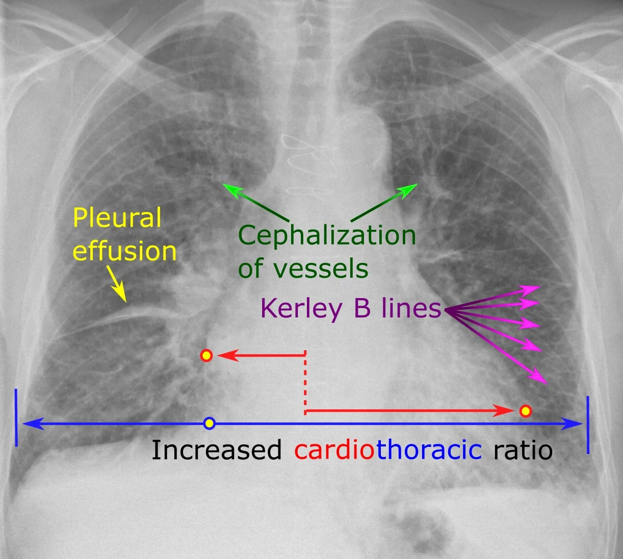
Long description
An annotated chest radiograph of an 83-year-old man with coronary artery bypass surgery and prior percutaneous coronary interventions presenting with laboured breathing, showing Kerley B lines, an enlarged heart, somewhat dilated pulmonary blood vessels and minor right-sided pleural effusion, consistent with stage II congestive heart failure.

- Natriuretic peptides. BNP and NT-proBNP are released when the myocardium is stretched. NICE advises NT-proBNP in everyone with suspected chronic HF. A level above 2000 ng/L (236 pmol/L) should prompt urgent specialist assessment and transthoracic echocardiography within 2 weeks; between 400 and 2000 ng/L, assessment within 6 weeks; below 400 ng/L in an untreated person makes HF less likely.[8] The ESC uses NT-proBNP 125 pg/mL (ng/L) or BNP 35 pg/mL as the cut-off in the non-acute (community) setting, and NT-proBNP 300 or BNP 100 in the emergency setting.[15] For new suspected acute HF, a single measurement with a BNP below 100 ng/L or NT-proBNP below 300 ng/L rules out the diagnosis (NICE).[9]
- Interpret with care. Levels are lower in obesity, in people of African or African-Caribbean ethnicity, and in those on diuretics, ACE inhibitors, ARNIs, ARBs, beta-blockers or MRAs. They are higher for other reasons: kidney disease, lung disease, sepsis, cirrhosis, AF, age.[8] Natriuretic peptides cannot distinguish HFrEF from HFpEF, and a normal value does not fully exclude HFpEF.[8][1]
- 12-lead ECG. Look for old infarction (Q waves), left ventricular hypertrophy, bundle branch block (a left bundle branch block with a wide QRS affects device choices; see bundle branch blocks), AF or other arrhythmia (see atrial fibrillation and flutter and ventricular arrhythmias), conduction block (bradyarrhythmias and AV block), and the QRS duration. The basics of reading it are in ECG interpretation.[8] A baseline ECG is also needed before a beta-blocker is prescribed.[8]
- Chest X-ray. May show an enlarged heart (cardiomegaly), diversion of blood to the upper-lobe veins (cephalisation, so the upper-zone vessels look larger than the lower-zone ones), Kerley B lines and other signs of interstitial oedema, perihilar "bat's wing" alveolar oedema, prominent hila, and pleural effusions, which can be unilateral and are often right-sided first.[8] A film can look normal in HF, so the X-ray supports rather than proves the diagnosis.
- Transthoracic echocardiography. The key test: it gives EF, chamber sizes, wall thickness, valve disease, intracardiac shunts, right ventricular function and pulmonary pressure, and diastolic function.[8]
- Blood tests: full blood count (anaemia), urea, creatinine and electrolytes (kidney function and potassium before RAAS drugs, sodium), liver function, thyroid function, lipids, HbA1c, and urinalysis; iron studies (ferritin and transferrin saturation).[8] Spirometry or peak flow if lung disease is suspected.[8] Troponin when ischaemia or myocarditis is possible.
- What the results tend to show. Natriuretic peptides are raised (thresholds above). A low haemoglobin or iron deficiency is common and worth treating (see intravenous iron). Creatinine may be raised, because kidney perfusion is low or kidney disease coexists, and it can rise further after RAAS drugs and diuretics. A low serum sodium usually means water retention (vasopressin) and marks more severe disease; NICE advises restricting fluid in dilutional hyponatraemia, and some guidelines mention tolvaptan for volume overload with sodium below 130 mmol/L in acute HF.[8][15] Thyroid function is checked because overactive and underactive thyroid can each cause or worsen HF, and amiodarone can disturb thyroid function.[8][20]
- Cardiac MRI gives the most accurate EF and volumes and tissue characterisation: scar or fibrosis, myocarditis, sarcoidosis, amyloid, iron overload, Fabry disease and non-compaction.[15]
- Coronary assessment: non-invasive stress imaging (stress echocardiography or radionuclide), CT coronary angiography, or invasive coronary angiography to find ischaemic disease, which may be treatable by revascularisation.[15]
- Right heart catheterisation measures filling pressures, cardiac output and pulmonary vascular resistance. It is not routinely done in acute HF.[15][9]
- Amyloid and genetic testing. In HF with suspected cardiac amyloidosis, ESC 2026 recommends serum and urine immunofixation, a serum free light-chain assay, and bone scintigraphy (DPD, PYP or HMDP); genetic testing is recommended when it enables diagnosis, treatment decisions or screening of relatives.[4][6] Endomyocardial biopsy is reserved for rapidly progressive disease or a specific suspected diagnosis.[15] Details of amyloid and cancer-therapy cardiotoxicity are covered in their own topics.
Diagnosing chronic heart failure in a patient with breathlessness or swelling
- Symptoms or signs suggest heart failure (breathlessness, orthopnoea, oedema, raised JVP, crackles)History, examination, 12-lead ECG, chest X-ray, blood tests (full blood count, kidney function, electrolytes, thyroid, HbA1c, iron studies). Think of other causes of the same picture.
- Measure NT-proBNP (or BNP)Take into account obesity, African ethnicity and diuretics or HF drugs (all lower the level) and kidney disease, AF, age and lung disease (raise it).
- Question: NT-proBNP (NICE)
Above 2000 ng/L
- Urgent specialist assessment and echocardiography within 2 weeksVery high levels carry a poor prognosis.
400 to 2000 ng/L
- Specialist assessment and echocardiography within 6 weeks
Below 400 ng/L
- HF less likely in an untreated personReview other causes of symptoms. If HF is still suspected (for example obesity with typical findings, HFpEF), discuss with a heart failure specialist.
- Echocardiogram: confirm a structural or functional abnormality and measure LVEFIf LVEF is not clearly reduced, supporting evidence of raised filling pressures is needed to confirm HF (diastolic function on imaging, natriuretic peptide, or invasive haemodynamics) (AHA/ACC 2022).
- Establish the cause and the trigger, assign group, stage and NYHA classCoronary assessment, cardiac MRI, valve review, arrhythmia, toxins, thyroid, iron, genetic and amyloid tests as indicated. Then start treatment.
United Kingdom (NICE)
NICE (2025) measures NT-proBNP in all suspected cases and sets referral times by level (above 2000 ng/L within 2 weeks; 400 to 2000 ng/L within 6 weeks). For acute presentations NICE CG187 uses BNP below 100 or NT-proBNP below 300 ng/L to rule HF out. The 2022 AHA/ACC/HFSA and ESC guidelines also use natriuretic peptides to diagnose HF.[8][9][7]
Treatment has three goals: reduce death and hospitalisation, relieve symptoms, and improve quality of life. The principle for modern therapy is to start all four foundational drug groups early and increase to target, rather than waiting for one to reach full dose first; the 2026 ESC guideline found no evidence favouring any particular sequence and accepts starting them together.[8][26]
"Guideline-directed medical therapy" (GDMT) is the older umbrella term. The 2026 ESC guideline splits it into foundational medical therapy (strongest evidence in unselected patients), additional medical therapy (symptom or subgroup benefit, such as ivabradine, vericiguat and digoxin) and guideline-directed interventional therapy (devices and procedures such as CRT and mitral valve repair).[3][5] The first of these, in HFrEF, is the "four pillars".
| Drug group | Drug | Start | Target |
|---|---|---|---|
| ACE inhibitor | Captopril | 6.25 mg three times daily | 50 mg three times daily[26] |
| Enalapril | 2.5 mg twice daily | 10 to 20 mg twice daily[26] | |
| Lisinopril | 2.5 to 5 mg once daily | 20 to 35 mg once daily[26] | |
| Ramipril | 1.25 to 2.5 mg twice daily[22] | 5 mg twice daily[26] | |
| ARB (if ACE inhibitor not tolerated) | Candesartan | 4 mg once daily | 32 mg once daily[26] |
| Losartan | (12.5) 25 to 50 mg once daily | 150 mg once daily[26] | |
| Valsartan | 40 mg twice daily | 160 mg twice daily[26] | |
| ARNI | Sacubitril-valsartan | 49/51 mg twice daily; 24/26 mg twice daily in selected patients, for example not on an ACE inhibitor or ARB, on a low dose of one, or systolic BP 100 to 110 mmHg (double every 2 to 4 weeks as tolerated) | 97/103 mg twice daily[26] |
| Beta-blocker | Bisoprolol | 1.25 mg once daily | 10 mg once daily[26] |
| Carvedilol | 3.125 mg twice daily | 25 mg twice daily (up to 50 mg twice daily if over 85 kg)[26] | |
| Metoprolol succinate (controlled release) | 12.5 to 25 mg once daily | 200 mg once daily[26] | |
| Nebivolol | 1.25 mg once daily | 10 mg once daily[26] | |
| MRA | Spironolactone | 12.5 to 25 mg once daily (12.5 mg where kidney function or potassium calls for caution) | 50 mg once daily[26] |
| Eplerenone | 25 mg once daily | 50 mg once daily[26] | |
| SGLT2 inhibitor | Dapagliflozin | 10 mg once daily | 10 mg once daily[26] |
| Empagliflozin | 10 mg once daily | 10 mg once daily[26] |
Doses marked with the 2026 ESC guideline come from its Table 11, read in the primary paper; captopril, lisinopril and ARB doses are the 2021 ESC doses and should be checked against the product information and your local formulary.
The evidence in numbers. These trial results (all from one review of landmark trials) are what justify the "pillars":
- ACE inhibitors: CONSENSUS (enalapril, severe HF) mortality 26% versus 44% at 6 months.[14]
- ARNI: PARADIGM-HF, sacubitril-valsartan versus enalapril, HR 0.80.[14]
- Beta-blockers: MERIT-HF, CIBIS-II and COPERNICUS, HR 0.65 to 0.66 for all-cause mortality.[14]
- MRAs: RALES (spironolactone) and EMPHASIS-HF (eplerenone) both reduced deaths or hospitalisation.[14]
- SGLT2 inhibitors: DAPA-HF HR 0.74 and EMPEROR-Reduced HR 0.75, with and without diabetes.[14]
ACE inhibitor, ARB or ARNI (the RAAS pillar).
- Which one first? Guidelines differ. The 2022 AHA/ACC/HFSA guideline recommends an ARNI as the preferred first-line RAAS inhibitor in HFrEF and recommends replacing a tolerated ACE inhibitor or ARB with an ARNI, using an ACE inhibitor if an ARNI is not feasible, and an ARB if ACE inhibitors are not tolerated. The ESC recommends starting an ACE inhibitor and switching to an ARNI if the patient stays symptomatic.[6][7]
- NICE (2025) offers an ACE inhibitor with a beta-blocker, an MRA and an SGLT2 inhibitor, and suggests switching the ACE inhibitor to an ARNI for people who remain symptomatic on the maximum tolerated dose of all four. If the patient cannot tolerate an ACE inhibitor (other than angioedema), offer an ARNI. After angioedema, or if ARNI is not tolerated, consider an ARB.[8]
- NICE says primary care prescribers should consider seeking heart failure specialist advice before starting an ARNI.[8]
- Monitoring. Check kidney function and electrolytes before starting, 1 to 2 weeks after starting, 1 to 2 weeks after each dose increase, and every 3 to 6 months once on the maximum tolerated dose. If creatinine rises by more than 50% or potassium rises above 5.5 mmol/L, follow local guidance rather than automatically stopping. Measure blood pressure before and after each dose increase.[8]
- Cautions. Hyperkalaemia, a rise in creatinine, low blood pressure, cough (ACE inhibitor) and angioedema. Never give an ACE inhibitor together with sacubitril-valsartan (angioedema risk). Start sacubitril-valsartan no sooner than 36 hours after the last ACE inhibitor dose; the same gap applies when switching back. Do not start sacubitril-valsartan if potassium is above 5.4 mmol/L or systolic BP is below 100 mmHg.[21] ACE inhibitors, ARBs, ARNIs, MRAs and SGLT2 inhibitors are not recommended in pregnancy because of fetal toxicity.[26]
Beta-blocker.
- Bisoprolol, carvedilol and metoprolol succinate have outcome data (MERIT-HF, CIBIS-II, COPERNICUS in the table above). Nebivolol was studied in patients aged 70 or over (SENIORS).[14] Use a drug that has outcome data in HF rather than another beta-blocker.
- Start when the patient is stable and euvolaemic, at a low dose; raise gradually. Assess heart rate and clinical status after each increase.[8] Congestion can transiently worsen when starting or raising a dose; adjust the diuretic.
- Do not give if the heart rate is below 50 beats per minute or there is second- or third-degree AV block without a pacemaker (NICE). A 12-lead ECG is needed before starting.[8]
- Do not withhold a beta-blocker solely because of age, peripheral vascular disease, erectile dysfunction, diabetes, interstitial lung disease or COPD.[8]
- In acute decompensation, continue an existing beta-blocker unless the heart rate is below 50, there is second- or third-degree AV block, or the patient is in shock. Do not start a new beta-blocker while the patient is still congested or unstable; start or restart it once stabilised and euvolaemic (for example when intravenous diuretics are no longer needed), and ensure stability for about 48 hours before discharge.[9]
Mineralocorticoid receptor antagonist (MRA). Spironolactone or eplerenone (the latter if gynaecomastia occurs). Do not start if potassium is above 5.0 mmol/L or there is severe renal impairment. Reduce the dose if potassium is above 5.5 (below 6.0) mmol/L, creatinine is above 220 micromol/L or eGFR is below 30; stop and seek specialist advice if potassium is above 6.0 mmol/L, creatinine is above 310 micromol/L or eGFR is below 20. Check creatinine or eGFR and electrolytes at 1 week, monthly for 3 months, 3-monthly for the first year, then 6-monthly.[23] The main danger is hyperkalaemia, especially when combined with an ACE inhibitor or ARB in a patient with kidney disease. Advise against potassium-containing salt substitutes.[8] NICE also lists potassium binders (patiromer, sodium zirconium cyclosilicate) as options for persistent hyperkalaemia.[8] The non-steroidal MRA finerenone is recommended by NICE for mildly reduced and preserved EF.[8] Its label for HF with LVEF 40% or more starts 20 mg once daily if eGFR is 60 or above and 10 mg if eGFR is 25 to below 60, with targets of 40 mg and 20 mg respectively; start only if potassium is 5.0 mmol/L or less.[25]
SGLT2 inhibitor. Dapagliflozin or empagliflozin, 10 mg once daily, regardless of diabetes.[26][14] They also slow the decline in kidney function. Common cautions: genital fungal infection, volume depletion when combined with high-dose diuretics (the diuretic may need a reduction), and rare ketoacidosis, which can occur with only moderately raised glucose (below 14 mmol/L); stop at once if ketoacidosis is suspected. Interrupt them for volume depletion, major surgery or acute serious illness. They are not for type 1 diabetes.[24]
Loop diuretics (furosemide, bumetanide, torasemide) remove fluid and relieve symptoms, but they have not been shown to improve survival. NICE advises using the lowest dose needed to control fluid retention and adjusting up and down according to need.[8] The Uganda Clinical Guidelines give oral or IV furosemide 20 to 40 mg once daily or every 12 hours, increased as required to 80 to 160 mg according to response.[20] Weigh daily; a gain of a few kilograms over a few days signals fluid. If a loop diuretic is inadequate, a thiazide-type diuretic can be added to produce sequential blockade of the nephron, with close monitoring of sodium, potassium and creatinine.[17][15] Do not routinely restrict sodium or fluid for everyone with HF, but ask about intake, restrict fluid in dilutional hyponatraemia and advise reduction in people with very high intake (NICE).[8] The Ugandan guideline advises reducing salt and limiting fluid to 1 to 1.5 litres a day.[20]
| Class | Examples | Main adverse effects |
|---|---|---|
| Loop | Furosemide, bumetanide, torasemide (torsemide) | Low potassium, sodium, magnesium and calcium; dehydration and low blood pressure; raised uric acid and gout; raised blood glucose; hearing loss and tinnitus, more likely with rapid intravenous injection or kidney failure[27] |
| Thiazide and thiazide-like | Bendroflumethiazide, hydrochlorothiazide, chlortalidone (chlorthalidone), indapamide | Low potassium and sodium, raised uric acid and lipids, raised glucose, and occasionally high calcium[28] |
| Potassium-sparing: aldosterone antagonists | Spironolactone, eplerenone | High potassium (can be fatal; risk rises with ACE inhibitors, ARBs, NSAIDs and kidney disease), and with spironolactone gynaecomastia[29] |
| Potassium-sparing: sodium channel blockers | Amiloride, triamterene | High potassium, especially in older people, diabetes and kidney disease; occasionally low sodium[30] |
| Carbonic anhydrase inhibitor | Acetazolamide | Metabolic acidosis, electrolyte disturbance, drowsiness and tingling in the hands and feet; avoid in sulphonamide hypersensitivity; diuretic effect fades with continuous use[31] |
| Osmotic | Mannitol | Can expand the circulating volume and worsen heart failure or pulmonary oedema, and can cause dehydration and electrolyte disturbance; contraindicated in anuria, severe heart failure and pulmonary oedema[32] |
- Ivabradine slows the heart by inhibiting the sinus node's "funny" current. In the SHIFT trial it was given to patients with LVEF 35% or less in sinus rhythm with a resting heart rate of 70 per minute or more.[18] It reduced cardiovascular death or HF hospitalisation (HR 0.82).[14] ESC 2026 Table 11: 5 mg twice daily rising to 7.5 mg twice daily.[26] NICE recommends it as an option under its technology appraisal TA267.[8] It is not for patients in AF. Use after the beta-blocker has been raised to the maximum tolerated dose, or if a beta-blocker is not tolerated.
- Hydralazine with isosorbide dinitrate. An option for patients who cannot take ACE inhibitors, ARBs or ARNIs; NICE advises seeking specialist advice about it, especially for patients of African or Caribbean ethnicity with NYHA class III or IV symptoms.[8] ESC 2026 Table 11: start 37.5 mg hydralazine with 20 mg isosorbide dinitrate three times daily, target 75 mg with 40 mg three times daily.[26]
- Digoxin. NICE: offer digoxin to people with worsening or severe HFrEF despite optimised four-drug treatment, with specialist advice before starting.[8] The old DIG trial found no effect on mortality (35% versus 35%); in the more recent DIGIT-HF trial of digitoxin, death or HF hospitalisation was lower (40% versus 44%, HR 0.82).[14] The 2026 ESC press release lists upgraded recommendations for digoxin or digitoxin.[3] ESC 2026 Table 11 for digoxin: 62.5 to 250 microgram once daily.[26] Digoxin is dosed by age, kidney function and interactions, not titrated to a target; use 62.5 microgram once daily in older adults, low body weight or kidney disease.[20] Do not routinely monitor levels; a level taken 8 to 12 hours after the last dose may help confirm toxicity or non-adherence, and toxicity can occur even within the "therapeutic range".[8] Use caution in older adults and kidney disease.[20] Digoxin toxicity can itself cause arrhythmias, classically atrial tachycardia with varying AV block, which can look like AF; supraventricular and ventricular arrhythmias also occur. Low potassium or magnesium and high calcium make the heart more sensitive to digoxin, and loop or thiazide diuretics promote hypokalaemia, so check electrolytes whenever they are combined (see bradyarrhythmias and AV block).[33]
- Vericiguat, a soluble guanylate cyclase stimulator, was studied in patients with recent worsening: in VICTORIA, death or first HF hospitalisation HR 0.90; in VICTOR, which enrolled stable outpatients, no significant benefit.[14] ESC 2026 Table 11: 2.5 mg once daily to 10 mg once daily.[26]
- Intravenous iron. Check haemoglobin, ferritin and transferrin saturation. NICE: consider iron sucrose, ferric carboxymaltose or ferric derisomaltose in HFrEF with haemoglobin below 150 g/L and iron deficiency (transferrin saturation below 20% or ferritin below 100 ng/mL); hypophosphataemia has been reported with ferric carboxymaltose and can be monitored. If iron-deficiency anaemia is found, look for another cause rather than assuming HF explains it.[8] Trials (AFFIRM-AHF, IRONMAN) showed fewer HF hospitalisations with intravenous iron in iron-deficient patients.[14]
- Anticoagulation is used for AF according to the stroke-risk rules, and in sinus rhythm in patients with previous thromboembolism, a left ventricular aneurysm or intracardiac thrombus (NICE). Full detail of AF management and anticoagulation is in atrial fibrillation and flutter.[8]
Devices are considered after at least 3 months of optimised drug therapy and when the patient has a reasonable expectation of meaningful survival for more than a year.[15] The rules are guideline-specific and change, so check the current document.
- Implantable cardioverter-defibrillator (ICD) for primary prevention of sudden cardiac death (see ventricular arrhythmias). Both the ESC and AHA/ACC give a strong recommendation for ischaemic cardiomyopathy with LVEF 35% or less despite optimal therapy (and at least 40 days after infarction); for non-ischaemic cardiomyopathy the AHA/ACC keeps the strong recommendation while the ESC downgraded it to a weaker one.[7][15] NICE recommends ICDs and CRT as options for HFrEF under its technology appraisal TA314. Discuss that the shock function can be switched off.[8]
- Cardiac resynchronisation therapy (CRT) is a biventricular pacemaker that coordinates the contraction of a ventricle delayed by left bundle branch block (see bundle branch blocks). It is recommended in symptomatic patients (NYHA II to III) in sinus rhythm with LVEF 35% or less despite optimal therapy, with left bundle branch block and QRS 150 ms or more (strong recommendation) or QRS 120 to 149 ms (weaker).[15] Devices are usually combined with a defibrillator (CRT-D).
Look for ischaemia in every new HFrEF patient. A 2026 review of HFrEF guidelines found recommendations to consider revascularisation where the coronary anatomy is suitable, whereas NICE advises not routinely offering revascularisation to people with HFrEF and coronary disease.[15][8] The heart team decides. If coronary disease is the cause, a statin is part of treatment, and antiplatelet therapy is not routine in HF unless there is coexisting arterial disease, such as a previous myocardial infarction; see chronic coronary syndrome.[15]
Patients in stage D with refractory symptoms should have early consultation with an advanced HF centre (ESC 2026).[26] Options include transplantation, durable left ventricular assist devices (LVAD; for advanced HFrEF with NYHA class IV symptoms despite optimal therapy, as a bridge to transplant or as long-term support), intermittent inotropes and palliative care, discussed in their own topics.[8][15]
Evidence is weaker here than in HFrEF, and recommendations differ between guidelines.
- HFimpEF. Continue the HFrEF treatment (see above).[6] Gradual withdrawal under frequent clinical, laboratory and imaging surveillance may be considered (class IIb, ESC 2026).[26]
- HFmrEF. SGLT2 inhibitors have a stronger recommendation (ESC class I, AHA/ACC class 2a); beta-blocker, ACE inhibitor/ARB/ARNI and MRA have a weaker recommendation in both.[7] NICE says to consider an ACE inhibitor, beta-blocker, MRA and SGLT2 inhibitor, and recommends finerenone as an option. ARNI was not cost-effective in this group.[8] Under ESC 2026 these patients are treated as HFrEF.[3]
- HFpEF. The mainstays are SGLT2 inhibitors (EMPEROR-Preserved HR 0.79; DELIVER HR 0.82) and a diuretic for congestion.[14] NICE advises considering an MRA and an SGLT2 inhibitor; finerenone reduced worsening HF events and cardiovascular death in FINEARTS-HF (rate ratio 0.84), whereas TOPCAT with spironolactone was neutral for its primary endpoint.[8][14] Sacubitril-valsartan did not significantly beat valsartan in PARAGON-HF (rate ratio 0.87, 95% CI 0.75 to 1.01); the AHA/ACC gives a weak recommendation for ARNI, ACE inhibitor/ARB or MRA, particularly with LVEF toward the lower end.[14][7]
- Obesity-related HFpEF. In SUMMIT, tirzepatide reduced cardiovascular death or worsening HF (10% versus 15%, HR 0.62) in patients with obesity and HFpEF.[14] The 2026 ESC guideline recommends that semaglutide or tirzepatide should be considered in symptomatic HF with LVEF 45% or more and BMI 30 kg/m2 or more, regardless of diabetes.[26][5]
- Look for the cause. Treat hypertension, AF, coronary disease, obesity, sleep apnoea and kidney disease. Specific causes such as cardiac amyloidosis and hypertrophic cardiomyopathy have their own treatments and are easy to miss under the label HFpEF.[1]
Acute (decompensated) HF is an emergency: new or worsening symptoms and signs of HF needing urgent treatment. It may be new-onset or an acute decompensation of chronic HF. Common presentations are acute pulmonary oedema, decompensated congestion, and cardiogenic shock (low blood pressure with poor organ perfusion despite adequate filling).
- Airway, breathing, circulation; sit the patient upright; attach monitors; measure oxygen saturation, blood pressure, heart rate and urine output.
- ECG (arrhythmia, ischaemia, STEMI), troponin, electrolytes, creatinine, blood gas if distressed or shocked, natriuretic peptide if the diagnosis is uncertain, chest X-ray, and early bedside echocardiography (within 48 hours in NICE).[9]
- Look for and treat a trigger: acute coronary syndrome, arrhythmia, infection, hypertensive crisis, valve rupture, medication change.
Acute pulmonary oedema and decompensated heart failure: a first-hours outline for adults
- Breathless patient with signs of pulmonary congestionSit upright, attach monitors, gain IV access, send bloods (electrolytes, creatinine, troponin), 12-lead ECG, chest X-ray, bedside echocardiography. Give oxygen only if hypoxic.
- Question: Shock or poor perfusion? (low blood pressure, cold peripheries, reduced urine output, confusion)
Yes: cardiogenic shock
- Move to a monitored unit (coronary care, high dependency or intensive care)Urgent echocardiography. Treat the cause: revascularisation if acute myocardial infarction, correct arrhythmia, surgery for acute valve rupture. Consider an inotrope or a vasopressor (noradrenaline (norepinephrine)) to support perfusion; avoid vasodilators. Discuss temporary mechanical circulatory support early with a specialist centre.
No: congested but perfused
- Intravenous loop diureticBolus or infusion. If already on an oral diuretic, use a higher dose than the oral dose. Check urine output and response in the first hours; escalate or add a thiazide-type diuretic or acetazolamide if the response is poor.
- Question: Severe hypertension, ongoing ischaemia or severe valve regurgitation?
Yes
- Consider an intravenous nitrate with close blood pressure monitoring (at least level 2 care)
No
- Diuretic alone; do not routinely give nitrates, opiates or inotropes (NICE)
- Question: Severe breathlessness with acidosis, or failing despite treatment?
Yes
- Non-invasive ventilation (CPAP or bilevel) without delay; invasive ventilation if exhaustion, reduced consciousness or progressive respiratory failure
No
- Reassess hourly: breathing, blood pressure, urine output, weight, creatinine, electrolytes
- After stabilisationStart or restart oral foundational therapy (beta-blocker once euvolaemic and stable; ACE inhibitor or ARNI; MRA; SGLT2 inhibitor). Plan follow-up within 2 weeks of discharge.
Give oxygen only when the patient is hypoxic. Guideline thresholds for starting oxygen range from a saturation below 90% to below 94%, with a target around 95%.[15]
East Africa
The Uganda Clinical Guidelines 2023 for pulmonary oedema advise sitting the patient up and high-concentration oxygen starting at 5 L/min aiming for SpO2 above 95%, which is more generous than international guidance to give oxygen only when hypoxic.[20]
Intravenous loop diuretic is the first-line treatment of fluid overload. In a patient already on oral furosemide, the intravenous dose should be at least equal to, and up to 2.5 times, the oral dose (the high-dose arm of the DOSE trial), then urine output and urinary sodium are reassessed at 1 to 2 hours.[16] NICE: start with either a bolus or an infusion; if the patient already takes a diuretic, consider a higher dose than the pre-admission dose; monitor kidney function, weight and urine output closely.[9] In a randomised trial of adding acetazolamide to loop diuretics, the intravenous loop-diuretic bolus was set at twice the oral maintenance dose and given in split doses.[17] A systematic review found that continuous infusion gave more diuresis than bolus dosing but inconsistent clinical benefit and a possible risk of renal harm, and trial designs differed widely.[17] Diuretic resistance means no incremental diuresis despite higher doses; options are sequential nephron blockade (adding a thiazide-type diuretic, acetazolamide or metolazone) and, if confirmed, ultrafiltration (NICE; not routinely).[9][17] Measuring urinary sodium 1 to 2 hours after the first loop-diuretic dose identifies poor responders: a cumulative sodium output of under 50 mmol has been used to define a poor response, and the 2026 ESC guideline says urinary sodium-guided diuretic therapy may be considered.[16][26]
East Africa
For acute pulmonary oedema the Uganda Clinical Guidelines give furosemide 40 to 80 mg by slow IV or IM injection, repeated as needed up to 2-hourly according to response, and glyceryl trinitrate 500 microgram sublingually every 4 to 6 hours at hospital level, only if systolic blood pressure is above 100 mmHg (some guidelines say above 110).[15] The guideline also lists morphine, which NICE advises against routine use of.[20][9]
- Nitrates. NICE: do not routinely offer; use IV nitrates only for concomitant ischaemia, severe hypertension or regurgitant aortic or mitral valve disease, with close blood pressure monitoring at level 2 care or above. Do not use sodium nitroprusside.[9] Other guidelines allow IV vasodilators as adjuncts when systolic blood pressure is above 100 to 110 mmHg.[15]
- Opiates. NICE: do not routinely offer opiates in acute HF.[9]
- Inotropes and vasopressors. Do not use routinely; consider for potentially reversible cardiogenic shock in a coronary or high-dependency unit with at least level 2 care.[9] Inotropes (for example dobutamine) raise output but also raise arrhythmia risk and myocardial oxygen demand, so they are given for the shortest time possible as a bridge to recovery or definitive therapy.
- Ventilatory support. Do not routinely use non-invasive ventilation (CPAP or bilevel positive pressure) in cardiogenic pulmonary oedema, but start it without delay if there is severe dyspnoea with acidaemia, either at presentation or if the patient does not respond to medical treatment. Consider invasive ventilation for respiratory failure, reduced consciousness or exhaustion.[9]
- Pulmonary artery catheterisation is not routine.[9]
Cardiogenic shock after myocardial infarction needs urgent revascularisation and a team approach. Temporary mechanical circulatory support (an intra-aortic balloon pump, microaxial flow pump or extracorporeal membrane oxygenation) can be considered at specialist centres. The 2026 ESC guideline recommends a multidisciplinary shock team in potential candidates for temporary mechanical support.[26] Discuss early with a specialist centre.[9]
Start or optimise foundational oral therapy before discharge and in the first weeks after (ESC), with review by the heart failure team within 2 weeks (NICE).[9][15] The 2026 ESC guideline recommends starting an SGLT2 inhibitor in hospital after initial stabilisation.[26]
- Non-dihydropyridine calcium-channel blockers (verapamil, diltiazem) and short-acting dihydropyridines in HFrEF.[8] Diltiazem and verapamil depress contraction.
- NSAIDs and salt-retaining steroids retain salt and water and blunt diuretic and ACE inhibitor effect; avoid them where possible, and if a course is unavoidable, use the shortest one and review fluid status and kidney function.
- Thiazolidinediones (pioglitazone) cause fluid retention and are not recommended where HF is present or at risk.[15]
- Antiarrhythmics: amiodarone only on specialist advice, with liver and thyroid tests at 6-monthly review.[8]
- Potassium-containing salt substitutes with RAAS drugs.[8]
- Cardiotoxic agents: alcohol, cocaine, amphetamines, anabolic steroids, and some cancer treatments.[1][4]
- Education and self-monitoring. Teach daily weighing, the symptoms of deterioration, when to call, drug names and purposes, and adherence. NICE: a specialist team offers extended first consultations, a care plan, and review at least every 6 months (sooner after changes).[8]
- Salt and fluid. International guidance does not recommend routine restriction (NICE); local guidelines such as the Ugandan one give practical limits.[8][20]
- Exercise and cardiac rehabilitation. NICE offers a personalised exercise-based rehabilitation programme at home, in the community or in hospital; ESC 2026 recommends exercise-based cardiac rehabilitation for all stable patients unless contraindicated.[8][26]
- Lifestyle. Stop smoking, avoid heavy alcohol, and manage weight.[26]
- Other advice. Offer pre-conception counselling (contraception and pregnancy) in women of childbearing age, and screen for depression, anxiety and frailty.[8][26]
- Palliative care. Begin when symptoms worsen despite optimal treatment, alongside, not instead of, disease-directed treatment. Do not offer long-term home oxygen for advanced HF; discuss goals of care, advance care planning and ICD deactivation.[8][4]
Prevention is organised by stage.
- Stage A (at risk). Treat hypertension (the ESC 2026 guideline recommends a systolic target below 130 mmHg for patients with hypertension or stage B HF), diabetes (SGLT2 inhibitors reduce HF hospitalisation in type 2 diabetes; ESC 2026 also supports a GLP-1 receptor agonist when there are other cardiovascular risk factors), obesity, lipids and smoking.[5][26][7]
- Stage B (pre-HF). An ACE inhibitor (or ARB) in asymptomatic left ventricular systolic dysfunction with LVEF 40% or less, and a beta-blocker with LVEF below 50%; eplerenone after myocardial infarction with LVEF 40% or less and signs of HF or diabetes.[26]
- Public health. Preventing recurrent streptococcal infection and rheumatic carditis, and controlling hypertension, address two of the leading causes of HF in sub-Saharan Africa.[12]
AF and HF cause and worsen each other. Rate and rhythm strategy, cardioversion and anticoagulation are covered in atrial fibrillation and flutter; see also approach to tachyarrhythmias. In HFrEF avoid verapamil and diltiazem for rate control.[8]
Peripartum cardiomyopathy (PPCM) is systolic HF that appears late in pregnancy or in the first months after delivery and has no other explanation.[19] Incidence varies widely by region.[19] It is a recognised cause of HF in Africa.[12] Presentation can mimic late pregnancy, so measure natriuretic peptides and obtain an echocardiogram. Treatment follows HF therapy adapted to pregnancy or breastfeeding: ACE inhibitors, ARBs, ARNIs, MRAs, ivabradine and SGLT2 inhibitors are not recommended during pregnancy because of fetal risk; if a beta-blocker is needed in pregnancy, switch a non-selective beta-blocker to a beta-1 selective one.[26] Bromocriptine has been studied as a specific treatment but carries a thromboembolic risk and needs larger trials.[19]
In high-income countries HF is mostly reported in people over 65.[13] Do not withhold beta-blockers because of age alone; tailor the drugs, starting doses and speed of titration to frailty, prognosis and preferences, and check postural blood pressure after each dose change because postural hypotension is a common cause of admission.[8] Screen for frailty and depression.[26]
Kidney disease complicates HF therapy and HF worsens kidney function. If eGFR is 45 ml/min/1.73 m2 or lower, consider lower starting doses and smaller increments; if below 30, the HF team should consider liaising with a renal physician.[8] A modest rise in creatinine after starting RAAS drugs is expected and should not by itself lead to stopping them.[8][7] SGLT2 inhibitors slow kidney function decline.[14]
- Arrhythmias and sudden cardiac death, which carry a risk at every stage; AF, ventricular arrhythmias and bradyarrhythmias also occur (see ventricular arrhythmias).[1]
- Thromboembolism (AF, left ventricular thrombus or aneurysm).[8]
- Kidney dysfunction (cardiorenal syndrome), hyponatraemia, anaemia and iron deficiency, hyperkalaemia on treatment.
- Recurrent hospitalisation and frailty, cachexia, depression, sleep-disordered breathing.[4]
- Progression to advanced HF.
Prognosis depends on cause, EF, age, comorbidity, natriuretic peptide level and treatment. In Europe, the one-year mortality of chronic HF in the ESC registry was 6.4% versus 23.6% for acute HF.[10] Prompt guideline therapy improves outcomes. NICE advises discussing prognosis sensitively and honestly, and frankly acknowledging the uncertainty.[8]
East Africa
The Uganda Clinical Guidelines 2023 describe a stepwise approach for health centres and hospitals: bed rest with the head up, salt and fluid reduction (1 to 1.5 L/day), furosemide 20 to 40 mg oral or IV daily or every 12 hours increased to 80 to 160 mg as needed, an ACE inhibitor started low (enalapril 2.5 mg daily, increased over 2 weeks to 10 to 20 mg, maximum 40 mg; or lisinopril 5 mg increased to 40 mg), and, when available and the patient is stable, carvedilol 3.125 mg every 12 hours increased every 2 weeks to a maximum of 25 mg 12-hourly (or bisoprolol 1.25 mg once daily to a maximum of 10 mg). Second- and third-line: spironolactone 25 to 50 mg daily (a higher start than the 25 mg of the ESC table) and digoxin 125 to 250 microgram daily. Use ACE inhibitors and beta-blockers with caution when systolic blood pressure is below 90 mmHg, monitoring kidney function.[20]
NotePractical points where echocardiography and drugs are scarce
- Diagnosis may rest on history, signs, ECG and chest X-ray when echocardiography or natriuretic peptides are unavailable. HF is under-diagnosed where these tests are scarce.[12] Remember that natriuretic peptide levels run lower in people of African ethnicity, so a borderline level should not be reassuring.[8]
- Treat the cause that is common locally: control hypertension, treat rheumatic valve disease (and arrange valve intervention where possible), think of peripartum cardiomyopathy, HIV, tuberculous pericarditis, thiamine deficiency and anaemia.[12][20]
- Use cheap generics. An ACE inhibitor, a beta-blocker, spironolactone and a loop diuretic cover three of the four pillars at low cost; SGLT2 inhibitors are the least available. Studies in sub-Saharan Africa have shown low use of beta-blockers (about 30% at discharge) compared with digoxin (about 60%), and ACE inhibitors or ARBs in about 80%, so improving beta-blocker use is a major gap.[12]
- New HF, to confirm the diagnosis, find the cause and start treatment (NICE: specialist assessment within 2 or 6 weeks depending on NT-proBNP).[8]
- Acute pulmonary oedema not responding to initial treatment, shock, or acute coronary syndrome.
- NYHA III to IV symptoms despite treatment, recurrent admissions, or a suspected specific cause (amyloid, sarcoid, myocarditis, hypertrophic cardiomyopathy, peripartum cardiomyopathy).
- Consideration of ICD, CRT, revascularisation, valve intervention, or advanced therapies (early referral to an advanced HF centre).[26]
- Pregnancy in a woman with HF or cardiomyopathy.
- HF is a syndrome of symptoms and signs due to a structural or functional cardiac abnormality, backed by raised natriuretic peptides or evidence of congestion. "Congestive" is outdated because congestion is not always present.
- Classify by EF (HFrEF 40% or less; HFmrEF 41 to 49%; HFpEF 50% or more; HFimpEF = previously reduced, now above 40%)[6], by stage (A at risk, B pre-HF, C symptomatic, D advanced) and by NYHA class (I to IV). The 2026 ESC guideline abolishes HFmrEF (HFrEF below 50%); the Second Universal Definition drops fixed cut-offs.
- Failing output activates the sympathetic system, RAAS and vasopressin. The result is salt and water retention, remodelling, congestion (S3, oedema, orthopnoea) and a vicious circle that drugs interrupt.
- NT-proBNP is the first test (NICE: above 2000 ng/L, echo within 2 weeks; 400 to 2000, within 6 weeks; below 400 makes HF unlikely). Echocardiography measures EF and finds the cause.
- HFrEF foundation: ACE inhibitor/ARB/ARNI, beta-blocker, MRA and SGLT2 inhibitor, titrated every 1 to 2 weeks to target doses; monitor potassium and creatinine 1 to 2 weeks after each change.
- Loop diuretics relieve congestion; ivabradine, hydralazine-nitrate, digoxin, vericiguat and IV iron are additions for defined situations; ICD and CRT depend on EF and QRS after at least 3 months of optimal drugs.
- HFpEF/HFmrEF: SGLT2 inhibitor first, MRA (including finerenone) and diuretics for congestion; semaglutide or tirzepatide in obesity (ESC 2026).
- Acute pulmonary oedema: sit up, oxygen only if hypoxic, IV loop diuretic, nitrates only in selected patients, non-invasive ventilation if severe distress with acidosis, inotropes only for reversible shock.
- Avoid verapamil and diltiazem in HFrEF, NSAIDs, thiazolidinediones, and potassium salt substitutes with RAAS drugs.
- In Africa HF is younger; hypertension, rheumatic heart disease and cardiomyopathies (including peripartum) lead; beta-blocker use is low and should be increased.
Answer each one in your head first, then open the answer.
Why is the term congestive heart failure no longer preferred?
Show answerHide answer
Because the syndrome is defined by congestion or a changed cardiac output that traces back to a heart problem. Congestion may be absent in treated, early or low-output patients, and the label delays recognition of stages A and B.
A 62-year-old man has an LVEF of 30%, is breathless on climbing one flight of stairs but comfortable at rest, and takes only furosemide. What are his HF group, NYHA class and the first drugs to add?
Show answerHide answer
HFrEF, NYHA class II or III (breathless on less than ordinary activity, comfortable at rest suggests II to III depending on how much effort). Add an ACE inhibitor (or ARNI), a beta-blocker, an MRA and an SGLT2 inhibitor, titrating every 1 to 2 weeks and checking potassium and creatinine 1 to 2 weeks after each change.
A patient is admitted with pulmonary oedema, a saturation of 97% on air, a blood pressure of 150/90 mmHg and basal crackles. What do you give?
Show answerHide answer
Sit him upright and give an intravenous loop diuretic, with no oxygen as he is not hypoxic. A nitrate is not routine and is reserved for ischaemia, severe hypertension or regurgitant valve disease. Non-invasive ventilation is for severe dyspnoea with acidosis.
A 28-year-old woman presents 6 weeks after delivery with orthopnoea and an LVEF of 28%. What is the likely diagnosis and which HF drugs are unsuitable if she were still pregnant?
Show answerHide answer
Peripartum cardiomyopathy. ACE inhibitors, ARBs, ARNIs, MRAs, ivabradine and SGLT2 inhibitors are not recommended in pregnancy because of fetal toxicity; beta-1 selective beta-blockers can be used.
Why does NT-proBNP below 400 ng/L not completely exclude HF, and in whom is it lower than expected?
Show answerHide answer
HFpEF can occur with normal natriuretic peptides despite raised filling pressures. Levels are lower in obesity, in people of African or African-Caribbean ethnicity, and in those taking diuretics, RAAS inhibitors, beta-blockers or MRAs.
Cardiac physical examination
How to examine the heart at the bedside: hands, pulses, blood pressure, JVP, precordium, heart sounds, murmurs and oedema, and what each sign means.Electrocardiogram (ECG) interpretation
What the 12-lead ECG records and a systematic way to read it: rate, rhythm, axis, intervals, ischaemia, chamber enlargement, artefacts and who needs one.Atrial fibrillation and atrial flutter
Atrial fibrillation and flutter: types, causes, ECG, emergency cardioversion, rate and rhythm control, ablation, and stroke prevention with CHA2DS2-VASc.Ventricular arrhythmias
PVCs, ventricular tachycardia, ventricular fibrillation and torsades de pointes: ECG recognition, cardiac arrest, drugs, ICDs and ablation.Bundle branch blocks
How bundle branch blocks and fascicular blocks arise, how to read them, and what they mean for ischaemia, syncope, pacing and resynchronisation.Bradyarrhythmias and atrioventricular block
Slow heart rhythms and conduction block: sinus node disease, first- to third-degree AV block, emergency treatment and pacemaker indications.Approach to tachyarrhythmias
The patient with a fast heart rate or palpitations: red flags, history, ECG, the unstable-patient rule, and the stable pathway by QRS width and regularity.Supraventricular tachycardias
Supraventricular tachycardias: sinus, atrial, AVNRT and AVRT types, the ECG approach, vagal manoeuvres, adenosine, cardioversion, ablation and special groups.
29 other topics link here.
- Acute pericarditis
- Aortic aneurysm
- Aortic dissection
- Approach to tachyarrhythmias
- Atrial fibrillation and atrial flutter
- Bradyarrhythmias and atrioventricular block
- Bundle branch blocks
- Cardiac physical examination
- Cardiac tamponade
- Carotid artery stenosis
- Chronic coronary syndrome and stable angina
- Deep vein thrombosis
- Dilated cardiomyopathy
- Dyslipidaemia
- Electrocardiogram (ECG) interpretation
- Essential (primary) hypertension
- Hypertensive crises
- Lymphoedema
- Peripheral arterial disease
- Pulmonary hypertension and cor pulmonale
- Restrictive cardiomyopathy
- Secondary hypertension
- ST-elevation myocardial infarction (STEMI)
- Supraventricular tachycardias
- Syncope
- Unstable angina and NSTEMI
- Valvular heart disease
- Vasospastic (Prinzmetal) angina
- Ventricular arrhythmias
- Bozkurt B, et al. AHA/ACC/ESC/WHF Expert Consensus Document: Second Universal Definition of Heart Failure (2026). Global Heart 2026;21(1). doi:10.5334/gh.1569. Accessed .
- Biegus J, Ponikowski P. The second universal definition of heart failure (2026). What's new?. ESC Heart Failure 2026;13(5). doi:10.1093/eschf/xvag240. Accessed .
- European Society of Cardiology. Major changes made to the ESC Guidelines on heart failure. Press release, Munich, 28 August 2026 (2026 ESC Guidelines for the management of heart failure, Eur Heart J, doi:10.1093/eurheartj/ehag100). Accessed .
- Guideline Central. New 2026 ESC Management of Heart Failure Guideline - Key Highlights. Guideline Central, published 1 September 2026, secondary report (new recommendations and 2021 versus 2026 evidence-based dose table). Accessed .
- AJMC. 5 Things to Know About the 2026 ESC Heart Failure Guidelines. The American Journal of Managed Care, 14 September 2026. Accessed .
- American College of Cardiology. 2022 AHA/ACC/HFSA Heart Failure Guideline: Key Perspectives. ACC Ten Points to Remember, 2022 (summary of J Am Coll Cardiol 2022 guideline). Accessed .
- American College of Cardiology. American and European HF Guideline Comparison: Key Points. ACC Ten Points to Remember, 2024 (summary of JACC Heart Fail 2024 guideline comparison). Accessed .
- National Institute for Health and Care Excellence. Chronic heart failure in adults: diagnosis and management (NICE guideline NG106). NICE, published 12 September 2018, last updated 3 September 2025. Accessed .
- National Institute for Health and Care Excellence. Acute heart failure: diagnosis and management (NICE guideline CG187). NICE, published 8 October 2014, last updated 17 November 2021. Accessed .
- Shahim B, Kapelios CJ, Savarese G, Lund LH. Global public health burden of heart failure: an updated review. Cardiac Failure Review 2023;9. doi:10.15420/cfr.2023.05. Accessed .
- Kapelios CJ, Shahim B, Lund LH, Savarese G. Epidemiology, clinical characteristics and cause-specific outcomes in heart failure with preserved ejection fraction. Cardiac Failure Review 2023;9. doi:10.15420/cfr.2023.03. Accessed .
- Bloomfield GS, Barasa FA, Doll JA, Velazquez EJ. Heart failure in sub-Saharan Africa. Current Cardiology Reviews 2013;9(2):157-173. doi:10.2174/1573403X11309020008. Accessed .
- Sliwa K, Stewart S, Viljoen C, et al. Generating important insights into the spectrum and outcomes of acute heart failure across the African continent: the Sub-Saharan Africa Survey of Heart Failure (THESUS-HF II). Global Heart 2025;20(1). doi:10.5334/gh.1449. Accessed .
- Tkaczyszyn M, et al. Landmark clinical trials shaping contemporary guideline-directed medical therapy for heart failure. ESC Heart Failure 2026;13(4). doi:10.1093/eschf/xvag141. Accessed .
- Datta S, Majumder S, et al. Heart failure with reduced ejection fraction: a systematic review of clinical practice guidelines and recommendations. European Heart Journal Quality of Care and Clinical Outcomes 2026;12(5):679-703. doi:10.1093/ehjqcco/qcag029. Accessed .
- George J, Tang WHW. Evolving decongestion strategies in the management of acute heart failure. Current Treatment Options in Cardiovascular Medicine 2025;27(1). doi:10.1007/s11936-025-01125-y. Accessed .
- Mohamed A, et al. Continuous infusion and sequential nephron blockade versus bolus furosemide in acute heart failure: a systematic review. Cureus 2026;18(6). doi:10.7759/cureus.111063. Accessed .
- Abdin A, Komajda M, Borer JS, et al. Efficacy of ivabradine in heart failure patients with a high-risk profile (analysis from the SHIFT trial). ESC Heart Failure 2023;10:2895-2902. doi:10.1002/ehf2.14455. Accessed .
- Biteker M, Kayatas K, Duman D, et al. Peripartum cardiomyopathy: current state of knowledge, new developments and future directions. Current Cardiology Reviews 2014;10(4):317-326. doi:10.2174/1573403X10666140320144048. Accessed .
- Ministry of Health, Republic of Uganda. Uganda Clinical Guidelines 2023, sections 4.1.3 Heart failure and 4.1.4 Pulmonary oedema. National Guidelines for Management of Common Health Conditions, Chapter 4 Cardiovascular Diseases, pp. 303-308. Accessed .
- Novartis Pharmaceuticals UK Ltd. Entresto (sacubitril/valsartan) Summary of Product Characteristics. electronic Medicines Compendium (UK), sections 4.2 to 4.4. Accessed .
- electronic Medicines Compendium. Ramipril Summary of Product Characteristics (heart failure dosing). electronic Medicines Compendium (UK), section 4.2. Accessed .
- NHS Specialist Pharmacy Service. Spironolactone monitoring. NHS SPS monitoring guidance, heart failure indication. Accessed .
- AstraZeneca UK Ltd. Forxiga (dapagliflozin) Summary of Product Characteristics. electronic Medicines Compendium (UK), sections 4.4 and 4.8. Accessed .
- Bayer plc. Kerendia (finerenone) Summary of Product Characteristics. electronic Medicines Compendium (UK), section 4.2, heart failure with LVEF 40% or more. Accessed .
- McDonagh TA, et al. ESC Scientific Document Group. 2026 ESC Guidelines for the management of heart failure (Table 11, evidence-based doses of disease-modifying drugs). Eur Heart J 2026;47(38):5346. doi:10.1093/eurheartj/ehag100. Accessed .
- electronic Medicines Compendium. Furosemide 40 mg Tablet Summary of Product Characteristics. electronic Medicines Compendium (UK), sections 4.3 to 4.5 and 4.8. Accessed .
- electronic Medicines Compendium. Hylaton (chlortalidone) 50 mg Tablets Summary of Product Characteristics. electronic Medicines Compendium (UK), sections 4.3, 4.4 and 4.8. Accessed .
- electronic Medicines Compendium. Spironolactone 12.5 mg Film-coated Tablets Summary of Product Characteristics. electronic Medicines Compendium (UK), sections 4.3, 4.4 and 4.8. Accessed .
- electronic Medicines Compendium. Amiloride 5 mg Tablets Summary of Product Characteristics. electronic Medicines Compendium (UK), sections 4.3, 4.4 and 4.8. Accessed .
- electronic Medicines Compendium. Acetazolamide 250 mg Tablet Summary of Product Characteristics. electronic Medicines Compendium (UK), sections 4.3, 4.4 and 4.8. Accessed .
- electronic Medicines Compendium. Mannitol 10% Solution for Infusion BP Summary of Product Characteristics. electronic Medicines Compendium (UK), sections 4.3 and 4.4. Accessed .
- electronic Medicines Compendium. Digoxin 0.125 mg Tablets Summary of Product Characteristics. electronic Medicines Compendium (UK), sections 4.4, 4.5, 4.8 and 4.9. Accessed .
Figure credits
Educational content for medical students and doctors. It is not a substitute for clinical judgement or local guidelines.