ST-elevation myocardial infarction (STEMI)
How coronary occlusion causes STEMI: ECG criteria and territories, reperfusion by PCI or fibrinolysis, antithrombotic drugs and post-infarct care.
- STEMI is sudden death of heart muscle from thrombotic occlusion of an epicardial coronary artery; the ECG shows ST-segment elevation or an accepted equivalent.
- The artery must be reopened fast. Primary PCI is preferred, with a safety goal of crossing the lesion from first medical contact (FMC) within about 120 minutes, and from hospital arrival within about 90 minutes.
- If PCI cannot be delivered inside that time where the patient is, give a fibrinolytic (target door-to-needle <30 minutes) and then either rescue or pharmacoinvasive PCI.
- Aspirin, a P2Y12 inhibitor and an anticoagulant are started early; beta-blockers, ACE inhibitors or ARBs, an MRA when appropriate and a high-intensity statin follow to protect the ventricle.
- Close early surveillance is needed for arrhythmias, heart failure, mechanical rupture, and pericarditis.
By the end of this page you should be able to:
- State the ECG criteria that define STEMI, including sex- and lead-specific cut-offs and the recognised STEMI equivalents.
- Predict the territory, clinical features and complications from the affected artery and the time since onset.
- Choose between primary PCI and fibrinolysis, and quote reperfusion time targets and the tenecteplase regimen.
- Prescribe immediate antithrombotic therapy with correct loading and maintenance doses.
- Recognise and manage right ventricular infarction and post-infarct complications such as shock, mechanical rupture and pericarditis.
- Outline post-MI care, secondary prevention, rehabilitation, work and driving, and how practice differs in low-resource settings.
Figure 112 lead generated inferior MI
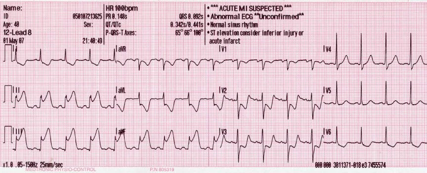
Long description
A 12-lead ECG showing pardee waves indicating acute myocardial infarction in the inferior leads II, III and aVF with reciprocal changes in the anterolateral leads.

DefinitionSTEMI
ST-elevation myocardial infarction (STEMI) is an acute myocardial infarction produced by sudden total thrombotic occlusion of an epicardial coronary artery, usually on a disrupted atherosclerotic plaque. The ischaemia is transmural, and irreversible loss of contractile tissue begins within minutes.
Myocardial infarction is defined (Fifth Universal Definition of Myocardial Infarction, 2026) as cardiac troponin rise with clinical, ECG, imaging or angiographic evidence of acute myocardial ischaemia. STEMI is the subset in which the ECG shows the characteristic elevation pattern; the diagnosis can often be made on ECG alone, without waiting for biomarker results, and treatment must not be delayed for a troponin. [1]
The following J-point cut-offs define STEMI in two anatomically contiguous leads. [2]
| Group | V2–V3 | Any other lead |
|---|---|---|
| Men <40 years | ≥2.5 mm | ≥1 mm |
| Men ≥40 years | ≥2.0 mm | ≥1 mm |
| Women (all ages) | ≥1.5 mm | ≥1.0 mm |
Additional high-risk presentations. [2]
- New or presumed new LBBB with modified Sgarbossa criteria: concordant ST elevation ≥1 mm; concordant ST depression ≥1 mm in V1–V3; or disproportionate discordant ST elevation with ST-to-S-wave ratio ≥0.25. Any single finding in one lead supports a STEMI-equivalent.
- Acute posterior MI: tall R wave with horizontal ST depression and upright T waves in V1–V4; posterior leads V7–V9 show ST elevation ≥0.05 mV (≥0.10 mV in men <40 years).
- Right ventricular infarction: right-sided lead V4R (and V3R) shows ST elevation, typically with an inferior infarct.
- Hyperacute T wave: very large, broad, asymmetric T wave in early STEMI (can precede obvious ST elevation by minutes).
- de Winter pattern: ST depression at the J point with upsloping ST segments blending into tall, symmetric T waves in V1–V4, with 0.5–1.0 mm ST elevation in aVR; usually proximal left anterior descending (LAD) occlusion.
- First diagonal occlusion: non-traditional, non-contiguous ST elevation in aVL and V2.
- Wellens syndrome: deeply inverted or biphasic T waves in V2–V3 with little or no ST elevation; critical proximal LAD stenosis, high risk but not labelled a STEMI equivalent.
- Left main/near-total left main obstruction: ST elevation in aVR >0.5 mm (often with aVL V1–V2) predicts a very high-risk lesion.
| Artery (typical) | Leads | Territory | Likely clinical picture |
|---|---|---|---|
| Proximal LAD | V1–V4 (±V5–V6, I, aVL) | Anterior/septal wall; large muscle mass | Greatest pump dysfunction; heart failure and arrhythmias common |
| Right coronary (RCA), proximal | II, III, aVF (±V4R) | Inferior wall ± right ventricle | Often RV involvement if proximal; haemodynamics depend on volume state |
| Circumflex / obtuse marginal | I, aVL, V5–V6 | Lateral wall | Smaller, but may be subtle on standard ECG |
| LCx / posterior descending territory | ST depression V1–V3 | Posterior wall | Frequently missed; posterior leads confirm |
Lateral MI (V5–V6, I, aVL) and posterior MI often accompany a large anterior or inferior infarct when multivessel disease is present. In most people the right coronary artery gives rise to the posterior descending artery; in a minority (left-dominant pattern) the circumflex artery does so. The clinical territory is therefore inferred from the whole ECG, not from any single lead.
A vulnerable plaque (thin fibrous cap over a lipid-rich core, inflamed, often with a large lipid pool) fissures or erodes. Platelets adhere, tissue factor is exposed, and an occlusive thrombus forms over minutes to hours. The myocardium distal to the occlusion is ischaemic almost immediately. Irreversible loss begins in the subendocardium before full-thickness necrosis develops over the following hours (the wavefront phenomenon). Collateral flow, pre-infarct angina (which can precondition the myocardium), metabolic demand and time therefore determine final infarct size.
Reperfusion restores flow but creates its own injury: reperfusion arrhythmias, calcium overload, reactive oxygen species and microvascular obstruction (the no-reflow phenomenon) can produce non-recoverable capillary damage even when the epicardial artery is open.
World Health Organization
Most concepts above are universal. The causes of coronary disease itself (smoking, hypertension, dyslipidaemia, diabetes, chronic renal disease) are the same worldwide; what differs by region is age of presentation, comorbidity (for example HIV-associated vascular disease in parts of sub-Saharan Africa) and access to reperfusion.
Patients report central or retrosternal crushing chest pain lasting longer than 20 minutes, often with radiation to the left arm, jaw, neck or back, and typically accompanied by sweating, nausea or vomiting, breathlessness and a sense of impending doom. Women, older adults and people with diabetes more often present with dyspnoea, fatigue, faintness, epigastric discomfort or no chest pain at all. Ask specifically about symptom onset time, the last time the patient was free of pain, prior angina or ischaemic heart disease, and any fibrinolytic or anticoagulant use.
Look for distress, sweating and sinus tachycardia or bradycardia; hypotension or a third or fourth heart sound suggests severe LV dysfunction; a new systolic murmur suggests mitral regurgitation from papillary muscle dysfunction or a ventricular septal defect (VSD); rales or an elevated JVP indicate pulmonary congestion or right heart failure; and cool, clammy peripheries with falling urine output define shock.
Killip classes describe current severity: class I has no clinical heart failure, class II has rales or crepitations with an S3 or elevated JVP, class III is frank pulmonary oedema, and class IV is cardiogenic shock. Class worsens prognosis stepwise.
| Mimic | Distinguishing feature |
|---|---|
| Acute (early) pericarditis | Diffuse ST elevation with PR depression; pain changes with position |
| Aortic dissection | Tearing pain to the back; pulse or BP discrepancy; mediastinal widening |
| Pulmonary embolism | Right heart strain, tachypnoea, pleuritic pain, hypoxia |
| Benign early repolarisation / LVH / Brugada | Stable pattern, no evolution, no symptoms |
| Hyperkalaemia | Peaked T waves, QRS widening, metabolic context |
| Myopericarditis | Elevated troponin with focal or diffuse ST changes; viral prodrome |
Figure 2AMI bloodtests engl
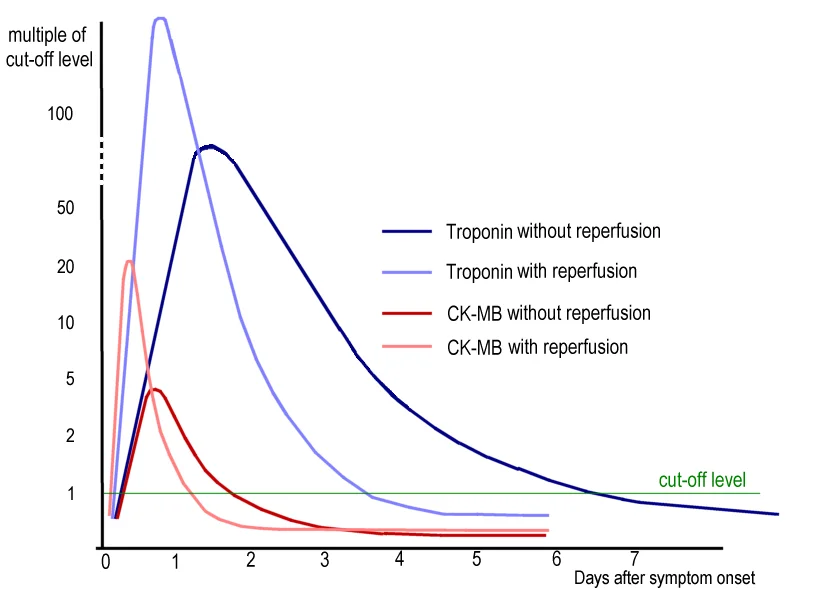
Long description
A self-made modification of the ACC/AHA Practice Guidelines 2005 (p. E32) graph described as showing typical changes in CK-MB and cardiac troponin in acute myocardial infarction.

- 12-lead ECG immediately (and repeat if the first tracing is non-diagnostic); add right-sided and posterior leads when inferior or suspected posterior involvement. Do not wait for pain-free status.
- Cardiac troponin (high-sensitivity if available) on presentation and serially; it rises early after symptom onset and remains elevated for days. A single negative test late in presentation does not exclude an acute evolving infarct.
- Bedside echocardiography for regional wall motion abnormality, valvular catastrophe, effusion or free-wall concerns, and to distinguish STEMI from dissection at the bedside.
- Coronary angiography is both diagnostic and therapeutic when primary PCI is available.
STEMI is diagnosed clinically by typical symptoms (or an equivalent silent presentation) plus either qualifying ST-segment elevation or an accepted equivalent on ECG. Biomarker confirmation supports but often follows the decision to reperfuse. Angiography confirms coronary occlusion.
- Record a 12-lead ECG (target <10 minutes after arrival). Call for reperfusion assessment.
- Give chewable (dispersible) aspirin 162–325 mg immediately unless contraindicated. [3]
- Analgesia: titrated intravenous morphine for persistent pain; monitor for respiratory depression.
- Oxygen only if hypoxic — give supplementary oxygen if arterial oxygen saturation is low or the patient is in acute heart failure or visibly dyspnoeic. Routine oxygen when saturation is normal is not helpful. [9]
- Sublingual glyceryl trinitrate (nitroglycerin) for pain and blood-pressure control; caution in inferior/RV infarcts.
- Definitive reperfusion decision using the algorithm below.
Reperfusion strategy in STEMI
- STEMI or accepted equivalent on ECGOnset of symptoms and haemodynamic stability
- Question: Time since symptom onset?
<12 hours (or >12 h with ongoing ischaemia or shock)
- Question: Can PCI be performed within the reperfusion window?
Yes – FMC to wire ≤120 min, door-to-balloon ≤90 min
- Primary PCICatheter lab activation, radial access, GPIIb/IIIa bailout only
No
- Give fibrinolytic immediately (door-to-needle <30 min)
- Then rescue PCI if reperfusion has failed, or pharmacoinvasive PCI 2–24 h if reperfusion succeeds
>12 hours and now pain-free, stable
- No routine immediate reperfusion; coronary angiography and PCI of the IRA as clinically indicated
Targets above are quoted from the current reperfusion literature and the major guidelines: FMC-to-wire 120 minutes and door-to-balloon 90 minutes are widely accepted goals; fibrinolysis should be given within 30 minutes of arrival. [6] [9] [10] [7]
Preferred when available. Radial access reduces bleeding. Key points: load aspirin and a P2Y12 inhibitor before/at the table; unfractionated heparin (or bivalirudin) with ACT guidance; thrombus aspiration only as a bail-out for large thrombus; intracoronary vasodilators for no-reflow; imaging (IVUS/OCT) to optimise stenting. Intracoronary imaging improves recognition of non-atherosclerotic causes (spontaneous dissection, vasospasm, embolism), which change management. [11]
Use when timely PCI is not achievable. Earlier treatment is better; each 30-minute delay reduces benefit.
Regimens quoted from major series and product literature. [5]
- Tenecteplase: single intravenous bolus, weight-adjusted:
- <60 kg: 30 mg; 60–69 kg: 35 mg; 70–79 kg: 40 mg; 80–89 kg: 45 mg; ≥90 kg: 50 mg.
- Streptokinase: 1.5 million units intravenously.
- (Alternative second-generation agents such as alteplase and reteplase follow local protocols.)
Adjunctive anticoagulation with fibrinolysis (unfractionated heparin, enoxaparin or fondaparinux) is continued to prevent re-occlusion; follow the chosen guideline or product dosing closely.
Contraindications include active or recent significant bleeding, intracranial or subarachnoid haemorrhage or known intracranial vascular abnormality, ischaemic stroke within the previous 3 months, aortic dissection, suspected pericarditis with pericardial effusion and marked, uncontrolled hypertension. Many are absolute.
Rescue PCI is indicated when pain, ST elevation or haemodynamic instability persist 60–90 minutes after fibrinolysis (ST resolution <50%). Pharmacoinvasive PCI (coronary angiography within 2–24 hours after successful lysis) is the standard follow-up after fibrinolysis in PCI-capable systems.
Antiplatelet and anticoagulant dosing below is standard guidance summarised in review sources. [3] [4]
| Drug | Loading | Maintenance |
|---|---|---|
| Aspirin | 162–325 mg chewed (325 mg if receiving ticagrelor) | 75–100 mg/day once stabilised ≤100 mg/day with ticagrelor |
| Clopidogrel | 300–600 mg | 75 mg once daily |
| Prasugrel | 60 mg (avoid if age ≥75 or body weight <60 kg) | 10 mg once daily (5 mg if ≥75 y or <60 kg) |
| Ticagrelor | 180 mg | 90 mg twice daily |
| UFH (parenteral) | 60–100 IU/kg IV (adjust for GPI or ACT) | Re-dose to a target ACT |
| Bivalirudin | 0.75 mg/kg IV bolus | 1.75 mg/kg/h infusion |
Bail-out GPIIb/IIIa inhibitors (e.g. tirofiban) are reserved for large thrombus or no-reflow, not used routinely. Ticagrelor is contraindicated with doses of aspirin above 100 mg/day.
If the patient already takes a vitamin K antagonist (INR >2.5) or a non-vitamin K anticoagulant, modify the parenteral anticoagulant accordingly.
- Beta-blocker: oral (or intravenous if tachycardic/hypertensive) early, once the patient is stable, unless in cardiogenic shock, high-grade AV block or acute heart failure. Avoid if hypotension.
- ACE inhibitor or ARB: start early after MI, especially with LV dysfunction, diabetes or an anterior infarct; improves ventricular remodelling. Avoid if hypotensive or hyperkalaemic.
- Mineralocorticoid receptor antagonist (spironolactone or eplerenone type): add when the LV ejection fraction is ≤40% with either heart failure or diabetes, after starting ACE inhibitor and beta-blocker.
- High-intensity statin: start at presentation, regardless of the arrival LDL, with a target LDL ≥50% reduction. [8]
- Nitrates: intravenous glyceryl trinitrate for recurrent pain or ischaemia; useful for hypertension and LV strain. Avoid or use carefully in inferior/RV infarcts where preload dependence matters.
- Oxygen: only if hypoxic, in heart failure or marked dyspnoea.
- Proton-pump inhibitor: co-prescription for gastric protection with dual antiplatelet therapy in high bleeding risk.
- Arrhythmias: the commonest early killer. Ventricular fibrillation or pulseless VT during the first hours, ventricular tachycardia during the first day, sinus bradycardia or heart block with inferior infarcts, new-onset AF, and accelerated idioventricular rhythm. Defibrillate VF/VT; treat reversible triggers (hypoxia, pain, hyperkalaemia). Ventricular arrhythmias Approach to tachyarrhythmias
- Heart failure and cardiogenic shock: treat with revascularisation (primary PCI remains first-line even in shock), fluids as indicated, norepinephrine and/or dobutamine, inotropes, and mechanical circulatory support if available.
- Mechanical complications (typically days 1–7): papillary muscle rupture causing severe acute mitral regurgitation; ventricular septal rupture (new harsh systolic murmur with biventricular failure — echocardiography/MRI and surgical or percutaneous closure); free-wall rupture causing tamponade and sudden collapse. Urgent echocardiography and surgical consultation. Cardiac tamponade Heart failure
- Recurrent ischaemia or re-infarction from incomplete reperfusion, stent thrombosis or residual disease.
- Pericarditis: early pericarditis a few days after a transmural MI (pericardial rub, diffuse ST elevation, pain eased leaning forward); Dressler syndrome (auto-immune) a few weeks later with fever, pleuritic pain and effusion.
- Right ventricular infarction: suspect with inferior infarct + hypotension + clear lung fields + distended JVP. Manage with fluids (careful), avoid nitrates/diuretics, inotropes if hypotensive, and early reperfusion.
- Left ventricular aneurysm (dyskinetic wall): risk of heart failure, recurrent VT, systemic embolism; anticoagulate if thrombus visible.
- LV thrombus and systemic embolism: clinical vigilance; anticoagulation when imaging shows thrombus; stroke symptoms need urgent stroke pathways (thrombolysis decision independent). Atrial fibrillation and atrial flutter
- Late arrhythmias: VT/VF late signals poor prognosis; strong indication for ICD assessment when reduced LVEF.
- Depression, fatigue and impaired exercise tolerance; structured rehabilitation.
- Inferior STEMI + hypotension and clear lungs → check V3R/V4R and echo.
- Fluids cautiously, with frequent reassessment; avoid nitrates/diuretics/preload reducers.
- Inotropes (dobutamine) if fluid alone does not restore pressure.
- Urgent reperfusion (primary PCI of the RCA/proximal occlusion).
- Vasopressors as needed; mechanical support in shock.
- Prognosis worse with RV involvement; intensive monitoring.
- Reperfusion confirmation: non-invasive (ST resolution, biomarker fall); planned angiography within 24 hours after fibrinolysis; recurrent symptoms demand re-investigation.
- Imaging: echocardiography before discharge (function, valve, thrombus); cardiac MRI if doubt about viability or size.
- Secondary prevention: aspirin indefinitely; P2Y12 for 12 months in most (shorter if high bleeding risk: 1–6 months with de-escalation); high-intensity statin with lipid target review; ACE inhibitor/ARB; beta-blocker; MRA where appropriate. Rivaroxaban 2.5 mg twice daily plus aspirin 100 mg after 12 months can be considered in high ischaemic/low bleeding risk (guidelines differ on class). [8]
- Blood pressure, glycaemic control, weight, tobacco cessation, alcohol reduction, and diet advice.
- Exercise-based cardiac rehabilitation improves outcomes and quality of life; start low level early in hospital and escalate under supervision.
- Psychosocial: screen for depression and cognitive change after MI.
- Immunisations: annual influenza vaccination recommended in most programmes.
Return to non-physically demanding work after an uncomplicated MI varies from a few days to a few weeks, and return to physically heavy work should be cleared by the cardiology team after a functional assessment. Commercial and occupational driving (heavy vehicles, public service vehicles, aircraft) requires longer recovery and formal medical review against national licensing standards. Tobacco use should be stopped; passenger and cargo roles may be restricted. Follow the treating cardiology team and national licensing authority rather than generic timetables.
Early successful reperfusion has transformed outcomes: most patients who survive the first hospital days do well long term. Prognosis is driven by age, LV ejection fraction, extent of coronary disease, arrhythmic substrate, renal function, diabetes and whether the post-MI treatment package (antithrombotic, statin, ACE inhibitor, beta-blocker, MRA, cardiac rehabilitation and smoking cessation) is delivered completely. Late mortality remains highest in those with reduced LVEF or recurrent ischaemia and in older adults.
In many low- and middle-income countries PCI is sparse and reperfusion may be delayed by cost, transport and infrastructure. Fibrinolysis is therefore the realistic first reperfusion method in much of the world, and its timely use (door-to-needle under 30 minutes) saves lives. A pharmacoinvasive approach — lyse early, then transfer for coronary angiography within 2–24 hours — captures most of PCI's benefit. Streptokinase is cheaper but antigenic (single use, plus anaphylaxis risk) and requires intravenous infusion; tenecteplase as a single bolus is easier in small centres without continuous infusion capacity. Cost analyses have found that an integrated package of aspirin, a beta-blocker, an ACE inhibitor and a fibrinolytic is more cost-effective in resource-constrained settings than high-cost PCI programmes. Delays remain the major problem: symptom-to-presentation intervals of hours to days, lack of EMS, lack of ECGs in the ambulance, shortage of trained providers and drug availability (for example streptokinase price and prioritisation on national drug lists). Public-awareness campaigns, ECG training, telemedicine support for spoke hospitals and clear referral protocols improve measures such as door-to-balloon times. [7] [6]
- STEMI is acute thrombotic occlusion of a coronary artery causing transmural ischaemia; diagnose on ECG and treat before biomarker results arrive.
- Reperfusion goals: from first medical contact to wire within about 120 minutes; door-to-balloon within about 90 minutes; fibrinolysis within about 30 minutes when PCI cannot meet those targets.
- Primary PCI is the preferred method; fibrinolysis plus rescue or pharmacoinvasive PCI is the practical alternative.
- Antithrombotic foundation: aspirin 162–325 mg then 75–100 mg/day; clopidogrel 300–600 mg then 75 mg; prasugrel 60 then 10 mg; ticagrelor 180 then 90 mg twice daily; UFH 60–100 IU/kg IV or bivalirudin 0.75 mg/kg bolus then 1.75 mg/kg/h.
- Anticoagulation with fibrinolysis uses a heparin, enoxaparin or fondaparinux schedule per the chosen guideline.
- Adjuncts: beta-blocker early if safe, ACE inhibitor or ARB early, MRA when LVEF ≤40 in heart failure or diabetes, high-intensity statin, PPI for high bleeding risk, oxygen only if hypoxic, nitrates with caution in RV infarct.
- Mechanical complications (papillary rupture, VSD, free-wall rupture) peak in the first post-MI week; urgent echo and surgery.
- Right ventricular infarction with an inferior MI: fluids, avoid nitrates, urgent reperfusion, consider inotropes.
- Long-term outcomes depend on complete delivery of secondary prevention and cardiac rehabilitation.
- In low-resource settings, timely fibrinolysis plus a pharmacoinvasive strategy is effective and cost-efficient.
Answer each one in your head first, then open the answer.
A 54-year-old man has 40 minutes of crushing central chest pain. ECG shows 3 mm ST elevation in V1-V4 and 1 mm in I, aVL. Which artery is involved and what emergency action is needed within the next 60 minutes?
Show answerHide answer
Proximal LAD occlusion causing an anterior STEMI. Give 325 mg aspirin, start oxygen only if hypoxic, arrange primary PCI (standard targets: FMC-to-wire within about 120 minutes, door-to-balloon under 90 minutes); if PCI cannot be delivered in that window, give tenecteplase at a weight-adjusted bolus within 30 minutes of arrival. [2] [6] [7]
A 68-year-old woman with an inferior STEMI is hypotensive (88/52) with clear lung fields and raised JVP. Her right-side lead V4R shows ST elevation. What is happening and how should management differ from a standard inferior infarct?
Show answerHide answer
Right ventricular infarction. Give cautious fluid boluses, stop nitrate and diuretic therapy, start inotropes if hypotension persists, and reperfuse urgently (primary PCI of the RCA). Mechanical support may be needed. Avoid preload reduction.
State the J-point ST-elevation cut-offs for a 70-year-old man in leads V2-V3 and for a 35-year-old woman in any lead.
Show answerHide answer
≥2.0 mm in V2-V3 for the man, and ≥1.0 mm in any other lead for the woman (her V2-V3 cut-off is ≥1.5 mm). [2]
A stable 80 kg patient arrives 3 hours after onset of anterior STEMI where the nearest cath lab is 3 hours away by road. What reperfusion strategy is chosen now, at what dose, and what must be arranged before the next day?
A patient has LBBB and concordant ST elevation of 1.2 mm in one lead during chest pain. Is this STEMI?
Show answerHide answer
Yes. Modified Sgarbossa criteria (concordant elevation ≥1 mm) overrule the usual LBBB appearance; an urgent STEMI pathway is indicated even though the finding is in a single lead. [2]
After an anterior STEMI treated with primary PCI, echo at day 2 shows LVEF 35%, no complications. Which drugs should be continued at discharge and what is the target LDL strategy?
Show answerHide answer
Dual antiplatelet therapy (duration to 12 months, earlier if bleeding risk), high-intensity statin with lipid review according to guideline, ACE inhibitor or ARB, beta-blocker, and (because LVEF ≤40 with heart failure) an MRA; add cardiac rehabilitation, smoking cessation support and influenza vaccination. [3] [8]
Cardiology topics: Heart failure, Electrocardiogram (ECG) interpretation, Cardiac physical examination, Atrial fibrillation and atrial flutter, Ventricular arrhythmias, Bradyarrhythmias and atrioventricular block, Supraventricular tachycardias, Approach to tachyarrhythmias. Other relevant topics (plain text): acute coronary syndrome distinction, valvular heart disease, cardiogenic shock, pericarditis and thrombolysis.
Each numbered citation in the text points to the corresponding entry in the front-matter reference list.
- Mills NL, Newby LK, Zaman S, et al. Fifth Universal Definition of Myocardial Infarction (2026). Glob Heart 2026;21(1):64. doi:10.5334/gh.1578 (PMC13523810). Accessed .
- Brady WJ, Muck AE, Moak JH. High-risk electrocardiogram presentations in the acute coronary syndrome patient – Beyond ST-segment elevation myocardial infarction. Turk J Emerg Med 2025;25(1):1–9. doi:10.4103/tjem.tjem_150_24 (PMC11774428). Accessed .
- Towashiraporn K, Krittayaphong R. Current Perspectives on Antithrombotic Therapy for the Treatment of Acute Coronary Syndrome. Int J Gen Med 2022;15:2397–2414. doi:10.2147/IJGM.S289295 (PMC8901254). Accessed .
- Zhai Y, Shang H, Li Y, et al. The efficacy and safety of bivalirudin and heparin in patients with acute coronary syndrome: a systematic review and meta-analysis. Syst Rev 2025;14:39. doi:10.1186/s13643-025-02782-7 (PMC11808951). Accessed .
- Karanth JB, Ramamoorthy A, Maribashetti K, et al. Thrombolysis in STEMI in the era of COVID – Holding fort in cardiologist deficit locales. Indian Heart J 2023;75(4):288–291. doi:10.1016/j.ihj.2023.05.002 (PMC10172153). Accessed .
- Hassan MO, Ahmed SA, Hassan MS, Köprülü D. Door-to-Balloon Time and Mortality Among Patients Undergoing Primary PCI, Challenges and Experience from Somalia's Largest PCI Center. Int J Gen Med 2024;17:237–244. doi:10.2147/IJGM.S448750 (PMC10812143). Accessed .
- Pool AJ, Smit PC, Slabber H, Stassen W. ST-Elevation Myocardial Infarction Systems of Care in Africa: A Scoping Review. Glob Heart 2026;21(1):11. doi:10.5334/gh.1524 (PMC12922679). Accessed .
- Kim S, Piserra-López A, Giordano S, et al. In Hospital Assessment and Management of High Bleeding Risk in Patients with ST-Elevation Myocardial Infarction (STEMI). J Cardiovasc Dev Dis 2026;13(5):183. doi:10.3390/jcdd13050183 (PMC13207790). Accessed .
- Byrne RA, Rossello X, Coughlan JJ, et al. 2023 ESC Guidelines for the management of acute coronary syndromes. Eur Heart J 2023;44(38):3720–3826. doi:10.1093/eurheartj/ehad191. Accessed .
- Rao SV, O'Donoghue ML, Ruel M, et al. 2025 ACC/AHA/ACEP/NAEMSP/SCAI Guideline for the Management of Patients With Acute Coronary Syndromes. Circulation 2025;151:e771–e862. doi:10.1161/CIR.0000000000001309. Accessed .
- Tamis-Holland JE, Abbott JD, Al-Azizi K, et al. SCAI Expert Consensus Statement on the Management of Patients With STEMI Referred for Primary PCI. J Soc Cardiovasc Angiogr Interv 2024;3(11):102294. doi:10.1016/j.jscai.2024.102294 (PMC11624394). Accessed .
Figure credits
Educational content for medical students and doctors. It is not a substitute for clinical judgement or local guidelines.