Cardiac tamponade
Fluid or blood around the heart raises pressure until the chambers cannot fill. Recognise tamponade, read the echo and drain before shock kills.
- Cardiac tamponade is compression of the heart by fluid, blood, pus, clot or gas in the pericardial sac. The pressure outside the chambers rises until they cannot fill.
- It is a clinical diagnosis. A large effusion is not the same thing as tamponade, and a small, rapidly accumulating collection can be fatal.
- The classic picture is hypotension, a raised jugular venous pressure and muffled heart sounds, but the full triad is often absent. Pulsus paradoxus and a jugular venous pulse without a y descent are more useful clues.
- Echocardiography shows the effusion and its effect: right atrial and right ventricular collapse, exaggerated respiratory variation in inflow, and a plethoric inferior vena cava.
- Drainage is the treatment. Fluids and the avoidance of diuretics, vasodilators and positive pressure ventilation buy time; they do not relieve the pressure.
By the end of this page you should be able to:
- Define cardiac tamponade and explain why the speed of fluid accumulation matters more than its volume.
- Relate the pressure-volume curve and ventricular interdependence to pulsus paradoxus and the other physical signs.
- Recognise tamponade from the bedside findings, including an incomplete Beck triad and a jugular venous pulse without a y descent.
- Interpret the electrocardiogram (ECG), chest radiograph and echocardiographic signs of tamponade, and know their limitations.
- Use a severity score to identify patients who need urgent drainage, while understanding its limits.
- Plan pericardiocentesis or surgical drainage, including the approach, the complications and the drugs and ventilatory settings to avoid.
- Adapt management for trauma, cardiac surgery, malignancy, tuberculosis, uraemia, aortic dissection and resource-limited settings.
Cardiac tamponade is one of the few cardiovascular emergencies in which a bedside procedure can restore a dying patient within minutes. The heart is not failing because the muscle is weak; it is failing because it cannot expand. Blood returning to the heart cannot fill chambers that are being squeezed from outside, so stroke volume falls and the patient develops obstructive shock.
The diagnosis is easily missed when the classic triad is incomplete, when the patient is hypovolaemic after trauma, or when a collection after cardiac surgery is loculated and hidden behind a drain. It is also easily overdiagnosed: many large effusions cause no haemodynamic compromise and need observation, not a needle.
This page follows the path from cause to pressure, from pressure to signs, and from signs to drainage. It also separates tamponade from the effusion that merely sits there, because that distinction decides who needs a procedure today.
DefinitionCardiac tamponade
Cardiac tamponade is the compression of the heart by accumulated pericardial contents under pressure, sufficient to impair diastolic filling and reduce cardiac output. It is a haemodynamic state, not a fluid volume: the same effusion can be harmless in one patient and lethal in another.
The 2015 European Society of Cardiology (ESC) guideline defines tamponade as a life-threatening, slow or rapid compression of the heart caused by accumulation of fluid, pus, blood, clots or gas after inflammation, trauma, rupture of the heart or aortic dissection.[1]
Tamponade can be classified in several ways, and each axis changes management:
- By speed. Acute tamponade develops over minutes to hours, typically after penetrating trauma, cardiac perforation or free-wall rupture. Subacute tamponade develops over days to weeks, usually from an effusion of malignant, tuberculous, uraemic or inflammatory origin.
- By composition. The fluid may be serous, sanguinous, purulent, chylous or gas. Purulent and bloody collections often need surgical drainage rather than a needle.
- By haemodynamic effect. An effusion may be present without tamponade, with tamponade, or with effusive-constrictive physiology, in which a scarred pericardium constricts the heart even after the fluid is removed.
- By size. Echocardiography grades a diastolic echo-free space as mild (less than 10 mm), moderate (10 to 20 mm) or large (more than 20 mm).[1] Size predicts the likelihood of a specific cause and of complications, but it does not define tamponade.
Two variants deserve names. Low-pressure tamponade occurs when hypovolaemia keeps the central venous pressure low despite significant compression; it is seen after trauma and after aggressive diuresis.[4] Regional tamponade occurs when a loculated collection compresses one chamber, often the left ventricle after cardiac surgery, so the usual right-sided echo signs are absent.[4]
Tamponade is uncommon in absolute terms but common enough that every clinician who drains a pericardium, inserts a pacemaker or manages chest trauma will meet it. Its frequency depends on the population: it is rare in uncomplicated viral pericarditis and much more common with malignancy, tuberculosis, purulent pericarditis, uraemia and recent cardiac procedures.[1]
Some useful anchors:
- Cancer accounts for 10 to 25% of pericardial effusions in developed countries, and tuberculosis is the dominant cause in developing countries, where it accounts for more than 60%.[1]
- Large idiopathic chronic effusions carry a 30 to 35% risk of progression to tamponade, and severe effusions may progress in up to one-third of cases.[1]
- After cardiac surgery, moderate to large effusions progress to tamponade in about 10% of cases at one month,[1] although the risk of a significant effusion or haematoma requiring treatment is approximately 1%.[7]
- In a real-world series of 81 patients drained for pericardial effusion, 77.8% had tamponade and in-hospital mortality was 14.8%.[6] That figure reflects a selected, often critically ill population and should not be applied to every effusion.
Almost any pericardial disease can cause tamponade. The cause matters because it determines whether a needle is enough, whether surgery is needed, and what fluid tests will change management.
| Cause | Typical setting | Clue or consequence |
|---|---|---|
| Malignancy | Lung, breast, melanoma, lymphoma and leukaemia; often subacute | Large effusion, frequent recurrence (40 to 70%), poor overall survival.[1] A malignant effusion may be the first sign of cancer.[1] |
| Pericarditis, including tuberculosis | Viral or idiopathic pericarditis; tuberculosis in endemic areas and in human immunodeficiency virus (HIV) infection | Tuberculosis often presents with chronic compression and a high risk of constriction.[1] Purulent pericarditis is heavily loculated and needs drainage.[1] |
| Post-cardiac surgery or percutaneous procedure | Coronary intervention, pacemaker or defibrillator lead, ablation, transseptal puncture, valve surgery | Can occur with chest drains in place and an open pericardium, because clot can compress a chamber focally.[2] Early tamponade after surgery is usually haemorrhage.[1] |
| Trauma | Penetrating chest injury, blunt injury, resuscitative procedures | Shock out of proportion to visible blood loss; the classic signs may be masked by hypovolaemia.[2] |
| Aortic dissection | Type A dissection with retrograde bleeding into the pericardium | Tamponade can be the presenting feature; drainage alone may worsen the tear.[1][3] |
| Uraemia | Advanced kidney disease, especially before or shortly after starting dialysis | Effusions are often bloody, so anticoagulation is risky; tamponade can occur with a modest collection.[1] |
| Post-myocardial infarction free-wall rupture | Several days after a large transmural infarct | A pericardial effusion larger than 10 mm should prompt a search for subacute rupture.[1] |
| Anticoagulation | Therapeutic heparin, warfarin or direct oral anticoagulants, especially after a cardiac procedure | Full anticoagulation may increase the risk of tamponade when the effusion is iatrogenic.[1] It is a contributor rather than a cause. |
| Other | Radiation, autoimmune disease, hypothyroidism, chylopericardium | Treat the underlying disease; the approach to drainage is the same.[1] |
The distinction between a malignant effusion and a malignant cause is important. In a patient with known cancer, an effusion may be due to radiation, chemotherapy, infection or heart failure, and cytology is needed before labelling it malignant.[1] Conversely, a malignant effusion can be the first sign of cancer.[1]
The normal pericardium contains 10 to 50 mL of fluid and is compliant enough to accommodate small changes in cardiac volume.[1] Its pressure-volume curve is J-shaped: an initial shallow portion allows some stretch, then the curve becomes almost vertical.[1][4] Once the reserve is used up, a small additional volume produces a large rise in pressure. This is why tamponade behaves like a "last-drop" phenomenon, in which the final increment causes critical compression and the first decrement during drainage produces the largest relative decompression.[1]
The clinical consequence is that rate matters more than volume. A rapid addition of 100 to 200 mL can raise pericardial pressure to 30 mmHg or more and cause severe tamponade, whereas an effusion that develops over weeks can reach litres with only a modest pressure rise.[4] A large, slowly growing effusion may therefore be tolerated until it is not; a small bleed after a pacemaker can kill within minutes.
As pericardial pressure rises, the heart compensates by raising systemic and pulmonary venous pressures until they match the pressure outside the chambers. Left and right ventricular diastolic, right atrial and pulmonary wedge pressures all rise and equalise, so the transmural distending pressure falls towards zero.[4] In tamponade, diastolic pressures throughout the heart are usually 15 to 30 mmHg, except in low-pressure tamponade.[1]
The equalisation explains the jugular venous findings. The venous pressure is high, but the ventricle cannot fill normally in diastole, so the usual rapid y descent is lost. The x descent is preserved or prominent because atrial relaxation still occurs.[3]
Because the total pericardial volume is fixed, the two ventricles compete for space. During inspiration, intrathoracic pressure falls and venous return to the right heart increases. The right ventricle fills, the interventricular septum bulges to the left, and left ventricular filling falls. Systemic arterial pressure therefore drops during inspiration. During expiration the reverse happens.[1][4]
That exaggerated respiratory variation is pulsus paradoxus, conventionally defined as an inspiratory fall in systolic arterial pressure of more than 10 mmHg during normal breathing.[1][3] The word "paradoxus" is historical: the pulse becomes weaker during inspiration even though the heart is still beating.
- Tachycardia and vasoconstriction are compensatory. Cardiac output is maintained partly by a faster heart rate and increased sympathetic tone, which is why a young patient can look deceptively stable until decompensation.[2]
- Hypotension appears when compensation fails. In moderate tamponade, blood pressure and cardiac output may be only slightly reduced, so a normal reading does not exclude the diagnosis.[4]
- Muffled heart sounds reflect the fluid layer between the chest wall and the heart, and are described in moderate to large effusions.[1]
- Low ECG voltage and electrical alternans reflect damping of the electrical signal by fluid and the swinging motion of the heart.[1]
The severity spectrum is continuous. When pericardial pressure exceeds about 10 to 12 mmHg, moderate tamponade is present; pulsus paradoxus is often absent at this stage, and the diagnosis may rest on the jugular pulse and echocardiography. When pressure exceeds about 25 mmHg, tamponade is severe, with a prominent pulsus, reduced stroke volume and usually sinus tachycardia.[4] Shock and death follow if the pressure is not relieved.[4]
Hypovolaemia can keep the central venous pressure low despite severe tamponade. This is low-pressure tamponade, and it is a particular trap after trauma and in patients who have been given diuretics for presumed heart failure.[4]
Dyspnoea is usually the first and most sensitive symptom.[2] It may be exertional at first and then present at rest or on lying flat. Chest discomfort is common, especially when the underlying process is inflammatory. Right upper quadrant pain and peripheral oedema can dominate when venous congestion is severe.[3] Fatigue, weakness, anorexia and palpitations are non-specific. In advanced cases the picture is one of obstructive shock: cool peripheries, confusion, oliguria and hypotension.[2]
A patient can also present with pulseless electrical activity. Tamponade should be on the differential for any patient in cardiogenic shock with a raised jugular venous pressure, particularly after a cardiac procedure.[3]
The classic triad described by Beck in 1935 is hypotension, a raised jugular venous pressure and muffled heart sounds.[2] It is memorable and worth knowing, but it is a poor screening test. In one emergency department series, none of the patients with tamponade had all three elements, and the sensitivity of any one element was only 50%.[2] A normal blood pressure does not exclude tamponade, and muffled sounds are subjective.
Pulsus paradoxus is the most useful bedside sign when it is present. Measure it with a sphygmomanometer: inflate the cuff above the systolic pressure, deflate slowly, and note the pressure at which Korotkoff sounds first appear during expiration and the pressure at which they become audible throughout the respiratory cycle. The difference is the paradoxus. A fall of more than 10 mmHg is abnormal.[1][3]
It is not specific. Severe asthma and exacerbations of chronic obstructive pulmonary disease can produce exaggerated swings in intrathoracic pressure and a pulsus.[2] It is also absent in some patients with severe tamponade:
- Conditions that keep left ventricular diastolic pressure high, atrial septal defect and aortic regurgitation can prevent the inspiratory fall.[4]
- Chronic kidney disease with a stiff, volume-loaded circulation often fails to show a pulsus.[4]
- Right heart failure or right ventricular hypertrophy can prevent right atrial and ventricular compression.[4]
- Hypovolaemia after trauma can make the sign difficult to detect.[2]
- In moderate tamponade, pulsus paradoxus is often absent even though tamponade is present.[4]
The jugular venous pressure (JVP) is almost always raised. The x descent is preserved, but the y descent is absent or attenuated because diastolic filling of the ventricle is blunted.[3] This is the finding behind the teaching "raised JVP without a y descent". In effusive-constrictive pericarditis, by contrast, the y descent may be prominent.[4] Kussmaul sign, a rise in jugular venous pressure on inspiration, can occur in tamponade but is more typical of constriction.[3]
Tachycardia is usual in severe tamponade.[4] The pulse may feel weak and vary in volume with respiration. Heart sounds are muffled in moderate to large effusions.[1] A pericardial friction rub suggests concomitant pericarditis.[3] Look for cool peripheries, prolonged capillary refill, low urine output and altered mental state, which indicate that compensation is failing.[2]
NoteA vignette
A 64-year-old man with metastatic lung cancer has two weeks of breathlessness that is worse lying flat. He is pale and anxious. Blood pressure is 96/70 mmHg, pulse 112 beats/min, and the jugular venous pressure is raised to the earlobe with no visible y descent. Heart sounds are quiet. The ECG shows sinus tachycardia and low voltage. A bedside echocardiogram shows a large circumferential effusion, late diastolic right atrial collapse and early diastolic right ventricular collapse. This is tamponade from a malignant effusion; he needs urgent drainage and an oncology plan.
Tamponade is one cause of obstructive shock, and the examination can be mimicked by other conditions that raise venous pressure and lower cardiac output. The table separates the most important alternatives.
| Condition | What it shares with tamponade | What separates it |
|---|---|---|
| Cardiac tamponade | Hypotension, raised JVP, tachycardia, pulsus paradoxus | Effusion on echo, right atrial and ventricular collapse, absent y descent, improvement after drainage.[1][3] |
| Constrictive pericarditis | Raised JVP, low output, fatigue and oedema | Prominent x and y descents, pericardial knock, usually no pulsus, thickened or calcified pericardium, no large effusion.[4] |
| Effusive-constrictive pericarditis | Tamponade plus a scarred pericardium | Prominent y descent; raised right atrial pressure persists after fluid is removed.[4] |
| Tension pneumothorax | Hypotension, raised JVP, tachycardia, shock | Unilateral reduced breath sounds, hyper-resonance and air in the pleural space; the signs can mimic tamponade.[2] |
The ECG can be normal, and a normal tracing does not exclude tamponade. The common findings are:
- Sinus tachycardia, which occurs in most patients as a compensatory response.[3]
- Low QRS voltage, defined as a maximum QRS amplitude below 0.5 mV in the limb leads.[3]
- Electrical alternans, a beat-to-beat change in QRS amplitude or axis caused by the heart swinging in a large effusion. It is specific but not sensitive.[3]
- Pericarditic changes, such as widespread ST elevation and PR depression, when the underlying process is pericarditis.[1]
Read the ECG alongside the clinical picture, and use it to look for alternative diagnoses such as myocardial infarction or pulmonary embolism. For a systematic approach, see ECG interpretation.
Echocardiography is the single most useful test: it confirms the effusion, estimates its size and distribution, and shows whether the chambers are compressed.[1] It is also the safest guide for pericardiocentesis.
| Sign | Timing or threshold | What it means and what can hide it |
|---|---|---|
| Right atrial collapse | Late diastole; persistence for more than one-third of the cardiac cycle is highly sensitive and specific | The atrial wall buckles when pericardial pressure exceeds atrial pressure. It may be absent when right heart pressures are high.[3] |
| Right ventricular collapse | Early diastole | The free wall indents as ventricular pressure falls below pericardial pressure. It is specific but less sensitive than atrial collapse.[3] |
| Exaggerated mitral inflow variation | More than 25% (ESC) or 30% (review) | Reflects ventricular interdependence. It may be inaccurate during mechanical ventilation and is reduced when left atrial pressure is high.[1][2] |
| Exaggerated tricuspid inflow variation | More than 40% | The reciprocal change on the right side; useful when mitral variation is equivocal.[5] |
| Inferior vena cava plethora | Less than 50% reduction in diameter with inspiration | A dilated, non-collapsing IVC reflects high right atrial pressure. It is sensitive but not specific.[3][5] |
| Swinging heart and septal motion | Any | The heart rocks in a large effusion; the septum shifts leftward in inspiration. These findings support the diagnosis but depend on image quality.[1] |
| Pulmonary vein diastolic flow | Inspiratory decrease and expiratory increase | A Doppler sign of raised filling pressure; helpful when other signs are subtle.[1] |
Two practical points follow. First, a large effusion without collapse or inflow variation is not automatically tamponade; treat the patient, not the number. Second, a loculated collection after cardiac surgery may compress one chamber and produce few classic signs, so transoesophageal echocardiography may be needed when transthoracic images are poor.[2]
Figure 1Tamponade
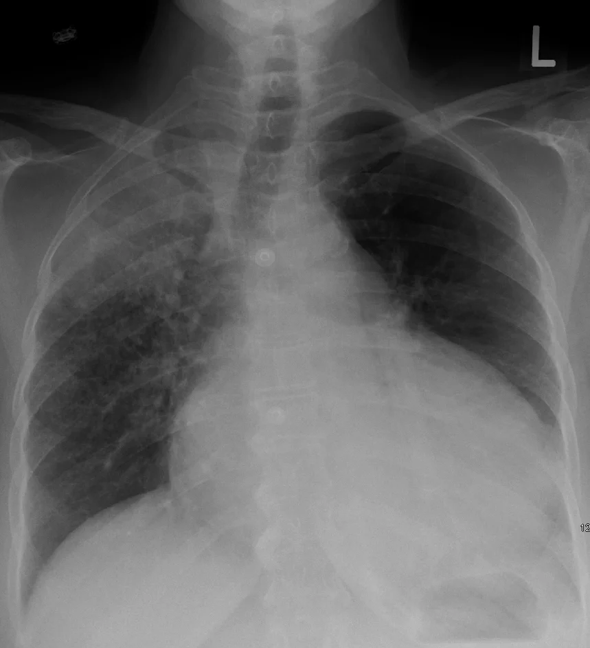
Long description
A chest X-ray showing a pericardial effusion due to malignancy with a bulbous heart and a primary lung cancer in the right upper lobe.

The chest radiograph is usually normal in acute tamponade because the pericardium has not had time to stretch. The cardiac silhouette enlarges only after a critical volume has accumulated; one review puts this at about 200 mL, while the ESC notes that an increased cardiothoracic ratio occurs only with effusions exceeding 300 mL.[3][1] A globular, water-bottle-shaped heart suggests a chronic large effusion, but a normal film never excludes tamponade.[2]
Computed tomography (CT) and magnetic resonance imaging (MRI) are not needed when echocardiography is available and the patient is unstable. They are useful when the effusion is loculated, when a pericardial mass or aortic dissection is suspected, or when transthoracic images are inadequate.[1] CT can show pericardial calcification; MRI characterises tissue and can show inflammation.[1]
No blood test diagnoses tamponade. Useful tests are guided by the suspected cause:
- Troponin and creatine kinase may be raised when there is myocardial involvement, but a normal troponin does not exclude tamponade.[1]
- Renal function and electrolytes are needed before drainage and in uraemic pericarditis.
- Full blood count, CRP and blood cultures help identify infection or inflammation.
- Coagulation studies matter before pericardiocentesis, although correction should not delay drainage in an unstable patient.
- Pericardial fluid should be sent for cell count, protein, glucose, lactate dehydrogenase, Gram stain, culture, acid-fast bacilli testing, cytology and, where available, polymerase chain reaction for tuberculosis.[3]
Catheterisation is rarely needed for diagnosis. It shows equalisation of diastolic pressures at 15 to 30 mmHg and the characteristic respiratory reciprocation of right and left heart pressures.[1] It is most useful when the diagnosis is unclear, when effusive-constrictive disease is suspected, or when pressures are needed during elective pericardiocentesis.[1]
There is no single diagnostic criterion for tamponade. The diagnosis is made when a pericardial effusion is present and the clinical picture shows impaired filling and reduced cardiac output, with echocardiographic support.[1][3] A practical definition is a patient with an effusion plus at least one of: hypotension or shock, a raised jugular venous pressure, pulsus paradoxus, or echocardiographic chamber collapse with exaggerated respiratory variation.
The ESC grades effusions by the diastolic echo-free space: mild (less than 10 mm), moderate (10 to 20 mm) or large (more than 20 mm).[1] Size is a risk marker, not a diagnosis. Haemodynamic severity is better described by the pericardial pressure: moderate tamponade when pressure exceeds about 10 to 12 mmHg, and severe tamponade when it exceeds about 25 mmHg.[4] These pressures are usually inferred from the clinical state and echo rather than measured.
A score developed and internally validated in emergency department patients with pericardial effusion predicts the need for drainage or death from tamponade within 24 hours.[5] It uses four bedside variables:
| Variable | Points |
|---|---|
| Systolic blood pressure less than 100 mmHg | 1.5 |
| Effusion diameter 1 to 2 cm | 0 |
| Effusion diameter 2 to 3 cm | 1.5 |
| Effusion diameter more than 3 cm | 2 |
| Right ventricular diastolic collapse | 2 |
| Mitral inflow velocity variation more than 25% | 1 |
The score stratifies the risk of drainage or death from tamponade within 24 hours as low (less than 2 points; 8.1%), intermediate (2 to 4 points; 63.8%) and high (4 or more points; 93.7%).[5] The area under the curve was 0.94 in the derivation cohort and 0.91 in the validation cohort.[5] The score was developed retrospectively at one centre, so it is a useful teaching and triage aid rather than a rule that overrides the bedside impression.[5]
Clinical pearlA large effusion is not the same as tamponade
Size alone does not decide drainage. A patient with a 25 mm effusion who is comfortable, normotensive and has no chamber collapse may be observed. A patient with a 12 mm effusion after a pacemaker who becomes hypotensive needs a needle. The rate of accumulation and the haemodynamic effect matter more than the measured depth.[1][4]
Tamponade is an emergency, but the sequence depends on stability. In a patient with shock or impending arrest:
- Call for help. Alert cardiology, cardiac surgery and, for trauma, the trauma team. Do not attempt drainage alone without a plan for complications.
- Give oxygen and monitor. Continuous ECG, blood pressure and urine output; secure intravenous access.
- Expand the circulation. Give intravenous crystalloid or blood products as needed to maintain preload.[3] In trauma, blood products are preferred when available.
- Avoid the things that make it worse. Do not give diuretics or vasodilators, and avoid positive pressure ventilation unless it is needed for oxygenation or surgery.[3][4]
- Confirm and drain. Perform bedside echocardiography while preparing for pericardiocentesis or surgical drainage. In an unstable patient, drainage should not wait for CT or a full laboratory work-up.[1]
In a stable patient, the priority is to confirm the diagnosis, identify the cause and decide whether drainage is needed now or can be planned. Careful monitoring with serial echocardiography and treatment of the underlying cause may be sufficient in selected patients.[3]
Indications. Pericardiocentesis is indicated for tamponade, for a symptomatic effusion, and for diagnosis when the cause is unclear and fluid analysis will change management.[1][3] In malignant disease, drainage of a large effusion is recommended even without tamponade because recurrence is common.[1]
Relative contraindications and cautions. Aortic dissection is the classic caution: needle drainage can worsen the tear, and definitive treatment is surgical.[3] If the patient is unstable and surgery is being arranged, controlled drainage of a small volume can be used as a bridge to maintain a blood pressure of about 90 mmHg.[1] Coagulopathy should be corrected when there is time, but it should not delay drainage in a dying patient. Loculated, posterior or very small (less than 10 mm) collections may be safer to approach surgically.[1]
Technique. The procedure is performed under local anaesthesia with echocardiographic or fluoroscopic guidance; blind needling should not be used except in the rare patient who is immediately dying.[1] The subxiphoid route is most common. The entry point is chosen where the effusion is closest to the transducer and the collection is largest, avoiding the liver, myocardium, lung, internal mammary artery and intercostal vessels.[1]
Technique details differ between sources, and both approaches are acceptable:
- The ESC describes a Tuohy-17 blunt-tip introducer advanced towards the left shoulder at a 30-degree angle to the skin for the subxiphoid route.[1]
- A perioperative review describes a 16 or 18 G Teflon-sheathed needle inserted 1 to 2 cm below and to the left of the xiphochondral junction at about 45 degrees, aiming towards the left shoulder under continuous aspiration and real-time ultrasound guidance.[2]
Once fluid is aspirated, a guidewire and a multi-hole pigtail catheter are usually placed, and the fluid is drained under controlled conditions.[1] Send the fluid for the tests listed above. As little as 50 mL can produce a dramatic haemodynamic improvement.[2] In a chronic large effusion, avoid removing a large volume rapidly, because decompression can cause acute ventricular dilatation and pulmonary oedema.[2]
Complications. The reported risk depends on how complications are defined. The ESC gives a range of 4 to 10%, depending on monitoring, operator skill and urgency.[1] A review reports major complications in about 1.5%.[2] In a large echocardiography-guided registry, the success rate was 97%, the total complication rate was 4.7%, and there was one procedural death among 1127 procedures.[3] The main complications are arrhythmia, puncture of a coronary artery or cardiac chamber, haemothorax, pneumothorax, pneumopericardium and liver injury.[1]
Surgical drainage is preferred when a needle is unlikely to be enough:[1][2]
- Purulent pericarditis, because the fluid is loculated and thick.
- Bleeding into the pericardium, especially after cardiac surgery or trauma.
- Clotted, loculated or posterior collections, and collections less than 10 mm deep where a needle is dangerous.
- Recurrent large effusions, especially malignant ones, when a pericardial window can provide sustained drainage.
- Aortic dissection or cardiac rupture, where the pericardium is not the primary problem.
A pericardial window creates a communication between the pericardial and pleural spaces. It is used for recurrent large effusions and for palliation in malignant disease when a more extensive operation would carry too much risk.[1] A pericardiectomy is reserved for constrictive pericarditis, not for acute tamponade.
The haemodynamic goals can be summarised as "full, fast and squeezed tight": maintain preload, avoid bradycardia and preserve systemic vascular resistance.[2]
- Fluids. Volume expansion with saline, blood, plasma or a colloid can be used as a temporary measure in hypotensive patients.[3] It raises filling pressures but does not relieve the pericardial pressure, so it is a bridge, not a treatment.
- Diuretics. Avoid them. Excessive diuresis can lower preload enough to produce low-pressure tamponade, particularly when a raised venous pressure has been mistaken for heart failure.[4]
- Vasodilators and negative inotropes. Avoid drugs that lower systemic vascular resistance or myocardial contractility. Induction of anaesthesia with vasodilating agents can cause precipitous collapse.[2]
- Positive pressure ventilation. Avoid it when possible. It reduces venous return to the right heart and can worsen hypotension; if ventilation is required, avoid high positive end-expiratory pressure and large tidal volumes.[2][3]
- Bradycardia. Treat it promptly. Cardiac output in tamponade is rate-dependent, so a slow heart rate can be catastrophic.[2]
- Anticoagulation. Do not start therapeutic anticoagulation for the effusion. In iatrogenic effusions, full anticoagulation may increase the risk of tamponade, and it should be avoided in uraemic patients with a bloody effusion.[1]
- Anti-inflammatory drugs. Non-steroidal anti-inflammatory drugs (NSAIDs) and colchicine treat pericarditis, not an isolated effusion. They are generally ineffective when there is no inflammation.[1]
Exam trapDo not treat tamponade as heart failure
A raised jugular venous pressure, dyspnoea and oedema look like heart failure, and it is tempting to give a diuretic. In tamponade, the high venous pressure is a compensatory response to compression. Diuresis lowers preload and can turn a compensated patient into a low-pressure tamponade.[4] The treatment is drainage, not diuresis.
After drainage, monitor in a critical care area with continuous ECG, arterial pressure, urine output and repeated echocardiography. Watch for reaccumulation, bleeding, pneumothorax and pulmonary oedema after rapid decompression.[2] Treat the underlying cause: antibiotics for purulent pericarditis, antituberculous therapy, anticancer treatment, dialysis or immunosuppression as appropriate.[1]
Refer immediately to a cardiac surgical centre for trauma, aortic dissection, cardiac rupture, purulent pericarditis, loculated or clotted collections, and recurrent effusions that need a window.[1] Refer to cardiology for any uncertain effusion, and to oncology for malignant effusions that may need intrapericardial therapy.[1]
Suspected cardiac tamponade: assessment and drainage
- Suspected tamponadeEffusion plus hypotension, raised JVP, pulsus paradoxus, muffled sounds or shock
- Bedside echocardiography and ECGConfirm effusion; look for RA/RV collapse, mitral inflow variation, IVC plethora, low voltage or alternans
- Question: Is the patient unstable or in shock?
Yes
- Resuscitate while preparing drainageOxygen, IV access, fluids or blood; avoid diuretics, vasodilators and positive pressure ventilation
- Question: What is the likely cause?
Aortic dissection
- Controlled small-volume drainage to about 90 mmHg, then urgent surgeryDo not attempt definitive pericardial drainage
Penetrating trauma or cardiac rupture
- Emergency thoracotomy and surgical repairPericardiocentesis is not the definitive treatment
Post-cardiac surgery bleeding
- Urgent surgical re-explorationClot may compress a chamber even with drains in place
Purulent or loculated
- Surgical drainage plus antibioticsA needle may not drain thick or loculated fluid
Other cause
- Urgent echo-guided pericardiocentesisSubxiphoid or apical route; place a pigtail catheter and send fluid
No
- Admit and monitorSerial examination and echocardiography; treat the underlying cause
- Question: Large, symptomatic or high-risk effusion?
Yes
- Plan drainagePericardiocentesis or surgical window depending on cause, loculation and recurrence risk
No
- Observe and follow upRepeat echo; advise return if breathlessness, syncope or shock develops
- Obstructive shock and cardiac arrest. Tamponade can progress to pulseless electrical activity and death within minutes.[3][4]
- Renal and hepatic congestion. Raised venous pressure reduces renal perfusion and causes right upper quadrant pain, hepatomegaly and peripheral oedema.[3]
- Pericardiocentesis complications. Arrhythmia, chamber or coronary puncture, haemothorax, pneumothorax, pneumopericardium and liver injury.[1]
- Reaccumulation. Common in malignant effusions, with a recurrence rate of 40 to 70%.[1]
- Constriction. Especially after tuberculous or purulent pericarditis; tuberculosis carries a high risk of progression to constriction.[1]
- Late tamponade after cardiac surgery. Associated with a higher mortality than early tamponade.[2]
Untreated tamponade is fatal.[4] With drainage, prognosis depends mainly on the cause, the haemodynamic state at presentation and how quickly the pressure is relieved.
In a single-centre series of 81 patients undergoing percutaneous pericardiocentesis, in-hospital mortality was 14.8% and mortality during a mean follow-up of 17.1 months was 44.4%.[6] In univariate analysis, haemodynamic instability was associated with in-hospital death (odds ratio 7.2), and a neoplastic cause was independently associated with medium-term mortality (hazard ratio 4.05).[6] These are selected patients from one centre, so the absolute figures should be interpreted with the local case mix in mind.
Cause-specific figures are more useful at the bedside:
- Malignant effusion. Overall survival is about 10 to 13 months in breast cancer and less than 6 months in other cancers; positive cytology worsens the outlook.[7]
- Tuberculous pericarditis. Mortality is 17 to 40% at 6 months, and constriction is a major late complication.[1]
- Purulent pericarditis. With comprehensive treatment, about 85% of patients survive the episode.[1]
- Post-cardiac surgery. Late tamponade, occurring more than 7 days after surgery, carries a higher mortality than early tamponade.[2]
Not every case can be prevented, but several categories are avoidable or detectable early:
- Procedure-related tamponade. Use echocardiographic monitoring during high-risk procedures, recognise hypotension or a new effusion early, and keep a low threshold for bedside echo when a patient deteriorates after a pacemaker, ablation or coronary intervention.[2]
- Post-cardiac surgery. Surveil patients with moderate to large effusions, especially in the first month, and investigate unexplained deterioration even when drains are present.[1][2]
- Tuberculosis. Early antituberculous treatment reduces the risk of constriction; adjunctive steroids reduce constriction further in selected patients.[1]
- Malignancy. Drain large effusions and consider intrapericardial therapy or a window to prevent recurrence.[1]
- Uraemia. Optimise dialysis and avoid anticoagulation when the effusion is bloody.[1]
- Anticoagulation. Weigh the bleeding risk before full anticoagulation in a patient with a new iatrogenic effusion.[1]
Penetrating chest injury is the commonest traumatic cause.[2] Shock is often out of proportion to the visible blood loss. Pulsus paradoxus may be missed when the patient is hypovolaemic, the jugular venous pressure may not be raised, and the Beck triad is present in only a minority.[2] A focused bedside ultrasound examination is rapid and accurate; in one review, a focused assessment with sonography for trauma (FAST) scan was 89% sensitive and 99% specific for pericardial tamponade.[2]
For life-threatening penetrating trauma, emergency thoracotomy is recommended rather than pericardiocentesis as a bridge to surgery.[1] The usual approach is a left anterolateral thoracotomy, which allows pericardiotomy, relief of tamponade and direct cardiac massage if needed.[1] Blunt trauma with a positive FAST and instability also needs urgent surgical assessment.
Tamponade after cardiac surgery can occur early or late, and it can occur with chest drains in place and an open pericardium because clot can compress a chamber focally.[2] Classic signs may be absent, especially when the collection is loculated or when the patient has underlying ventricular dysfunction.[2] Transthoracic images may be limited by drains, dressings and pacing wires; transoesophageal echocardiography is often better.[2]
Early tamponade in the first hours after surgery is usually due to bleeding, and surgical reintervention is mandatory.[1] Late tamponade, more than 7 days after surgery, carries a higher mortality.[2] The same principles apply after percutaneous coronary intervention, pacemaker or lead insertion, ablation and transseptal procedures.[1]
An effusion without haemodynamic compromise is common and usually managed conservatively, with clinical and echocardiographic surveillance.[7] Drainage is considered when the effusion is symptomatic, large and persistent, or associated with right-sided collapse. Some experts recommend draining effusions larger than 20 mm that persist for more than a month or show right-sided collapse.[3]
NSAIDs and colchicine are not indicated for an asymptomatic effusion in the absence of inflammation, and these drugs should not be used simply to "treat the fluid".[1] Stable patients with an effusion and no tamponade do not need drainage before elective non-cardiac surgery, but volume depletion during the perioperative period can precipitate decompensation.[2]
Tuberculosis is the dominant cause of pericardial effusion in many endemic countries, and it is often associated with HIV.[1] It can present as an effusion, effusive-constrictive disease or constriction.[1] A definite diagnosis requires tubercle bacilli in pericardial fluid or tissue; a probable diagnosis rests on a compatible clinical picture with a lymphocytic exudate and raised adenosine deaminase or unstimulated interferon-gamma, especially in an endemic area.[1]
Treatment is a standard six-month regimen: rifampicin, isoniazid, pyrazinamide and ethambutol for at least two months, followed by isoniazid and rifampicin.[1] Adjunctive prednisolone reduces the incidence of constrictive pericarditis by 46%, but it has a neutral effect on the combined outcome of death, tamponade requiring pericardiocentesis and constriction, and it increases the risk of HIV-associated malignancy.[1] It may be reasonable in patients without HIV and should be avoided in those with HIV, because of the increased risk of malignancy.[1] Pericardiocentesis is used for tamponade, and surgical drainage is used for loculated or thick fluid.
Tamponade can be the presenting feature of a type A aortic dissection.[1] Pericardiocentesis is relatively contraindicated because relieving the pressure can increase bleeding and worsen the tear.[3] If the patient is unstable while surgery is being arranged, controlled drainage of a very small volume can be used to maintain a systolic pressure around 90 mmHg.[1] Definitive treatment is urgent aortic surgery.
Cancer causes 10 to 25% of pericardial effusions.[1] Tamponade is a class I indication for pericardiocentesis.[1] For a large effusion without tamponade, the sequence is systemic anticancer treatment, drainage to relieve symptoms and establish a diagnosis, and intrapericardial cytostatic or sclerosing therapy to prevent recurrence.[1] Recurrence is common (40 to 70%), so a pericardial window may be preferable for recurrent effusions when surgery is appropriate.[1]
Uraemic effusions are often bloody, so anticoagulation should be avoided or used with great caution in patients starting dialysis.[1] Intensified dialysis and drainage are the mainstays when tamponade develops.[1] After myocardial infarction, an effusion larger than 10 mm should prompt a search for subacute free-wall rupture, which requires urgent surgery.[1]
- Diagnose clinically and with whatever ultrasound is available. A portable or point-of-care echo can confirm the effusion and show chamber collapse. If echo is unavailable, use the history, jugular venous pressure, pulsus paradoxus and signs of shock, and refer urgently.[1][2]
- Do not delay drainage in an unstable patient. Pericardiocentesis can be life-saving with a needle, syringe, local anaesthetic and sterile technique. Blind needling should be avoided except in an immediately life-threatening situation.[1]
- Send fluid for tuberculosis testing. Where available, use Xpert MTB/RIF, adenosine deaminase and culture; start empirical antituberculous treatment when the picture fits and the diagnosis cannot be excluded.[1]
- Refer early for surgery. Penetrating trauma, aortic dissection, purulent pericarditis, loculated collections and recurrent effusions need a surgical centre.[1]
- Treat the cause. Tuberculosis, malignancy and uraemia are common in low-resource settings, and each has a specific treatment that determines survival.[1]
East Africa
In east Africa and other tuberculosis-endemic regions, tuberculosis is the leading cause of pericardial effusion, and HIV co-infection is common. A patient with a subacute effusion and a raised jugular venous pressure should be investigated for tuberculosis, and treatment should not be delayed while waiting for pericardial fluid confirmation when the clinical picture fits.[1]
- Cardiac tamponade is a clinical diagnosis: an effusion plus impaired filling and reduced cardiac output. A large effusion alone is not tamponade.
- Rate of accumulation, not volume, determines the pressure rise. A rapid 100 to 200 mL can cause severe tamponade; a slow effusion can reach litres.
- Pulsus paradoxus is an inspiratory systolic fall of more than 10 mmHg. It is useful when present but often absent in moderate tamponade and in several comorbid conditions.
- The JVP is raised with an absent or attenuated y descent; the x descent is preserved.
- The Beck triad is often incomplete, and a normal blood pressure does not exclude tamponade.
- ECG: sinus tachycardia, low voltage and electrical alternans. Echo: RA and RV collapse, mitral inflow variation over 25%, IVC plethora, swinging heart.
- Chest radiograph is often normal in acute tamponade; it becomes abnormal only after a few hundred millilitres.
- Drainage is definitive. Pericardiocentesis is echo- or fluoroscopy-guided; surgery is preferred for purulent, bleeding, clotted, loculated, traumatic and dissecting collections.
- Fluids, avoidance of diuretics and vasodilators, and avoidance of positive pressure ventilation are temporary measures.
- Prognosis depends on the cause and haemodynamic state. Malignant and tuberculous effusions do worst; early drainage saves lives.
Answer each one in your head first, then open the answer.
A 58-year-old woman with breast cancer has a large pericardial effusion. She is breathless but her blood pressure is 128/82 mmHg, JVP is mildly raised, and there is no pulsus paradoxus. Echo shows a 25 mm effusion with no chamber collapse. Does she have tamponade, and does she need a needle tonight?
Show answerHide answer
She has a large effusion but not tamponade at this moment: the blood pressure is maintained and there is no chamber collapse or pulsus. She needs admission, monitoring and a plan for drainage, because large malignant effusions recur and can decompress suddenly. In malignant disease, drainage of a large effusion is recommended even without tamponade, but it can be planned rather than done as an emergency.
Why does pulsus paradoxus occur in tamponade, and why can it be absent in a patient with severe tamponade?
Show answerHide answer
During inspiration, venous return to the right heart increases. Because the pericardial volume is fixed, the right ventricle expands at the expense of the left ventricle, the septum shifts left, and left ventricular filling and stroke volume fall, so systolic pressure drops. It can be absent when left ventricular diastolic pressure is already high, in atrial septal defect or aortic regurgitation, in chronic kidney disease, in right heart failure or right ventricular hypertrophy, and in moderate tamponade before the pressure becomes severe.
A 72-year-old man becomes hypotensive 6 hours after coronary artery bypass grafting. He has chest drains in place, the JVP is raised, and the heart sounds are quiet. The transthoracic echo is technically difficult. What is the likely diagnosis and what should happen next?
Show answerHide answer
Post-operative tamponade, probably from bleeding and clot, which can compress a chamber even with drains in place. Transthoracic images are often limited by drains, dressings and pacing wires, so transoesophageal echocardiography should be arranged. If he is unstable, urgent surgical re-exploration is needed; early tamponade after cardiac surgery is usually haemorrhage, and reintervention is mandatory.
Why should a patient with tamponade not be given a diuretic or a vasodilator, and why is positive pressure ventilation a problem?
Show answerHide answer
A diuretic lowers preload and can produce low-pressure tamponade, especially when a raised venous pressure has been mistaken for heart failure. A vasodilator lowers systemic vascular resistance and can precipitate collapse in a patient who is depending on vasoconstriction to maintain blood pressure. Positive pressure ventilation raises intrathoracic pressure and reduces venous return to the right heart, further impairing filling. All three can worsen the haemodynamics before drainage is achieved.
A 30-year-old man is stabbed in the left chest. He is hypotensive, the JVP is not raised, and the FAST scan shows fluid around the heart. Why might the classic signs be misleading, and what is the definitive management?
Show answerHide answer
He is likely hypovolaemic from bleeding, so the JVP may not be raised and pulsus paradoxus may be difficult to detect. Penetrating trauma is the commonest traumatic cause of tamponade, and the Beck triad is present in only a minority. For life-threatening penetrating trauma, emergency thoracotomy is recommended rather than pericardiocentesis as a bridge, because it allows direct relief of tamponade, repair of the injury and cardiac massage if needed.
A 40-year-old man in a tuberculosis-endemic area has a subacute effusion and a raised JVP. What is the likely cause, how is it treated, and what complication should be watched for?
Show answerHide answer
Tuberculous pericarditis. Treatment is a six-month regimen of rifampicin, isoniazid, pyrazinamide and ethambutol for at least two months, followed by isoniazid and rifampicin. Adjunctive prednisolone reduces the risk of constriction by 46%, but it has a neutral effect on the combined outcome and increases HIV-associated malignancy, so it is avoided in HIV. Watch for constrictive pericarditis, which usually develops within six months, and for tamponade, which needs drainage.
Read next: Heart failure, which is the common mimic of a raised jugular venous pressure and breathlessness; Cardiac physical examination, for the jugular venous pulse and pulsus paradoxus; and ECG interpretation, for low voltage, electrical alternans and pericarditic changes.
Also related: Atrial fibrillation and flutter, because atrial arrhythmias can precipitate decompensation in a patient with a marginal cardiac output.
Planned topics that will complete the picture include pericardiocentesis, constrictive pericarditis, tuberculous pericarditis and malignant pericardial effusion.
Another topic links here.
- Adler Y, Charron P, Imazio M, et al. 2015 ESC Guidelines for the diagnosis and management of pericardial diseases. European Heart Journal 2015;36(42):2921-2964. doi:10.1093/eurheartj/ehv318. Accessed .
- Madhivathanan PR, Corredor C, Smith A. Perioperative implications of pericardial effusions and cardiac tamponade. BJA Education 2020;20(7):226-234. doi:10.1016/j.bjae.2020.03.006. Accessed .
- Khandaker MH, Espinosa RE, Nishimura RA, et al. Pericardial disease: diagnosis and management. Mayo Clinic Proceedings 2010;85(6):572-593. doi:10.4065/mcp.2010.0046. Accessed .
- Shabetai R. Pericardial effusion: haemodynamic spectrum. Heart 2004;90(3):255-256. doi:10.1136/hrt.2003.024810. Accessed .
- Duanmu Y, Choi DS, Tracy S, et al. Development and validation of a novel prediction score for cardiac tamponade in emergency department patients with pericardial effusion. European Heart Journal: Acute Cardiovascular Care 2021;10(5):542-549. doi:10.1093/ehjacc/zuaa023. Accessed .
- Pennacchioni A, Nanni G, Sgura FA, et al. Percutaneous pericardiocentesis for pericardial effusion: predictors of mortality and outcomes. Internal and Emergency Medicine 2021;16(7):1771-1777. doi:10.1007/s11739-021-02642-x. Accessed .
- Ivens EL, Munt BI, Moss RR. Pericardial disease: what the general cardiologist needs to know. Heart 2007;93(8):993-1000. doi:10.1136/hrt.2005.086587. Accessed .
Figure credits
Educational content for medical students and doctors. It is not a substitute for clinical judgement or local guidelines.