Dilated cardiomyopathy
Left ventricular dilation with systolic dysfunction not explained by load or coronary disease: causes, tests, family screening, and HFrEF-based treatment.
- Dilated cardiomyopathy (DCM) is left ventricular (LV) dilatation with systolic dysfunction that is not explained by abnormal loading conditions (such as hypertension or valve disease) or coronary artery disease.
- It is the most common non-ischaemic cause of heart failure and is a leading reason for heart transplantation in younger patients.
- Causes are many: inherited variants, especially in TTN and LMNA; viral myocarditis; alcohol; peripartum cardiomyopathy; anthracycline and other cancer therapy; incessant tachycardia; Chagas disease; and thyroid, iron, HIV and cocaine-related injury.
- Echocardiography is the first imaging test; cardiac MRI characterises tissue and risk; genetic testing and family screening are now standard; endomyocardial biopsy is reserved for selected situations.
- Treatment follows the heart-failure pathway, with drug therapy as for heart failure with reduced ejection fraction (HFrEF), plus implantable cardioverter-defibrillator (ICD) and cardiac resynchronisation therapy (CRT) when criteria are met. Some causes are partly reversible (alcohol abstinence, restoring rhythm, peripartum recovery).
By the end of this page you should be able to:
- Define DCM and separate it from ischaemic, hypertensive, valvular and other "secondary" heart muscle disease.
- List the common causes and recognise the clinical and ECG clues that point to one rather than another.
- Explain how dilatation and systolic dysfunction arise from myocardial injury, inflammation and fibrosis.
- Plan the investigation of a patient, including echocardiography, cardiac MRI, biopsy and genetic testing.
- State the device, anticoagulation and advanced-therapy indications, and the cause-specific treatments.
- Counsel families about genetic screening and prognosis.
DefinitionDilated cardiomyopathy
Dilated cardiomyopathy is a myocardial disorder in which the left ventricle is dilated and its systolic function is globally or regionally reduced, and this is not explained solely by abnormal loading conditions (such as hypertension or valve disease) or by coronary artery disease.
The dilatation and dysfunction may extend to the right ventricle, but right-sided involvement is not required for the diagnosis. When dysfunction is confined to the right ventricle, think first of arrhythmogenic right ventricular cardiomyopathy. A patient can also have LV dilatation with a normal ejection fraction and no athletic remodelling; this is "isolated left ventricular dilatation," considered an early manifestation rather than a cardiomyopathy in its own right. Non-dilated LV cardiomyopathy (hypokinetic non-dilated cardiomyopathy in older terms) is the parallel phenotype in which systolic dysfunction and often scar are present without dilatation; the ESC cardiomyopathy guidelines address both together.
In practice the left ventricle is usually considered dilated when the end-diastolic diameter is above 58 mm in men or 52 mm in women, or the indexed end-diastolic volume is at least 75 ml/m² in men or 62 ml/m² in women, and systolic dysfunction means an LVEF below 50%.[5] These are guideline reference values, not rules; athletic remodelling, small body size and stage of disease all matter.
DCM is the most frequent non-ischaemic cause of heart failure and a principal indication for heart transplantation in younger patients.[5] Estimated prevalence is roughly 1 in 250 to 1 in 400 adults.[4][5] That makes DCM common as heart muscle disease goes, yet it is under-recognised because early disease can be silent.
Family history matters more than many expect. About 30–50% of cases are familial, and in those families an identifiable genetic cause is found in roughly 30–40%.[4] The rest are gene-elusive or acquired. DCM affects both sexes; many cohorts include fewer women, and penetrance of some genetic forms appears lower in women, but sex-specific evidence is limited.[5]
DCM is an umbrella diagnosis. Once loading and coronary disease are excluded, the cause list is long, and more than one factor can act together (the "two-hit" idea: a person with a titin variant may tolerate it for years until a toxin or pregnancy unmasks disease).
| Cause | Typical clues |
|---|---|
| Idiopathic | No identified cause; diagnosis of exclusion; still a large minority of cases. |
| Genetic (e.g. TTN, LMNA, MYH7, TNNT2, DSP, FLNC) | Family history; young age; conduction disease with LMNA; ring-like scar on CMR with DSP/FLNC; arrhythmic family. |
| Viral myocarditis (enteroviruses, parvovirus B19, HHV6, others) | Recent viral prodrome; chest pain; raised troponin; biopsy when clinically necessary. |
| Alcohol | Heavy intake, usually >80 g daily for ≥5 years; liver, neurological or social clues. |
| Peripartum cardiomyopathy | Onset from late pregnancy to postpartum; previously healthy woman; hypertension in pregnancy. |
| Anthracyclines and other cancer therapy | Prior chemotherapy or chest radiotherapy; surveillance echo; trastuzumab and immune checkpoint inhibitors. |
| Tachycardia-induced cardiomyopathy | Mean heart rate >100–120 beats/min or high extrasystole burden; small LV and minimal scar. |
| Chagas disease | Latin American origin or travel; right bundle branch block, apical aneurysm. |
| Thyroid disease | Hypo- or hyperthyroidism; often reversible. |
| Iron overload (haemochromatosis) | Diabetes, skin pigmentation, hepatomegaly; ferritin/transferrin saturation. |
| HIV | Untreated HIV; nutritional deficiency; opportunistic infection. |
| Cocaine | Recreational use; catecholamine-driven injury and arrhythmia. |
| Autoimmune and storage disease | Sarcoidosis, amyloidosis, Fabry and other infiltrative phenocopies. |
Important cause-specific points:
- Genetic DCM. TTN (titin) truncating variants are the single most common genetic cause, in roughly 10–20% of patients with DCM and a quarter of familial cases; sarcomeric gene variants account for up to 10% of genetic forms; LMNA causes disease in up to about 6%.[5] LMNA and variants such as DSP, FLNC, RBM20 and PLN carry a distinctly higher arrhythmic and sudden-death risk.[4][5]
- Alcohol. Classically defined as more than about 80 g (8 units) of alcohol daily for at least 5 years. It occurs in an estimated 1–2% of people with heavy alcohol use and predominantly in men aged 30–55 years, but case series use a wide range of thresholds and women may be susceptible at lower doses.[5][7] Genetic susceptibility (especially TTN variants) and concomitant disease such as hypertension often act together.[7]
- Peripartum cardiomyopathy. An idiopathic cardiomyopathy with heart failure toward the end of pregnancy or in the first month after delivery in a previously healthy woman; LVEF is nearly always below 45%.[6] Risk factors include hypertensive disorders of pregnancy, older maternal age, multifetal pregnancy, multiparity and African ancestry. About 15% of patients carry variants in genes associated with DCM, which supports a shared genetic background.[6]
- Anthracyclines. Cardiotoxicity is dose-dependent and cumulative. The risk of clinical heart failure rises with cumulative doxorubicin exposure: about 3%, 7% and 18% at 400, 550 and 700 mg/m² in the original series, with pooled analyses reporting higher rates. This is why lifetime doxorubicin-equivalent exposure is conventionally limited to ≤450 mg/m² in patients without prior chest radiotherapy.[9] Dexrazoxane is licensed as cardioprotection with anthracycline doses above 300 mg/m² in breast cancer protocols.[9] Epirubicin is less cardiotoxic dose-for-dose (cardiotoxicity roughly 0.7 that of doxorubicin) but still produces dose-related heart failure, about 1.9%, 4% and 15% at 800, 900 and >1000 mg/m².[9]
- Tachycardia-induced cardiomyopathy. Suspect it when the mean heart rate is >100 beats/min or the premature-ventricular-contraction burden is high, especially with a small, non-scarred ventricle. It is a diagnosis made after the function improves with rhythm or rate control, usually within 1–6 months.[8]
- Chagas disease. About 30% of people with chronic Trypanosoma cruzi infection develop clinical disease, and chronic Chagas cardiomyopathy is the most common and severe complication; it develops 10–30 years after acute infection.[10] Antitrypanosomal therapy treats acute or indeterminate disease but has not shown mortality benefit once cardiomyopathy is present.[10]
The chain from cause to signs is reasonably consistent across causes. A genetic or acquired insult injures cardiomyocytes and endothelium; inflammation follows, both innate and adaptive, with cytokines and immune-cell infiltration; collagen is laid down; and the ventricle remodels—dilating, thinning and becoming spherical—while contractility falls. TTN-truncating variants are a model of the "two-hit" idea: the variant predisposes, but exposure such as chemotherapy, heavy alcohol or pregnancy often determines whether disease becomes overt.[5]
Three consequences dominate:
- Systolic dysfunction reduces stroke volume and raises filling pressures, producing the heart-failure syndrome.
- Arrhythmia arises from scar, fibrosis and channel dysfunction; high-risk genotypes (LMNA, DSP, FLNC, RBM20, PLN) are disproportionately arrhythmogenic even before the LVEF falls far.
- Thromboembolism can follow LV stasis and atrial fibrillation.
Recovery is also informative. Guideline-directed therapy produces LV reverse remodelling in up to about 40% of patients within 2 years of diagnosis, and patients with TTN variants recover more often (over half) than those with LMNA variants.[5] Viral myocarditis may progress at a variable rate, and endomyocardial biopsy is the gold standard when invasive assessment is needed, but its use is mainly reserved for life-threatening or refractory situations or specific "hot-phase" presentations.[5]
History usually reflects heart failure of gradual onset: exertional breathlessness, orthopnoea, paroxysmal nocturnal dyspnoea, fatigue, reduced exercise tolerance and palpitations. Syncope, stroke or systemic embolus can be the first presentation. Some patients are picked up incidentally with an abnormal ECG or echo.
On examination expect the general signs of HFrEF: a displaced, thrusting apex beat, a third heart sound, functional mitral and tricuspid regurgitation murmurs, elevated jugular venous pressure with hepatojugular reflux, pulmonary crackles, peripheral oedema and cool extremities. Arrhythmia clues are valuable: bradycardia or atrioventricular block with LMNA; atrial or ventricular arrhythmias with DSP/FLNC.
The crucial distinction is not DCM versus normality but DCM versus the many conditions that produce a similar phenotype. Rule out ischaemic disease (history, troponin, angiography or CT when indicated), longstanding hypertension, valve disease and congenital lesions before accepting the diagnosis. Among cardiomyopathies, hypertrophic cardiomyopathy shows thick walls and a small cavity, restrictive cardiomyopathy shows normal or small ventricles with stiff filling, arrhythmogenic right ventricular cardiomyopathy is right-sided, and takotsubo syndrome is acute and regional.
| Feature | Dilated (DCM) | Hypertrophic (HCM) | Restrictive (RCM) |
|---|---|---|---|
| What defines it | LV dilatation with global or regional systolic dysfunction. | Increased LV wall thickness (or mass). | Restrictive filling physiology of the left and/or right ventricle. |
| LV size | Dilated. | Not part of the definition. | Diastolic volumes normal or reduced. |
| Wall thickness | Not part of the definition. | Increased. | Normal in the definition. |
| Systolic function and ejection fraction | Reduced by definition; dilatation with a normal EF is very rare and is not itself a cardiomyopathy. | Not part of the definition. | Systolic volumes normal or reduced; function can be preserved, but contractility is rarely completely normal. |
| Relaxation and filling | Not part of the definition. | Not part of the definition. | Restrictive physiology (stiff, poorly filling ventricle). |
| Exclusion clause | Not explained solely by abnormal loading conditions or coronary disease. | Not explained solely by abnormal loading conditions. | Restrictive physiology can also appear late in end-stage DCM and HCM. |
All three definitions come from the 2023 ESC guideline.[11] Restrictive cardiomyopathy has its own page: Restrictive cardiomyopathy. Hypertrophic cardiomyopathy is not yet written up here.
The ECG is usually abnormal but never diagnostic. Common findings are sinus tachycardia, bundle-branch block (especially left), atrioventricular block, non-specific ST/T changes, pathological Q waves from scarring and arrhythmias (atrial fibrillation, non-sustained ventricular tachycardia). Conduction disease should raise suspicion of LMNA disease.
The chest X-ray typically shows cardiomegaly and pulmonary venous congestion or oedema once heart failure is present; it also excludes other causes of breathlessness such as infection or malignancy.
Figure 1LVNC3
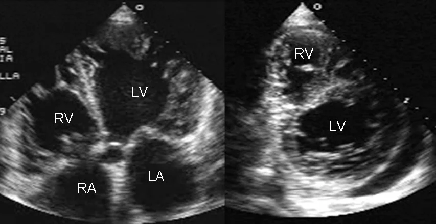
Long description
A transthoracic two-dimensional echocardiogram in apical four-chamber and parasternal short-axis views shows dilatation of both ventricles with multiple trabeculae and intertrabecular recesses in a case of non-compacted cardiomyopathy.

Transthoracic echocardiography is the first-line test. It measures chamber dimensions, wall thickness, global and regional systolic function and diastolic function; identifies valvular disease, intracavitary thrombus and pericardial effusion; and excludes loading and coronary explanations (regional wall-motion abnormalities from ischaemia). A global, dilated, hypokinetic ventricle is the hallmark.
Cardiac magnetic resonance is recommended at baseline and during follow-up when it will change management. It is the best test for tissue characterisation: late gadolinium enhancement marks fibrosis, "ring-like" scar suggests DSP or FLNC disease, native T1 and extracellular volume quantify diffuse disease, and the pattern helps distinguish ischaemic from non-ischaemic scarring and phenocopies such as amyloidosis or sarcoidosis.
Coronary CT angiography or invasive angiography is used when CAD must be excluded. Endomyocardial biopsy is reserved for suspected myocardial inflammation, infiltration or storage that other tests have not explained, and where it might change treatment (for example suspected giant-cell myocarditis or sarcoidosis, or discussion before immunosuppression).[4]
Genetic testing is now standard in DCM. All societies agree on careful genetic counselling before and after testing; a three- to four-generation family history and pedigree is essential. Testing the proband is recommended when it aids diagnosis, prognosis, treatment, reproductive planning or cascade testing, with the specific disease-causing genes tested first.[4]
Useful specificity from comparative data: LMNA variants are sought especially when conduction disease is present, and TTN truncating variants when the phenotype is otherwise typical.[4] Non-carrier relatives can be discharged from periodic cardiac screening once a confident variant is identified in the family; carrier relatives need multiparametric evaluation (ECG, imaging) and long-term follow-up.[4][5] Where no variant is found, first-degree relatives still need an initial and periodic evaluation, because gene-elusive disease is common.[4]
Diagnostic approach to suspected dilated cardiomyopathy
- Heart failure symptoms or abnormal ECGHistory and examination; exclude acute coronary syndrome and other immediate causes
- Transthoracic echocardiographyLV size and systolic function; valves; RV; thrombus
- Question: Dilated LV with reduced LVEF, unexplained by load or CAD?
No
- Look for ischaemic, hypertensive, valvular or secondary cardiomyopathyTreat the specific cause
Yes
- Baseline bundle: ECG, natriuretic peptide, troponin, renal, liver, thyroid, iron, HIV where relevant, chest X-ray
- Cardiac MRIScar, inflammation, infiltration, phenocopy
- Decide on endomyocardial biopsyIf myocarditis, infiltration or storage is suspected and may change treatment
- Genetic testing with counsellingFamily history; disease-gene panel; cascade testing
- Aetiology now clearerChoose cause-specific and HFrEF therapy; assess device need
There is no single numerical cut-off that defines DCM on its own. The diagnosis rests on dilated ventricle plus reduced function, by more than one modality where possible, in the absence of an explanatory cause. Guidelines differ on how LV dilatation is quantified, which is why the European guideline dimensional cut-offs (see Definition) are best read as reference values.
Severity is tracked with standard heart-failure tools: NYHA class, natriuretic peptides, LVEF, RV function and the presence of arrhythmia or scar. For sudden-death risk, the genotype matters as much as the LVEF, and several genotype-specific risk estimators are in use.
Decompensated patients are managed as acute heart failure: oxygenation, diuretics for congestion, vasodilators if blood pressure allows, and inotropes or temporary mechanical support only when needed. Precipitants (arrhythmia, ischaemia, infection, alcohol binge) should be sought and treated.
Advise against heavy alcohol, avoid cardiotoxic exposures where possible, optimise blood pressure, lipids, weight and sleep apnoea, vaccinate (influenza and pneumococcal), and individualise exercise. Supervised, moderate-intensity exercise totalling about 150 minutes per week is encouraged; high-intensity exercise is avoided in symptomatic disease, marked systolic dysfunction, exercise-induced arrhythmia or high-risk genotypes such as LMNA and TMEM43.[1]
Drug therapy is that of heart failure with reduced ejection fraction. The 2023 ESC cardiomyopathy guideline states plainly that pharmacological treatment of DCM does not differ from that of chronic heart failure.[1] That means the usual HFrEF classes—renin-angiotensin inhibition (ACE inhibitor, ARB or ARNI), a beta-blocker, a mineralocorticoid receptor antagonist, an SGLT2 inhibitor and diuretics for congestion—titrated to evidence-based or maximally tolerated doses with monitoring of renal function, potassium and blood pressure. An important first-time caveat: ivabradine is considered when the patient is in sinus rhythm with an inadequate heart rate on maximally tolerated beta-blocker therapy. Drug doses and titration schemes are covered in the heart-failure topic and are not repeated here.
Secondary prevention. An ICD is recommended after cardiac arrest from ventricular fibrillation or ventricular tachycardia, or with sustained VT causing syncope or haemodynamic compromise, once reversible causes are excluded.[3][4]
Primary prevention ICD. In DCM, an ICD should be considered in patients with symptomatic heart failure and LVEF ≤35% despite more than 3 months of optimal medical therapy, provided the patient is expected to survive substantially longer than 1 year with good functional status; the 2023 ESC DCM guideline review rates this indication IIa-A.[3][4] Note that the DANISH trial did not show a mortality benefit from prophylactic ICDs in non-ischaemic HFrEF overall, though sudden death was reduced, so the decision should be shared.[3] A reduced LVEF is not the whole story: with a genotype associated with high sudden-death risk (e.g. LMNA, FLNC-truncating, TMEM43, PLN, DSP, RBM20), an ICD can be considered even when the LVEF is above 35% when additional risk factors are present (IIa-C in the DCM guideline review) and may still be considered when they are absent (IIb-C); the 2023 ACC summary states more broadly that an ICD should be considered for certain genetic forms even with an LVEF above 35%.[1][4]
CRT. Cardiac resynchronisation is recommended for symptomatic patients in sinus rhythm with LVEF ≤35% and LBBB with a QRS duration of at least 150 ms despite optimal therapy; it is considered for LBBB QRS of 130–149 ms, for non-LBBB QRS ≥150 ms, and for patients with LVEF ≤35% in NYHA III–IV atrial fibrillation with intrinsic QRS ≥130 ms provided biventricular capture can be ensured (with AV-node ablation if capture is incomplete).[2] Upgrading a pacemaker or ICD patient who develops worsening heart failure with a high proportion of right-ventricular pacing is also a CRT indication.[4]
Anticoagulate for a reason, not because the ventricle is dilated. OAC is indicated in DCM with atrial fibrillation or flutter according to stroke-risk score: with the CHA₂DS₂-VASc score, ≥2 in men or ≥3 in women is a strong recommendation and 1 in men or 2 in women a consideration; the 2024 ESC atrial-fibrillation guideline (summarised in its key-point document and in Sorella's footnote) replaces this with the sex-free CHA₂DS₂-VA score, recommending anticoagulation at a score of ≥2 and considering it at 1. Anticoagulation is also always indicated in the presence of LV thrombus.[4] Direct oral anticoagulants are generally preferred to vitamin K antagonists in AF unless there is moderate-to-severe mitral stenosis or a mechanical valve; exact choice follows the atrial-fibrillation topic and local practice.
During bromocriptine treatment, at least prophylactic anticoagulation is recommended because of the thrombotic risk.
Alcohol. Abstinence is the single most important intervention in alcohol-induced cardiomyopathy; guideline-concordant HFrEF therapy is added. Some patients recover ventricular function even with controlled low intake (<80 g/day), but heavy intake must stop. Arrhythmia and sudden-death management follows the DCM rules, with additional caution when high-risk gene variants coexist.[7]
Peripartum cardiomyopathy. Treat as HFrEF with pregnancy-specific restrictions (ACE inhibitors, ARBs and MRAs are avoided during pregnancy; after delivery standard therapy is introduced). Prognosis is better than in many other DCMs: about half of women recover fully (LVEF >55% and NYHA I) and a further 35–40% partially recover, usually within 3–6 months.[6] Bromocriptine, a prolactin-release inhibitor used with standard therapy and at least prophylactic anticoagulation, is backed by a multicentre randomised protocol (2.5 mg daily for 7 days versus 2.5 mg twice daily for 14 days then once daily for 42 days in women with LVEF <35%) that showed LVEF rises of roughly 21–24 points over 6 months and full recovery in 52% and 68% of the two arms, with no deaths or transplants and no serious adverse events.[6] Recovery is better when the baseline LVEF is at least 30% and the LV is not too dilated. Subsequent pregnancy is high-risk, particularly if LV function has not normalised.
Cancer-therapy cardiotoxicity. Cardiology and oncology should decide together. Anthracycline-related LV dysfunction is often detected in the first 12 months after treatment; starting ACE inhibitors and beta-blockers promptly (e.g. when LVEF falls or troponin rises) improves recovery.[9] Treatment must balance cancer control with cardiac monitoring (serial LVEF, biomarkers), and neurohormonal therapy and diuretics are used as for HFrEF. Dexrazoxane and liposomal/epirubicin formulations reduce cardiotoxicity in selected protocols.
For refractory, end-stage disease or intractable ventricular arrhythmia, cardiac transplantation is a Class I indication in ESC terms for NYHA III–IV patients without absolute contraindications; LV assist devices are used as bridge to transplant or destination therapy in selected patients with advanced HFrEF.[4] Refer early, before irreversible organ dysfunction.
Stable patients need a multiparametric review with ECG and imaging every 1–2 years, and sooner if symptoms change.[4]
- Progressive heart failure, cardiogenic shock, arrhythmic death.
- Thromboembolism (LV or atrial thrombus, stroke, systemic emboli).
- Arrhythmias: AF, ventricular tachycardia, sudden cardiac death.
- Reverse remodelling failure or progression despite therapy.
- Transplant- or device-related complications in advanced disease.
- Psychosocial burden and impact on family.
Prognosis in DCM varies greatly by cause. General HFrEF prognosis applies, with the modifiers above: LMNA and other high-risk genotypes, persistent heavy alcohol use, advanced NYHA class and severe RV dysfunction all worsen outcome. Many patients improve substantially on modern therapy and recover LV function. Specific observations supported by the literature:
- About half of women with peripartum cardiomyopathy recover fully and 35–40% partially, mostly within 3–6 months.[6]
- Alcohol-induced cardiomyopathy carries a worse prognosis if drinking continues, but LV function recovers in abstainers; in modern series about 42% of patients improved LVEF, with no recovery among those persisting with heavy intake.[7]
- Anthracycline cardiotoxicity occurs in about 9% of treated adults, most within the first 12 months; early treatment with ACE inhibitors and beta-blockers produces full or partial LVEF recovery in most patients.[9]
- Tachycardia-induced cardiomyopathy often recovers within 2–3 months, and usually not beyond 6 months, once the arrhythmia is controlled, although LV volumes may remain increased.[8]
- About 30% of chronic T. cruzi infections progress to Chagas cardiomyopathy over 10–30 years, a complication with poor prognosis.[10]
- Gene-carriers of TTN recover LVEF more often than carriers of LMNA.[5]
Primary prevention means treating the causes: avoiding heavy alcohol, managing cancer therapy with cardioprotection, controlling thyroid disease, treating HIV and iron overload, and not letting tachyarrhythmia persist. Secondary and cascade prevention means family screening: a multiparametric evaluation (ECG and cardiac imaging, plus genetics when a variant is known) of first-degree relatives, then discharge for non-carriers and long-term follow-up for carriers.[4][5]
Pregnancy. Pregnant women with known DCM need a multidisciplinary assessment; ACE inhibitors, ARBs and MRAs are contraindicated in pregnancy, but beta-blockers (with foetal monitoring), hydralazine, diuretics and careful delivery planning are used. Evidence for device decisions in pregnancy is limited; specialist teams decide.
Children. Childhood DCM is often familial and may be more severe in the first year of life. Paediatric rhabdomyoma, fibroma or other infiltrative causes should be considered. Genetic referral is important.
Older adults and multimorbidity. Coronary disease, hypertension and renal impairment are common comorbidities and confound both diagnosis and therapy; polypharmacy and frailty affect drug tolerance.
A transthoracic echocardiogram is the one test that makes the diagnosis in almost any setting, and natriuretic peptides help when available. Where echo is scarce, a patient with unexplained heart failure and a globally dilated, hypokinetic ventricle plus bundle-branch block or apical aneurysm in someone from Latin America should raise Chagas disease. About one-third of chronic infections develop cardiac disease over decades, so screening blood donors and treating acute or indeterminate infection early matters.[10] Antitrypanosomal therapy does not reverse established cardiomyopathy, so prevention and early diagnosis dominate.[10]
In low-resource practice, focus on what changes outcomes: treat heart failure with whichever evidence-based drugs are available and affordable; insist on alcohol abstinence; control rate and rhythm with the tools at hand; anticoagulate for atrial fibrillation when safe; and refer for devices or transplantation where services exist. WCDs, SCD risk tools and advanced imaging may be unavailable; a careful clinical history and an ECG plus echo still go a long way.
East Africa
In East Africa, rheumatic heart disease, HIV, tuberculosis, severe malaria and anaemia are frequent alternative explanations for dilated, hypokinetic hearts; excluded them before diagnosing idiopathic DCM. Many drug and device services are concentrated in referral centres, so early referral for progressive disease is important.
- DCM is LV dilatation with systolic dysfunction not explained by loading or coronary disease; it is the commonest non-ischaemic cause of heart failure.
- Causes span genetic (TTN commonest), infection (myocarditis, Chagas, HIV), alcohol, peripartum, cancer therapy, tachycardia, endocrine, iron and cocaine; more than one often acts together.
- Presentation is heart failure; ECG and chest X-ray are non-specific; echo is first-line; MRI guides tissue diagnosis and risk.
- Genetic testing and family screening are standard; about 30–50% of cases are familial and 30–40% of those have an identifiable variant.
- Management follows HFrEF drug therapy; devices (ICD/CRT) have specific LVEF and QRS thresholds; anticoagulate only for AF or thrombus.
- Alcohol-induced disease needs abstinence; peripartum cardiomyopathy has a better recovery rate and may use bromocriptine with prophylactic anticoagulation; cancer-therapy disease needs early cardiology-oncology co-management.
- Arrhythmia risk is as important as LVEF in LMNA and similar genotypes, which may warrant device consideration above 35% LVEF.
- Advanced disease: consider transplant or mechanical support early.
Answer each one in your head first, then open the answer.
What two findings define dilated cardiomyopathy?
Show answerHide answer
Left ventricular dilatation plus global or regional systolic dysfunction, once abnormal loading conditions and coronary artery disease are excluded.
A 45-year-old woman develops dyspnoea two weeks after delivery. Echocardiography shows an LVEF of 35% and LV dilatation; coronary disease is excluded. What is the most likely diagnosis and what is her outlook?
Show answerHide answer
Peripartum cardiomyopathy. About half of women recover fully (LVEF >55% and NYHA I) and 35–40% partially, most within 3–6 months; bromocriptine added to standard therapy with prophylactic anticoagulation may improve recovery.
Which gene variants carry a particularly high arrhythmic risk even when the LVEF is above 35%?
Show answerHide answer
LMNA, DSP, FLNC, RBM20 and PLN variants, among others. These patients may warrant ICD consideration despite a higher LVEF.
Why is anticoagulation not given routinely to every patient with DCM?
Show answerHide answer
In chronic HFrEF without atrial fibrillation or LV thrombus there is no evidence of net benefit; anticoagulation is reserved for AF/flutter by stroke-risk criteria or documented thrombus.
What heart-rate findings should make you suspect tachycardia-induced cardiomyopathy, and how does its course differ from other DCM?
Show answerHide answer
A mean heart rate >100 beats/min (often 110–120) or a high extrasystole burden, with recovery of LV function within 2–3 months and usually not beyond 6 months after rhythm or rate control. Confirmation is made a posteriori when function normalises.
A patient resumes heavy drinking after a diagnosis of alcohol-induced cardiomyopathy. What are you most worried about?
Show answerHide answer
Worsening prognosis and lack of LVEF recovery: modern series report no recovery among those persisting with heavy intake, so complete abstinence is essential alongside standard HFrEF therapy.
- Heart failure
- ECG interpretation
- Cardiac physical examination
- Atrial fibrillation and flutter
- Ventricular arrhythmias
- Bradyarrhythmias and AV block
- Supraventricular tachycardias
- Approach to tachyarrhythmias
Hypertrophic cardiomyopathy and restrictive cardiomyopathy also live under the cardiomyopathy family and can be confused with DCM; myocarditis and cardiac sarcoidosis are important differential and treatment considerations.
- Arbelo E, Protonotarios A, Gimeno JR, et al. 2023 ESC Guidelines for the management of cardiomyopathies: key points. Eur Heart J 2023;44:3503–3626 (summarised on ACC.org, 30 Aug 2023). Accessed .
- Glikson M, Nielsen JC, Kronborg MB, et al. 2021 ESC Guidelines on cardiac pacing and cardiac resynchronisation therapy: key points. Eur Heart J 2021 (summarised on ACC.org, 1 Sep 2021). Accessed .
- Liang B, Zhao Y-X, Zhang X-X, et al. Reappraisal on pharmacological and mechanical treatments of heart failure. Cardiovasc Diabetol 2020;19:55. Accessed .
- Sorella A, Galanti K, Iezzi L, et al. Diagnosis and management of dilated cardiomyopathy: a systematic review of clinical practice guidelines and recommendations. Eur Heart J Qual Care Clin Outcomes 2025;11(2):206–222. Accessed .
- Gigli M, Stolfo D, Merlo M, et al. Pathophysiology of dilated cardiomyopathy: from mechanisms to precision medicine. Nat Rev Cardiol 2025;22(3):183–198. Accessed .
- Koenig T, Hilfiker-Kleiner D, Bauersachs J. Peripartum cardiomyopathy. Herz 2018;43(5):431–437. Accessed .
- Dominguez F, Adler E, Garcia-Pavia P. Alcoholic cardiomyopathy: an update. Eur Heart J 2024;45(26):2294–2305. Accessed .
- Kim D-Y, Kim S-H, Ryu K-H. Tachycardia induced cardiomyopathy. Korean Circ J 2019;49(9):808–817. Accessed .
- McGowan JV, Chung R, Maulik A, et al. Anthracycline chemotherapy and cardiotoxicity. Cardiovasc Drugs Ther 2017;31(1):63–75. Accessed .
- Cutshaw MK, Sciaudone M, Bowman NM. Risk factors for progression to chronic Chagas cardiomyopathy: a systematic review and meta-analysis. Am J Trop Med Hyg 2023;108(4):791–800. Accessed .
- Arbelo E, Protonotarios A, Gimeno JR, et al. 2023 ESC Guidelines for the management of cardiomyopathies (definitions of DCM, HCM and RCM, Section 3.2). Eur Heart J 2023;44(37):3503–3626. Accessed .
Figure credits
Educational content for medical students and doctors. It is not a substitute for clinical judgement or local guidelines.