Hypertrophic cardiomyopathy (HCM)
A thick, stiff heart muscle that can obstruct outflow, cause breathlessness and fainting, and cause sudden death — and how treatment changes the outlook.
- Hypertrophic cardiomyopathy (HCM) is an inherited disease of the heart muscle in which the left ventricle, usually the septum, becomes thick without a cause such as high blood pressure or a diseased valve.
- It affects roughly 1 in 500 people and often runs in families. Many carriers never develop symptoms.
- The thick septum can narrow the outlet of the left ventricle. The mitral valve leaflet is then pulled into the stream (systolic anterior motion, SAM), which both obstructs flow and leaks.
- The classic murmur becomes louder when the ventricle is emptied (standing, Valsalva) and softer when it is filled or faced with more resistance (squatting, handgrip).
- Most people have a near-normal lifespan. A minority have breathlessness, chest pain, fainting, atrial fibrillation or sudden cardiac death. Risk scores, an implantable defibrillator and drugs that relax the sarcomere have changed what can be offered.
By the end of this page you should be able to:
- Define HCM, distinguish obstructive from non-obstructive disease, and explain why "HOCM" is only one part of the spectrum.
- Trace the path from a sarcomere variant to hypertrophy, diastolic dysfunction, ischaemia, obstruction and arrhythmia.
- Recognise the history and bedside signs, and explain every change in the murmur produced by Valsalva, standing, squatting and handgrip.
- Choose and interpret the core investigations: ECG, echocardiography, cardiac MRI and genetic testing.
- Separate HCM from athlete's heart, hypertensive heart disease, aortic stenosis, amyloidosis, Fabry disease, Friedreich ataxia and Noonan syndrome.
- Estimate sudden cardiac death (SCD) risk with the ESC model and the AHA/ACC risk markers, and state when an ICD is indicated.
- Treat symptoms with the right drug in the right patient, avoid drugs that worsen obstruction, and know when to refer for septal reduction.
- Manage atrial fibrillation, pregnancy and family screening, and adapt care when resources are limited.
HCM is a disease of the sarcomere, the molecular motor that makes heart muscle contract. A variant in one of the genes that encodes its proteins makes the muscle work inefficiently. The heart responds by thickening, but the thickening is maladaptive: the ventricle becomes stiff, the septum bulges into the outflow tract, and the muscle's architecture breaks down into disarray and scar.
That single process explains almost everything a patient experiences. A stiff, small ventricle raises filling pressures, so the patient is breathless. A narrow outflow tract produces a murmur and, during exertion, a fall in output that causes fainting. Damaged muscle and scar create the substrate for ventricular arrhythmia, which is why HCM can kill a young person who seemed perfectly well. The clinical skill is to recognise the pattern, measure the obstruction and the risk, and then match treatment to the individual.
DefinitionHypertrophic cardiomyopathy
HCM is a primary disease of the heart muscle defined by left ventricular hypertrophy that cannot be explained by abnormal loading conditions (such as hypertension or aortic stenosis) or by an infiltrative or storage disease. In an adult, the usual diagnostic threshold is a maximal end-diastolic wall thickness of at least 15 mm in any left ventricular segment. A thickness of 13 to 14 mm can be diagnostic when there is a family history of HCM or a pathogenic sarcomere variant.
The 2023 European Society of Cardiology (ESC) guideline defines HCM broadly, as unexplained left ventricular hypertrophy with or without right ventricular involvement. The 2024 American Heart Association/American College of Cardiology (AHA/ACC) guideline uses a narrower, genetic definition: a primary myocardial disease caused by sarcomere variants, with the same 15 mm threshold.[6][1] In everyday practice the two definitions meet at the bedside: a thick ventricle with no loading cause, often with a family history, and sometimes with a pathogenic variant.
Several classifications are useful.
- By distribution. Asymmetric septal hypertrophy is a common pattern. Apical, concentric, mid-ventricular, lateral and right ventricular forms occur.[4] The pattern matters because apical disease can hide from a standard view and mid-ventricular disease can cause an apical aneurysm.
- By obstruction. Obstruction is dynamic. A resting peak gradient of at least 30 mmHg defines resting obstruction; a gradient below 30 mmHg at rest that rises to at least 30 mmHg with provocation is "provocable" or latent obstruction.[3] A gradient of at least 50 mmHg is the level at which septal reduction is considered.[12]
- By stage. Most patients are stable. A minority progress to a dilated, "end-stage" phenotype with systolic dysfunction, usually defined as an ejection fraction below 50%.[1]
The older term "hypertrophic obstructive cardiomyopathy" (HOCM) is still heard. It describes only the obstructive subset, and it can mislead by implying that obstruction is the whole disease. About one third of patients referred to specialist centres have resting obstruction and another third have provocable obstruction; the remainder have no obstruction at rest or on provocation.[12]
HCM is an important inherited heart muscle disease. Population studies put the prevalence of unexplained left ventricular thickening at 0.16% to 0.23% of adults,[12] and the familiar figure of 1 in 500 is a reasonable approximation.[4] Because genotype-positive, phenotype-negative carriers also exist, some estimates reach 0.6%.[12]
The absolute numbers are large. About 20 million people worldwide are thought to be affected,[4] and in the United States more than 750,000 people are estimated to have HCM.[5] Most are never diagnosed: estimates range from about 10% of cases recognised clinically worldwide to roughly 100,000 of an estimated 750,000 cases in the United States, and only about 6% have symptoms.[4][3] The disease occurs in every ancestry and does not appear to differ in frequency between them, although access to diagnosis does.[5]
HCM can present at any age, from infancy to old age. Many people are found through family screening or because a murmur is heard during a routine examination. Others present in the third to fifth decade with exertional symptoms.
Roughly 40% to 60% of patients with HCM carry a pathogenic or likely pathogenic variant in a gene encoding a sarcomere protein, and the proportion depends on how strictly the phenotype is defined and on the population studied.[5][6] The inheritance is autosomal dominant: one altered copy is enough to cause disease, and each child of an affected parent may inherit it.
Two genes account for most identified variants. About 70% of patients with a sarcomere variant have it in MYBPC3 (myosin-binding protein C3) or MYH7 (beta-myosin heavy chain).[5] The rest cluster in genes such as TNNT2, TNNI3, TPM1, MYL2, MYL3 and ACTC1.[5] MYBPC3 variants tend to appear later and with lower penetrance.[2]
Carrying a variant is not the same as having the disease. Penetrance is incomplete and age-dependent: some carriers never develop hypertrophy, while relatives with the same variant can differ widely in wall thickness, obstruction and outcome.[5] This is why a negative echocardiogram in a young carrier does not end surveillance, and why a negative genetic test in a patient with a typical phenotype does not exclude HCM.
The explanation is not fully known. Modifier genes, epigenetic effects, sex, body mass and environment all probably contribute. Obesity, for example, is associated with higher outflow gradients and worse outcomes, although whether it is causal is unclear.[5]
Not every thick heart is sarcomeric HCM. Important phenocopies include:
- Fabry disease, an X-linked lysosomal storage disorder caused by alpha-galactosidase A deficiency.
- Cardiac amyloidosis, usually transthyretin (ATTR) or light-chain (AL).
- PRKAG2 and LAMP2 glycogen storage disease, which can produce hypertrophy with pre-excitation.
- Noonan syndrome and other RASopathies, often with pulmonary stenosis and dysmorphic features.
- Friedreich ataxia, a mitochondrial disorder with neurological signs and concentric hypertrophy.
- Danon disease and Pompe disease.
These matter because some have specific treatments (enzyme replacement in Fabry, tafamidis in ATTR amyloidosis) and because they change family counselling.
Genetic testing is recommended for patients who meet diagnostic criteria, because a result can confirm the diagnosis, inform prognosis, guide reproductive choices and allow cascade testing of relatives.[1] Testing is most useful when the phenotype is typical and there is a family history; it is less useful in isolated, atypical hypertrophy, where a variant of uncertain significance can create more anxiety than clarity.
The sequence is straightforward.
- Test the proband (the first affected person in the family) with a sarcomere gene panel.
- If a pathogenic or likely pathogenic variant is found, offer predictive cascade testing to adult first-degree relatives, with pre- and post-test counselling. Testing then cascades outwards to cousins and beyond.[1]
- Relatives who carry the variant need cardiac surveillance even if their echocardiogram is normal, because penetrance is age-dependent.
- Relatives who do not carry the variant can usually be discharged from cardiac screening.
- If no variant is found in the proband, first-degree relatives still need clinical screening with echocardiography and ECG, because the family may have a variant that current testing cannot detect.
Clinical pearlThe family is the patient
A diagnosis of HCM is also a diagnosis about a family. Ask about sudden death, heart failure, pacemakers and defibrillators in relatives, and draw a full family pedigree. A relative who died suddenly in their twenties may be the reason a 19-year-old cousin needs an echocardiogram.
From sarcomere variant to the clinical syndrome
- Sarcomere variantAltered myosin, myosin-binding protein C or other motor protein; inefficient force generation
- Compensatory hypertrophyThickened myocytes, asymmetric septal growth, small ventricular cavity
- Question: Where does the hypertrophy sit?
Septum bulges into the outflow tract
- LVOT narrowing and SAMMitral leaflet is drawn into the septum; obstruction plus mitral regurgitation
- Exertional dyspnoea, angina, syncopeHigh intracavity pressure, ischaemia, falling cardiac output
Diffuse or apical hypertrophy
- Stiff, small ventricleRaised filling pressures, diastolic dysfunction, atrial enlargement
- Dyspnoea and atrial fibrillationPulmonary congestion; atrial stretch and fibrosis
- Disarray, fibrosis and ischaemiaThe arrhythmia substrate; re-entry around scar
- Ventricular arrhythmia or progressive failureSyncope, cardiac arrest, or end-stage systolic dysfunction
The earliest structural change is myocyte hypertrophy, driven by the mutant sarcomere. The septum is usually affected more than the free wall, producing asymmetric septal hypertrophy. The ratio of septal to posterior wall thickness is often abnormal, although the absolute thickness matters more for diagnosis. The hypertrophied septum bulges into the left ventricular outflow tract (LVOT), the channel between the ventricle and the aorta.
Obstruction in HCM is dynamic, not fixed. As blood accelerates through the narrowed outflow tract, the anterior mitral leaflet is drawn towards the septum by a combination of suction (the Venturi effect) and drag from the flow stream. This is systolic anterior motion (SAM).[4] Once the leaflet touches the septum, two things happen at once: the outflow tract narrows further, and the mitral valve fails to close properly, producing a posteriorly directed mitral regurgitation jet.
The result is a self-reinforcing loop. Faster flow pulls the leaflet further in, which narrows the tract more, which speeds the flow again. Anything that reduces ventricular volume or increases contractility worsens the loop; anything that fills the ventricle or reduces contractility softens it. That is the physiological basis of the bedside manoeuvres and of drug treatment.
The thickened, fibrotic ventricle is stiff. It relaxes slowly and cannot fill at low pressure, so left atrial pressure rises and the patient becomes breathless, especially on exertion.[4] A small cavity makes this worse: even a modest increase in volume produces a large rise in pressure. Over time the left atrium dilates, which is the substrate for atrial fibrillation.
The hypertrophied muscle has a higher oxygen demand and a compromised supply. Microvascular dysfunction, reduced capillary density, compression of small vessels during systole and a mismatch between wall thickness and blood supply all contribute. The result is exertional angina even with normal coronary arteries, and patchy subendocardial injury that feeds fibrosis.[4] This is why chest pain in HCM is not automatically angina from atheroma, and why coronary angiography may be normal.
The arrhythmia substrate is built from three elements: myocyte disarray, replacement fibrosis and ischaemia. Disarray disrupts normal conduction, fibrosis creates slow zones and barriers, and ischaemia makes membranes unstable. Ventricular premature beats and non-sustained ventricular tachycardia (NSVT) are common, and sustained ventricular tachycardia or fibrillation is the mechanism of sudden death. In the atria, stretch and fibrosis produce atrial fibrillation, which is both a symptom and a risk factor for stroke.
A small proportion of patients lose systolic function and develop a dilated ventricle, usually after years of disease. The ejection fraction falls below 50%, and the physiology shifts from obstruction and diastolic dysfunction towards heart failure with reduced ejection fraction. This is often called the dilated or end-stage phase and carries a worse prognosis.[1]
Gross. The heart is heavy, with a thickened septum and a small left ventricular cavity. The hypertrophy may be focal, asymmetric or concentric, and the outflow tract can be narrowed by a bulging septal bulge. In apical disease the apex is thickened and the cavity is shaped like a spade.
Microscopic. The hallmark is myocyte disarray: bundles of muscle fibres run in different directions instead of in parallel, so that the tissue looks disorganised even at low power. There is also myocyte hypertrophy, nuclear enlargement, interstitial and replacement fibrosis, and abnormal intramural coronary arteries with thickened walls. These changes are not specific to HCM, but their combination in a young patient with unexplained hypertrophy is characteristic.[4]
Fibrosis can be quantified on cardiac MRI as late gadolinium enhancement (LGE). It is present in up to 65% of patients and is often patchy and mid-wall, favouring the insertion points of the right ventricle and the hypertrophied segments.[4] Extensive fibrosis is associated with a higher risk of ventricular arrhythmia and adverse outcome.
Many people with HCM have no symptoms. When symptoms occur, they usually reflect obstruction, diastolic dysfunction or arrhythmia.[3]
- Exertional breathlessness is a common and often dominant symptom. It comes from high left atrial pressure and, when obstruction is present, from a failure of cardiac output to rise with exercise.
- Chest pain is often exertional and can mimic angina. It may be caused by ischaemia, obstruction or diastolic dysfunction.
- Syncope or presyncope, especially during or immediately after exertion, is worrying. It may reflect a fall in output from obstruction, an arrhythmia or abnormal vascular responses.
- Palpitations suggest atrial fibrillation, ventricular ectopy or sustained arrhythmia.
- Fatigue and exercise intolerance are easily dismissed but often the most limiting symptom.
Symptoms can vary from day to day. Breathlessness often worsens after a large meal, alcohol, dehydration or heat, because these change preload and afterload.[3][12]
Red flagExertional syncope
Syncope during exertion, or a cardiac arrest in a young person, is never "just a faint" until HCM and other structural heart diseases have been excluded. A normal resting ECG and a normal physical examination do not exclude HCM.
HCM can cause sudden cardiac death in young people, including competitive athletes.[12] The typical victim may have had no symptoms, or only vague warning signs such as exertional dizziness. This is why pre-participation screening, a careful family history and a low threshold for echocardiography matter. It is also why an athlete with a thick ventricle needs a structured assessment rather than an automatic diagnosis of "athlete's heart".
The examination is normal in many patients, especially those without obstruction. When signs are present, they are best elicited in a quiet room with the patient supine, then standing.
Pulse and impulse. The carotid pulse may be brisk and then fall in mid-systole before rising again, giving a jerky or bisferiens quality.[4] The apical impulse is sustained and displaced outwards. A palpable fourth heart sound (S4) can add a second component, producing a double apical impulse. An S4 is common and reflects a vigorous atrial contraction against a stiff ventricle.[4]
Murmur. The classic murmur is a harsh, mid-to-late systolic crescendo–decrescendo murmur, loudest at the left lower sternal border or apex, and it does not radiate to the carotids.[12] When SAM causes mitral regurgitation, a separate holosystolic murmur may be heard at the apex and radiate to the axilla.[3] The murmur of HCM is dynamic: its intensity changes with manoeuvres that alter ventricular volume, afterload or contractility.
| Manoeuvre | What it does to the gradient and murmur | Why |
|---|---|---|
| Valsalva (strain phase) | Gradient rises; murmur becomes louder | Increased intrathoracic pressure reduces venous return, so the ventricle is smaller; a smaller cavity worsens SAM and obstruction.[4][12] |
| Standing | Gradient rises; murmur becomes louder | Blood pools in the legs, preload falls, and the ventricle empties; obstruction increases.[4] |
| Squatting | Gradient falls; murmur becomes softer | Squatting increases venous return and afterload, so the ventricle is fuller and the obstruction is reduced.[4] |
| Handgrip | Gradient falls; murmur becomes softer | Isometric contraction raises afterload, which opposes the ejection gradient and reduces the outflow velocity.[4] |
| Post-premature beat | Gradient rises; murmur becomes louder | The pause after an ectopic beat increases filling and contractility, so the next beat is more forceful and more obstructive.[4] |
Exam trapWhich way does the murmur move?
The HCM murmur gets louder when the ventricle is under-filled (standing, Valsalva) and softer when it is full or facing high resistance (squatting, handgrip). Mitral regurgitation behaves in the opposite direction with handgrip: it gets louder. Aortic stenosis radiates to the carotids, HCM usually does not.
A thick left ventricle has many causes. The history, the ECG, the pattern of hypertrophy and the presence of extracardiac features usually separate them, but occasionally a cardiac MRI, genetic test or biopsy is needed.[12]
| Condition | Clues that point away from HCM | Confirmatory clue |
|---|---|---|
| Athlete's heart | Intense training; balanced four-chamber dilatation; normal diastolic function and strain; no LGE; hypertrophy regresses with deconditioning.[12] | Normal ECG or isolated voltage; echo after detraining |
| Hypertensive heart disease | Long-standing or resistant hypertension; concentric hypertrophy; no SAM; regression with blood pressure control.[12] | Blood pressure history; serial imaging |
| Aortic stenosis | Calcified, restricted aortic valve; fixed ejection murmur radiating to the carotids; reduced carotid upstroke; no SAM. | Echocardiography with valve gradient and area |
| Cardiac amyloidosis | Low QRS voltage relative to wall thickness; apical sparing of longitudinal strain; granular sparkling; subendocardial or transmural LGE; carpal tunnel syndrome, macroglossia, neuropathy.[12] | Bone scintigraphy, serum free light chains, biopsy |
| Fabry disease | Neuropathic pain, angiokeratomas, albuminuria, hypohidrosis, cornea verticillata; short PR interval or pre-excitation; basal-inferolateral LGE.[12] | Alpha-galactosidase A activity and genetic testing |
| Friedreich ataxia | Progressive ataxia, scoliosis, diabetes, absent reflexes; concentric hypertrophy. | Neurological examination and FXN genetic testing |
| Noonan syndrome | Short stature, webbed neck, dysmorphic facies, pulmonary valve stenosis; hypertrophy often concentric. | Clinical genetics; PTPN11, RAF1 and related genes |
Two practical points follow. First, hypertension and HCM commonly coexist, and hypertension alone does not exclude HCM; the pattern of hypertrophy and the presence of SAM or a sarcomere variant decide.[12] Second, a patient with "hypertrophy and heart failure" who has low voltage on the ECG should be investigated for amyloidosis before being labelled as HCM, because the treatment is entirely different.
The ECG is abnormal in up to 95% of patients, but there is no single diagnostic pattern.[4] Common findings are:
- left ventricular hypertrophy with strain;
- deep, narrow Q waves in the inferior or lateral leads, reflecting septal hypertrophy;[4]
- T-wave inversion, which may be giant and symmetrical in apical HCM;
- left atrial enlargement;
- pre-excitation, which suggests a glycogen storage disease such as PRKAG2 or Fabry disease.[4]
A normal ECG is reassuring but does not exclude the diagnosis, especially in young carriers and in apical disease.
Figure 1Echocardiography in hypertrophic cardiomyopathy
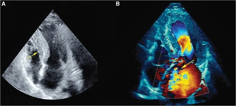
Long description
Two transthoracic echocardiogram panels from a patient with hypertrophic cardiomyopathy. Panel A is a two-dimensional apical four-chamber view in which the septum is clearly thicker than the lateral wall and narrows the left ventricular cavity, marked by a yellow arrow. Panel B is the same view with colour Doppler, showing an anteriorly directed mitral regurgitation jet marked by a yellow arrow.
Transthoracic echocardiography (TTE) is the main imaging test and the backbone of diagnosis and follow-up.[1] It answers five questions.
- Is the ventricle thick, and where? Measure the maximum end-diastolic wall thickness in all segments. A thickness of at least 15 mm, or at least 13 mm in a relative of an affected patient, supports the diagnosis.[1]
- Is there obstruction? Measure the peak LVOT gradient at rest and with provocation (Valsalva, standing, exercise). A resting gradient of at least 30 mmHg defines obstruction; a gradient of at least 50 mmHg is significant enough to consider septal reduction.[12]
- Is there SAM and mitral regurgitation? SAM is visible as the anterior leaflet moving towards the septum in systole.[4]
- Is the ventricle stiff? Assess diastolic function, left atrial size and filling pressures.
- Is there an apical aneurysm? Look especially in mid-ventricular and apical phenotypes; an aneurysm with scar is a risk marker for sudden cardiac death.[1]
Provocation is essential. A resting study alone will miss latent obstruction in a substantial proportion of patients, and exercise echocardiography is used when symptoms are exertional and the resting or provoked gradient does not explain them.
Cardiac MRI (CMR) is useful at initial evaluation in many patients, and it is particularly useful when the echocardiogram is inconclusive, when apical or focal hypertrophy is suspected, or when another diagnosis is possible.[1] It measures wall thickness accurately, shows the distribution of hypertrophy, identifies apical aneurysms, and characterises tissue.
Late gadolinium enhancement (LGE) marks replacement fibrosis. It is present in up to 65% of patients and is often patchy and mid-wall.[4] Extensive LGE, usually defined as 15% or more of left ventricular mass, is associated with a higher risk of sudden cardiac death and is used as a risk modifier; some reviews quantify this increase as about twofold, while others describe a continuous association with no fixed multiplier.[4][12] CMR also helps to distinguish HCM from athlete's heart, amyloidosis and Fabry disease.[12]
Genetic testing is recommended when it can confirm the diagnosis, inform prognosis or reproductive decisions, or enable cascade testing.[1] A pathogenic or likely pathogenic variant in a sarcomere gene supports the diagnosis; a variant of uncertain significance should not be used to label a patient or to justify invasive treatment. Genetic testing does not replace clinical screening of relatives, because no test detects every cause.
- Exercise testing with ECG and blood pressure monitoring: assess functional capacity, provocable obstruction, exercise-induced arrhythmia and the blood pressure response, all of which inform the overall assessment.
- Ambulatory ECG monitoring: detect NSVT and paroxysmal atrial fibrillation. The 2024 AHA/ACC guideline gives more weight to longer, faster and more frequent NSVT episodes.[1]
- Blood tests: natriuretic peptides and troponin are often mildly raised and help with prognosis, but they do not diagnose HCM.
- Coronary assessment: needed before myectomy and when chest pain could be coronary disease.
- Cardiopulmonary exercise testing: useful for assessing functional limitation and for transplant assessment.
Approach to a patient with suspected HCM
- SuspicionExertional dyspnoea, chest pain or syncope; murmur that changes with manoeuvres; family history; abnormal ECG; athlete with LVH
- History and examinationSymptoms, family history of SCD, drugs, blood pressure; pulse, apical impulse, S4, murmur with Valsalva/standing/squat/handgrip
- 12-lead ECG and transthoracic echoWall thickness, distribution, SAM, LVOT gradient at rest and with provocation, mitral regurgitation, diastolic function
- Question: Is the diagnosis clear?
Yes: unexplained LVH ≥15 mm (or ≥13 mm with family history)
- Cardiac MRI and genetic testingLGE, apical aneurysm, alternative diagnoses; sarcomere panel and counselling
- Assess symptoms and SCD riskNYHA class, gradient, Holter, exercise test, ESC HCM Risk-SCD or AHA/ACC markers
- Treat and follow upBeta-blocker or alternative; myosin inhibitor or septal reduction if refractory; ICD if high risk
No or uncertain
- Look for a phenocopyAmyloid, Fabry, hypertensive heart disease, aortic stenosis, athlete's heart, Noonan, Friedreich
- Cardiac MRI ± biopsy or specialist referralTissue characterisation, genetic testing, multidisciplinary review
- Screen first-degree relativesECG and echo; cascade genetic testing if a pathogenic variant is found
The diagnosis rests on demonstrating unexplained left ventricular hypertrophy. In an adult, a maximal wall thickness of at least 15 mm is diagnostic; 13 to 14 mm is borderline and needs supporting features such as a family history, a pathogenic variant or an abnormal ECG.[1] In a first-degree relative of a patient with definite HCM, a thickness of at least 13 mm is enough.[1] In children, the threshold is a wall thickness more than two standard deviations above the predicted value for body size.[6]
Risk stratification is central to management, because an ICD can prevent death but carries a lifelong burden of complications. Two approaches are in use, and they do not always agree.
The ESC HCM Risk-SCD model. This calculator estimates the five-year risk of sudden cardiac death for patients aged 16 years or older. It uses seven variables: age at evaluation, maximum left ventricular wall thickness, left atrial diameter, maximum LVOT gradient, family history of SCD, non-sustained ventricular tachycardia, and unexplained syncope.[11] The result is a percentage:
- 6% or more: an ICD should be considered.
- 4% to less than 6%: an ICD may be considered.
- Less than 4%: the patient is in the low-risk category; extensive LGE or a low ejection fraction can still enter shared decision-making.[1]
The AHA/ACC approach. The 2024 AHA/ACC guideline uses a set of major risk factors. An ICD is reasonable when at least one is present:
- sudden death in a first-degree or close relative at age 50 years or younger;
- maximum left ventricular wall thickness of 30 mm or more in any segment;
- one or more recent episodes of syncope that are thought to be arrhythmic;
- a left ventricular apical aneurysm with transmural scar or LGE;
- left ventricular systolic dysfunction with an ejection fraction below 50%.[1]
Non-sustained VT and extensive LGE are treated as risk modifiers rather than major factors, and the guideline emphasises shared decision-making.[1]
NoteThe guidelines differ
The ESC model gives a continuous five-year risk and uses thresholds of 6% and 4%. The AHA/ACC guideline uses categorical major risk factors and a lower threshold for offering an ICD in some patients. Both agree that a survivor of cardiac arrest or sustained ventricular tachycardia with haemodynamic compromise needs an ICD.[1] A patient can therefore be "low risk" by one approach and "high risk" by the other. The conversation should acknowledge that uncertainty rather than hide it.
An ICD is recommended for survivors of cardiac arrest caused by ventricular fibrillation or ventricular tachycardia, and for patients with spontaneous sustained ventricular tachycardia and haemodynamic compromise.[1] For primary prevention, the decision follows the risk assessment above. An ICD is not a treatment for symptoms, and it should not be implanted solely to allow competitive sport.[1]
Regular low-to-moderate intensity exercise is recommended for all able patients, because it improves fitness, symptoms and quality of life.[1] The old advice to avoid all exercise was too broad.
Competitive sport is a more individual decision. The 2024 AHA/ACC guideline states that universal restriction from vigorous activity or competitive sports is not indicated for most patients with HCM.[1] Athletes with a mild phenotype, no resting or provocable obstruction and no exercise-induced complex arrhythmia may be considered for competitive sport after specialist assessment, whereas those with high-risk features, significant obstruction or exercise-induced arrhythmia should not compete.[1] Shared decision-making, with a cardiologist experienced in sports cardiology, is essential.
Other lifestyle points:
- Avoid dehydration, heavy alcohol binge drinking and sauna use if they provoke symptoms.
- Treat hypertension, sleep apnoea and obesity, which add to the load on a stiff ventricle.
- Discuss driving and insurance according to local rules.
- Screen and treat atrial fibrillation, because stroke risk is high.
Drug treatment is aimed at symptoms and obstruction, not at curing the disease. The table summarises the main options.
| Drug | Who it is for | Practical dosing | Key cautions |
|---|---|---|---|
| Beta-blocker (e.g. metoprolol, bisoprolol, atenolol, nadolol) | First-line for exertional dyspnoea, angina or palpitations, with or without obstruction.[3] | Titrate according to resting heart rate and symptoms. | Bradycardia, hypotension, fatigue; caution in asthma and decompensated heart failure. |
| Verapamil or diltiazem | Alternative when beta-blockers are ineffective or not tolerated.[12] | Standard oral doses, titrated to heart rate and symptoms. | Avoid in severe obstruction (the Austrian consensus sets the cut-off at a resting gradient of 100 mmHg or more; other reviews caution against use in severe obstruction without an exact threshold), pulmonary hypertension or congestion, because vasodilation can precipitate pulmonary oedema.[12][3] |
| Disopyramide | Add-on for persistent obstruction despite a beta-blocker or calcium-channel blocker.[10] | Initiate as an inpatient in a specialist centre; titrate with ECG monitoring. | Anticholinergic effects, QT prolongation, proarrhythmia; use with an AV-nodal blocking drug to control the ventricular rate if atrial fibrillation develops.[6] |
| Mavacamten | Symptomatic obstructive HCM in adults; added to a beta-blocker or used alone if intolerant.[1] | Start 5 mg once daily; allowable doses 2.5, 5, 10 or 15 mg; maximum 15 mg daily.[8] | Reduces ejection fraction; do not start if LVEF is below 55%, interrupt if below 50%; CYP2C19 and CYP3A4 interactions; REMS programme; may cause fetal harm and is contraindicated in pregnancy.[8][1] |
| Aficamten | Symptomatic obstructive HCM in adults.[9] | Start 5 mg once daily; increase by 5 mg every 2 to 8 weeks to a maximum of 20 mg.[9] | Reduces ejection fraction; do not start if LVEF is below 55%; reduce dose if LVEF is 40–49%, interrupt if below 40%; CYP2C9 interactions.[9] |
Beta-blockers remain the first-line drug because they slow the heart rate, lengthen diastole and reduce contractility, all of which lessen obstruction and improve filling.[3] Non-vasodilating beta-blockers are preferred, because vasodilating agents can worsen the gradient.[6] Verapamil and diltiazem are useful alternatives, but they are negative inotropes and vasodilators, so they must be used carefully in severe obstruction.
Disopyramide is a class IA antiarrhythmic with a negative inotropic effect. It reduces the resting gradient and improves symptoms in some patients, but the evidence comes from small, largely retrospective studies, and it should be started in hospital with QTc monitoring.[10]
Mavacamten and aficamten are a new class of drugs that partially inhibit cardiac myosin, reducing the number of force-generating cross-bridges and thereby relaxing the sarcomere. In obstructive HCM they lower the LVOT gradient, improve symptoms and functional capacity, and can reduce the need for septal reduction.[7][6]
They are not harmless. Both reduce systolic function and can cause heart failure, so ejection fraction must be measured before treatment and monitored during titration.[8][9] Mavacamten is metabolised through CYP2C19, with a terminal half-life of 6 to 9 days in normal metabolisers and 23 days in poor metabolisers, and is available only through a restricted programme; strong CYP2C19 inhibitors are contraindicated.[8][7] Aficamten has a shorter, more predictable half-life and is metabolised through several pathways, but it still requires echocardiographic monitoring.[6]
Exam trapMyosin inhibitors are not for everyone
These drugs are licensed for symptomatic obstructive HCM, not for non-obstructive disease or for asymptomatic patients. Mavacamten is contraindicated in pregnancy. Neither should be started when the ejection fraction is already below 55%. A patient who becomes breathless, gains weight or develops oedema on treatment needs an urgent ejection fraction check.
In obstructive HCM, anything that reduces preload or afterload, or increases contractility, can worsen the gradient.
- Vasodilators (including vasodilating beta-blockers, ACE inhibitors, angiotensin receptor blockers and dihydropyridine calcium-channel blockers such as amlodipine) can worsen obstruction and should be avoided when possible.[12]
- High-dose diuretics can reduce preload excessively and worsen obstruction; low-dose loop diuretics may be used cautiously when there is congestion.[6]
- Nitrates should be used only when obstruction has been excluded, because they can precipitate syncope in obstructive disease.[6]
- Digoxin has positive inotropic effects and is best avoided in obstructive HCM; it may be used cautiously for rate control in non-obstructive disease.[12]
Red flagThe breathless patient with HCM who is given a vasodilator
A patient with obstructive HCM and pulmonary congestion can look like ordinary heart failure. Giving an ACE inhibitor, a nitrate or a large intravenous diuretic can drop preload and afterload, increase the gradient and cause profound hypotension. Ask about the murmur and check the ejection fraction and gradient before treating "heart failure" in a thick ventricle.
When symptoms persist despite optimal drug treatment and the gradient is high, septal reduction can relieve obstruction. The usual criteria are a resting or maximally provoked LVOT gradient of at least 50 mmHg with NYHA class III–IV symptoms despite maximal tolerated therapy, or exertional syncope with a high gradient.[1]
- Surgical myectomy removes a wedge of the hypertrophied septum through the aortic valve, often with mitral valve repair if there is SAM-related regurgitation. It is the preferred operation when there is another surgical lesion, such as mitral valve disease, and at experienced centres it relieves obstruction in most patients.[1] Operative mortality is below 1% at experienced centres, and higher when myectomy is combined with mitral valve repair.[4][3]
- Alcohol septal ablation injects ethanol into a septal perforator artery, causing a controlled myocardial infarction in the hypertrophied septum. It is an alternative when surgery is contraindicated or the risk is unacceptable, and it requires suitable septal anatomy.[1]
Both procedures require a multidisciplinary team, a clear gradient, and a patient who understands the trade-offs. Myectomy is generally preferred in younger patients and in those with complex mitral disease; ablation is less invasive but creates a scar and has a higher chance of heart block requiring a pacemaker.
Atrial fibrillation is common in HCM, affecting about one in five patients.[12] It causes symptoms, worsens heart failure and substantially increases the risk of stroke.
- Anticoagulation is recommended for all patients with HCM and clinical atrial fibrillation, regardless of the CHA2DS2-VASc score. Direct-acting oral anticoagulants are first-line, with vitamin K antagonists as the second-line option.[1]
- Rate control with a beta-blocker or a non-dihydropyridine calcium-channel blocker is preferred. Digoxin is an alternative only in non-obstructive disease.[12]
- Rhythm control with cardioversion, antiarrhythmic drugs or catheter ablation can be considered for symptoms, but the decision is individual and should involve a cardiologist.
- Subclinical atrial fibrillation detected by a device or monitor also warrants consideration of anticoagulation.[1]
Clinical pearlAnticoagulate the disease, not the score
In HCM, atrial fibrillation is a stroke risk even when the CHA2DS2-VASc score is low. The guideline recommendation is anticoagulation independent of the score.[1] Do not withhold it because the calculator says "low risk".
Pregnancy is usually tolerated well, but it needs planning.
- Pre-conception counselling should cover inheritance, medication review and the risk of worsening symptoms.[1]
- Beta-blockers can be continued for symptoms or arrhythmia, with monitoring of fetal growth.[1]
- Mavacamten is contraindicated in pregnancy because it may cause fetal harm; it should be stopped before conception.[1][8]
- Anticoagulation for atrial fibrillation should use low-molecular-weight heparin or vitamin K antagonists, depending on the stage of pregnancy.[1]
- Vaginal delivery is recommended for most women unless there is an obstetric indication, severe heart failure or severe outflow obstruction.[1]
A pregnancy heart team, including cardiology, obstetrics and anaesthesia, should manage women with significant obstruction or previous arrhythmia.
First-degree relatives should be offered clinical screening with ECG and echocardiography, and cascade genetic testing if a pathogenic variant is known.[1] Screening usually begins in childhood or adolescence in families with early disease, and continues into adult life. The interval depends on age and findings; many centres repeat echocardiography every few years in genotype-positive, phenotype-negative relatives.
Patients with HCM need lifelong follow-up. The frequency depends on symptoms, obstruction, arrhythmia and risk; regular review with ECG and echocardiography is usual, with more frequent visits when titrating myosin inhibitors or after septal reduction.
- Sudden cardiac death from ventricular arrhythmia, especially in young people and athletes.[12]
- Heart failure from diastolic dysfunction, obstruction or end-stage systolic dysfunction.
- Atrial fibrillation and stroke, with a high thromboembolic risk.[12]
- Infective endocarditis, which can occur on a damaged mitral valve, especially after valve surgery; assess and treat according to local endocarditis guidance.
- Apical aneurysm and its complications, including thrombus and arrhythmia.[1]
- Complications of treatment: heart block after septal ablation, ventricular septal defect or aortic regurgitation after myectomy, and heart failure from myosin inhibitors.[4]
Most people with HCM have a near-normal life expectancy.[4] Contemporary cohorts report annual mortality of about 0.9% to 1.5%, falling to around 0.5% per year with modern treatment.[4] Sudden cardiac death is a feared complication, but its absolute risk in an individual is often low and can be estimated with the risk tools above.
Poor prognostic features include:
- young age at diagnosis and a family history of early sudden death;
- massive hypertrophy (30 mm or more);[1]
- extensive LGE on MRI;
- left ventricular systolic dysfunction;
- apical aneurysm;
- atrial fibrillation and progressive heart failure.
There is no way to prevent the genetic variant, but the consequences can be reduced.
- Family screening finds affected relatives before symptoms or sudden death.[1]
- Pre-participation screening of athletes, with history, examination and ECG where the health system supports it, can identify HCM and other causes of sudden death.
- Blood pressure control prevents hypertensive remodelling from being mistaken for, or adding to, HCM.
- Vaccination and general cardiovascular prevention apply as for the general population.
Children and adolescents. HCM in childhood is often genetic, and a RASopathy or metabolic disease should be considered. Growth affects the normal range of wall thickness, so paediatric thresholds use body-surface-area-adjusted values. Competitive sport and school activities need individual advice. Genetic testing of parents is important because a child's diagnosis may reveal an affected parent.
Older adults. Hypertension and aortic stenosis become common, and the combination can mask or mimic HCM. Amyloidosis should be considered when hypertrophy is accompanied by low voltage, carpal tunnel syndrome or heart failure with preserved ejection fraction. Treatment should account for frailty, polypharmacy and the higher risk of drug-induced hypotension.
Pregnancy. See above.
Athletes. See the lifestyle section. The key is not to label a physiological adaptation as disease, and not to clear a patient with definite HCM and high-risk features for competitive sport.[1]
HCM can be diagnosed and managed with relatively little technology, but access to specialist care determines outcomes.
- Echocardiography is the key test. A transthoracic study can measure wall thickness, detect SAM and estimate the gradient. Where echo is unavailable, a loud murmur that changes with Valsalva, a family history of sudden death and an abnormal ECG should prompt referral.
- ECG is cheap and widely available. It is abnormal in most patients, but a normal ECG does not exclude the disease.
- Beta-blockers are widely available and effective for symptoms and obstruction.[12] Verapamil is an alternative where beta-blockers are unavailable or contraindicated.
- Genetic testing and cardiac MRI are often unavailable. Do not delay diagnosis or treatment waiting for them; use family history and echo, and refer when possible.
- Septal reduction and ICDs require specialist centres. Refer patients with refractory symptoms, high gradients, syncope or high-risk features. Until then, optimise medical therapy and counsel about exercise and family screening.
- Anticoagulation for atrial fibrillation is essential and can be done with vitamin K antagonists where direct-acting agents are unaffordable.
- HCM is unexplained left ventricular hypertrophy, usually asymmetric septal, with a diagnostic threshold of 15 mm (13 mm in relatives of affected patients).
- It affects about 1 in 500 people, is inherited in an autosomal dominant pattern in 40% to 60%, and is most often caused by MYBPC3 or MYH7 variants.
- Penetrance is incomplete and age-dependent, so carriers need surveillance even when the echocardiogram is normal.
- The pathophysiology is a chain: sarcomere variant → hypertrophy and disarray → SAM and dynamic obstruction → diastolic dysfunction, ischaemia and fibrosis → arrhythmia and sudden death.
- The murmur is harsh, mid-to-late systolic, and does not radiate to the carotids; it gets louder with Valsalva and standing, and softer with squatting and handgrip.
- A jerky pulse, sustained apical impulse, double impulse and S4 are useful bedside clues.
- The ECG is abnormal in up to 95% of patients but is not diagnostic; echocardiography is the main imaging test, and cardiac MRI adds tissue characterisation and risk information.
- Sudden death risk is estimated with the ESC HCM Risk-SCD model (seven variables, five-year risk) or the AHA/ACC major risk factors; an ICD is indicated after cardiac arrest or sustained ventricular tachycardia, and considered for high-risk primary prevention.
- Beta-blockers are first-line for symptoms; verapamil or diltiazem are alternatives; disopyramide is an add-on; mavacamten and aficamten are cardiac myosin inhibitors for obstructive disease.
- Vasodilators, high-dose diuretics, nitrates and digoxin can worsen obstruction and should be avoided or used with great caution.
- Septal reduction (surgical myectomy, or alcohol ablation when surgery is unsuitable) is for refractory symptoms with a gradient of at least 50 mmHg.
- Atrial fibrillation requires anticoagulation regardless of the CHA2DS2-VASc score.
- Most patients live a near-normal lifespan; contemporary annual mortality is about 0.9% to 1.5%.
- Family screening and genetic counselling are part of treating the patient.
Answer each one in your head first, then open the answer.
A 19-year-old footballer collapses during a match and is resuscitated. His ECG shows deep lateral Q waves and left ventricular hypertrophy, and echocardiography shows a septal thickness of 22 mm with systolic anterior motion. What is the diagnosis, and what should happen next?
Show answerHide answer
Hypertrophic cardiomyopathy with obstructive physiology. He has survived a cardiac arrest, so he needs a comprehensive assessment, including cardiac MRI, genetic testing, family screening and an implantable cardioverter-defibrillator. He should not return to competitive sport. His first-degree relatives need ECG and echocardiography, with cascade genetic testing if a pathogenic variant is found.
Why does the murmur of HCM become louder during the strain phase of the Valsalva manoeuvre, while the murmur of mitral regurgitation becomes louder with handgrip?
Show answerHide answer
Valsalva reduces venous return, so the left ventricle is smaller; a smaller cavity worsens systolic anterior motion and obstruction, increasing the gradient and the murmur. Handgrip raises afterload, which increases the pressure against which the left ventricle ejects and reduces the outflow gradient, so the HCM murmur softens; the same afterload rise increases mitral regurgitation, so that murmur becomes louder.
A 55-year-old woman has breathlessness and a thick ventricle. Her ECG shows low voltage, and echocardiography shows concentric hypertrophy with a sparkling appearance. What diagnosis should you consider, and what test would you do?
Show answerHide answer
Cardiac amyloidosis, not sarcomeric HCM. The combination of low voltage despite thick walls, a sparkling myocardium and concentric hypertrophy is a red flag. Investigate with serum free light chains, protein electrophoresis, bone scintigraphy and, if needed, biopsy. The treatment is entirely different, and a misdiagnosis of HCM can delay disease-modifying therapy.
A 40-year-old man with obstructive HCM has a resting gradient of 70 mmHg and NYHA class III symptoms despite a beta-blocker. What are the options, and what would you offer first?
Show answerHide answer
Options are a cardiac myosin inhibitor (mavacamten or aficamten), disopyramide, or septal reduction. Because he has a high gradient and severe symptoms despite a beta-blocker, septal reduction with surgical myectomy at an experienced centre is the definitive option; a myosin inhibitor is a reasonable alternative if he prefers drug therapy and has a normal ejection fraction. The choice should be shared, and he should be reviewed by a specialist HCM team.
A 28-year-old woman with HCM is planning a pregnancy. She takes mavacamten and has paroxysmal atrial fibrillation. What should change before conception?
Show answerHide answer
Mavacamten must be stopped before conception because it may cause fetal harm. Her atrial fibrillation needs anticoagulation, which should be planned with low-molecular-weight heparin or a vitamin K antagonist depending on the stage of pregnancy. Beta-blockers can often be continued with fetal growth monitoring, and delivery is usually vaginal unless there is an obstetric or cardiac indication for caesarean section. Pre-conception genetic counselling should be offered.
A 32-year-old man has a septal thickness of 14 mm, no symptoms, a normal blood pressure and a father with HCM. Does he have HCM, and what would you do?
Show answerHide answer
He probably does. In a first-degree relative of a patient with definite HCM, a wall thickness of at least 13 mm is diagnostic. He needs a cardiac MRI, genetic testing if his father has a known pathogenic variant, and lifelong surveillance. He should be advised about exercise and symptoms, and his siblings should be screened.
Read next: cardiac physical examination, ECG interpretation, atrial fibrillation and flutter, aortic stenosis, cardiac amyloidosis, Fabry disease, and genetic counselling. These are covered elsewhere in the library; the principles above are the ones to carry into them.
- Sanghvi MM, Dhall E, Anwar A Chahal C, O'Mahony C, Mohiddin SA, Savvatis K. Hypertrophic cardiomyopathy management: a systematic review of the clinical practice guidelines and recommendations. European Heart Journal – Quality of Care and Clinical Outcomes 2025;11(7):919–933. doi:10.1093/ehjqcco/qcae117. Accessed .
- Gill R, Siddiqui A, Yee B, DiCaro MV, Houshmand N, Tak T. Advancements in the diagnosis and treatment of hypertrophic cardiomyopathy: a comprehensive review. Journal of Cardiovascular Development and Disease 2024;11(9):290. doi:10.3390/jcdd11090290. Accessed .
- Banthiya S, Check L, Atkins J. Hypertrophic cardiomyopathy as a form of heart failure with preserved ejection fraction: diagnosis, drugs, and procedures. US Cardiology Review 2024;18:e17. doi:10.15420/usc.2023.21. Accessed .
- Antunes MO, Scudeler TL. Hypertrophic cardiomyopathy. International Journal of Cardiology. Heart & Vasculature 2020;27:100503. doi:10.1016/j.ijcha.2020.100503. Accessed .
- Litt MJ, Ali A, Reza N. Familial hypertrophic cardiomyopathy: diagnosis and management. Vascular Health and Risk Management 2023;19:211–221. doi:10.2147/VHRM.S365001. Accessed .
- La Franca E, Parisi F, Lucchino A, Sade LE, Wong T, Loddo I, et al. Hypertrophic cardiomyopathy: state of the art on diagnosis and current therapeutic approach. Cardiovascular Ultrasound 2026;24:21. doi:10.1186/s12947-026-00382-6. Accessed .
- Braunwald E, Saberi S, Abraham TP, Elliott PM, Olivotto I. Mavacamten: a first-in-class myosin inhibitor for obstructive hypertrophic cardiomyopathy. European Heart Journal 2023;44(44):4622–4633. doi:10.1093/eurheartj/ehad637. Accessed .
- MyoKardia, Inc. (a Bristol Myers Squibb company). CAMZYOS (mavacamten) capsules: full prescribing information. DailyMed, US National Library of Medicine. Revised September 2026. Accessed .
- Cytokinetics Incorporated. MYQORZO (aficamten) tablets: full prescribing information. DailyMed, US National Library of Medicine. Published January 2026. Accessed .
- Corriveau S, Heydari B, Garceau P. Does disopyramide still have a place in the management of obstructive hypertrophic cardiomyopathy?. CJC Open 2024;6(6):811–817. doi:10.1016/j.cjco.2024.03.006. Accessed .
- Oliveira Antunes M, Fernandes F, Arteaga-Fernandez E, Alvarez Ramires FJ, Machado Correia V, Novaes Cardoso J, et al. Validation of ACC/AHA and ESC sudden cardiac death risk guidelines in a diverse hypertrophic cardiomyopathy cohort: the Stratification HCM study. Global Heart 2024;19(1):94. doi:10.5334/gh.1380. Accessed .
- Verheyen N, Auer J, Bonaros N, Buchacher T, Dalos D, Grimm M, et al. Austrian consensus statement on the diagnosis and management of hypertrophic cardiomyopathy. Wiener Klinische Wochenschrift 2024;136(Suppl 15):571–597. doi:10.1007/s00508-024-02442-1. Accessed .
Figure credits
Educational content for medical students and doctors. It is not a substitute for clinical judgement or local guidelines.