Restrictive cardiomyopathy
Stiff ventricles that cannot relax: how restricted filling causes heart failure, from amyloid to fibrosis, and how to tell it from constrictive pericarditis.
- Restrictive cardiomyopathy is a stiff, non-dilated myocardium that empties normally but cannot relax and fill; the ventricles depend on atrial kick and heart rate, so even small changes in volume or rate cause big changes in output.
- It is the least common of the classical cardiomyopathies and is really a shared haemodynamic endpoint for many diseases, above all transthyretin and light-chain amyloidosis, sarcoidosis, iron overload, endomyocardial fibrosis of the tropics, and idiopathic cases.
- The patient usually presents with right-sided or biventricular heart failure — breathlessness, elevated jugular venous pressure, hepatomegaly, ascites and oedema — often with a normal-sized heart on the chest radiograph and a normal or near-normal ventricular ejection fraction.
- The two traps are missing cardiac amyloidosis, where disease-modifying therapy exists, and confusing restriction with constrictive pericarditis, which is cured by pericardiectomy rather than drugs.
- Echocardiography, serum and urine light-chain studies, bone scintigraphy, cardiac magnetic resonance imaging and, when needed, biopsy and genetic testing separate the causes and guide treatment.
By the end of this page you should be able to:
- Explain the haemodynamic definition of restriction and why stroke volume is relatively fixed.
- List the major causes of restrictive cardiomyopathy and link each to the test that confirms it.
- Recognise the clinical picture of right-sided or biventricular failure with a preserved ejection fraction, including the warning signs that point to cardiac amyloidosis.
- Plan the stepwise investigation of a patient with suspected restriction and interpret the key results.
- Distinguish restriction from constrictive pericarditis on clinical, haemodynamic and imaging grounds.
- Outline cause-directed treatment, including modern transthyretin amyloid therapy, light-chain amyloid chemotherapy, iron removal, safe diuretic use, and when to consider transplantation.
Restrictive cardiomyopathy is best understood as a pattern, not a single disease. The ventricles are non-dilated and often of normal wall thickness, systolic function is usually preserved, but the myocardium is stiff, so diastolic filling is restricted and the ventricles cannot accept blood without a steep rise in pressure. A large review proposes that restriction should only be called permanent when the pattern persists on repeated echocardiograms over months, because congestion relieved by diuretics can mimic it.[1] That caution matters: treating a transient filling problem as irreversible restriction leads to wrong conclusions.
The reader is assumed to know basic heart failure physiology. What follows is specific to the stiff ventricle.
DefinitionRestrictive cardiomyopathy
Restrictive cardiomyopathy is a heart muscle disease in which the ventricles are non-dilated and systolic function is normal or only mildly reduced, but the myocardium is stiff, so filling is restricted. The defining feature is persistent restrictive ventricular physiology: a rapid rise in ventricular pressure for only a small increase in filling volume, with atrial dilatation. Diagnosis requires that this pattern is persistent, not a single observation, and that common causes such as coronary artery disease, hypertension, valvular disease and congenital heart disease are excluded.[1]
The ventricle obeys a biphasic filling curve. In early diastole blood rushes in (the E wave); in late diastole the atrium contracts and adds a second volume (the A wave). In restriction the stiff ventricle lets little in during early diastole, the early deceleration time shortens, and the atrium has to work harder, so the A wave is prominent. Chronically the atria dilate, end-diastolic volume falls relative to the atrial size, and the atria become the main driver of output. This is why the ventricle is described as depending on atrial contraction.
Four broad mechanisms produce this stiffness: interstitial fibrosis with intrinsic myocardial dysfunction; infiltration of the extracellular space, as in amyloidosis; accumulation of material inside cardiomyocytes, as in storage diseases; and endomyocardial fibrosis of tropical regions.[1] Genetic disease is an increasingly recognised cause, with variants in genes coding for sarcomeric, cytoskeletal and Z-line proteins (for example TNNI3, TNNT2, MYH7, MYBPC3, DES and FLNC) producing autosomal-dominant disease that can look identical to hypertrophic or dilated forms in relatives.[1] [3]
Restrictive cardiomyopathy is the least common cardiomyopathy phenotype in most series, and the exact figure depends on how restriction is defined. Endomyocardial fibrosis, by contrast, is a leading cardiac disease in some regions: it is commonly seen in equatorial countries and accounts for roughly 20% of heart failure cases and 15% of cardiac deaths in equatorial Africa.[1] This is a good example of why regional epidemiology is not a footnote in this topic — the same phenotype has very different causes in different parts of the world.
Think of the causes in five groups: infiltration, storage, fibrosis, endomyocardial disease, and idiopathic.
- Infiltration of the extracellular space. Cardiac amyloidosis is the most common identifiable cause in adults in high-income settings. Light-chain (AL) amyloidosis arises from a plasma cell clone and carries the highest cardiac morbidity; transthyretin (ATTR) amyloidosis can be wild-type (mainly older men) or variant (inherited). Cardiac sarcoidosis can present with restriction early, though it more often evolves into a dilated, hypokinetic ventricle. Haemochromatosis and other iron-overload states deposit iron in cardiomyocytes and the reticuloendothelial system; the restriction is partly reversible if iron is removed early.
- Accumulation inside cardiomyocytes. Anderson–Fabry disease (a lysosomal storage disease from GLA variants), glycogen storage diseases (including PRKAG2), Danon disease (LAMP2) and other lysosomal or mitochondrial disorders.
- Intrinsic myocardial disease. Sarcomeric mutations (especially troponin I) and desminopathy can produce restriction, sometimes with overlap with hypertrophic cardiomyopathy in the same family.
- Endomyocardial disease. Endomyocardial fibrosis (EMF or tropical endomyocardial fibrosis) is a chronic fibrotic and often calcific process of the apical endocardium and underlying myocardium, strongly associated with eosinophilia; it is the classic restrictive disease of the African and other tropical belts. Hypereosinophilic (Löffler) endocarditis is an eosinophil-driven acute fibrotic endocarditis that can destroy the valves; it is rare. Carcinoid heart disease produces right-sided fibrotic plaques from vasoactive substances released by neuroendocrine tumours.
- Radiation, drugs and systemic disease. Mediastinal or left-breast radiation can damage any cardiac structure and often produces a mixture of restriction, pericardial disease and valvular disease many years later; contemporary dose and delivery have reduced but not eliminated this risk. Drugs such as anthracyclines, and systemic diseases such as scleroderma and pseudoxanthoma elasticum, are recognised causes.
- Metabolic and miscellaneous. Metastatic cancer involving the heart can restrict filling. Idiopathic restriction is a diagnosis of exclusion, usually made in children or young adults, and often turns out to be genetic.
East Africa
Endomyocardial fibrosis remains an important cause of heart failure in equatorial Africa, but the infrastructure to confirm every case — echocardiography, endomyocardial biopsy, eosinophil counts — is unevenly available. A practical approach is to suspect it in young patients with apical obliteration, atrioventricular valve regurgitation and fibrosis on imaging, and to treat the eosinophilic phase aggressively where it can be identified.
The history already narrows the differential. A family history of cardiomyopathy, neurological features, carpal tunnel syndrome and neuropathy, skin or eye findings, hepatosplenomegaly, and exposure to radiation all point to specific causes.
The key mechanical fact is that a stiff ventricle has a fixed upper limit to its volume. In early diastole the pressure rises for almost no volume change, so the usual suction-like early filling is lost. The atrium contracts and produces a late diastolic volume, which is why atrial size increases and why the ECG often shows biatrial abnormality. Because stroke volume changes little, cardiac output is governed mainly by heart rate, and the patient cannot increase output on effort. Myocardial contractility, chronotropic reserve and inotropic reserve are reduced even when the ejection fraction is still normal.[1]
This physiology has direct therapeutic consequences. The ventricle cannot tolerate either large volumes or abrupt falls in preload, so over-diuresis collapses output and congestion resists under-diuresis. The heart rate must be maintained, because bradycardia cannot be compensated by increased stroke volume. Anything that slows contraction or reduces rate — beta-blockers, non-dihydropyridine calcium channel blockers, and digoxin in the wrong setting — can be poorly tolerated.[1]
Diastolic pressure in the ventricles and, eventually, the atria rises, pulmonary pressures follow, and the right heart is put under load. In amyloidosis the infiltrate extends into the atrial wall, which impairs atrial contractility, promotes stasis and thrombus formation, and raises cardioembolic risk out of proportion to the ventricular disease.[4]
In many causes the pattern is not static. Iron overload and sarcoidosis may start with restriction and become hypokinetic and dilated later; AL amyloid progresses with continuing organ damage. Recognising the early phase is therefore time-critical when a disease-modifying treatment exists.[1]
Breathlessness on exertion is the usual first complaint. Because the stiff ventricle cannot accept increased venous return on effort, pulmonary congestion appears early. Orthopnoea and paroxysmal nocturnal dyspnoea develop, then ankle swelling, ascites and abdominal discomfort. Syncope on effort occurs in some patients when rate or output cannot rise to meet demand. Atypical chest pain is reported, particularly in amyloidosis. Fatigue from low cardiac output is common. Arrhythmia symptoms — palpitations from atrial fibrillation in particular — are a frequent first presentation of cardiac amyloidosis.[1] [3]
Use the approach in cardiac physical examination. The jugular venous pressure is elevated, often with a prominent x and y descent, and reflects the high right atrial pressure. Kussmaul sign — a paradoxical rise in the jugular venous pressure on inspiration — may be seen in cardiac amyloidosis and contributes to a picture that otherwise resembles constrictive pericarditis.[3] The precordium is quiet; an S3 or S4 gallop may be present. Hepatomegaly, ascites and peripheral oedema reflect the right-sided failure. Mitral and tricuspid regurgitation murmurs appear as the atria dilate. The chest radiograph can look surprisingly normal, with little cardiac enlargement but prominent venous engorgement.
The ECG may look completely normal early on, so a normal tracing does not exclude the diagnosis. Biatrial enlargement and non-specific ST–T changes are common. In cardiac amyloidosis the voltage of the QRS complexes is low relative to the thick ventricle on imaging, which is an important but not specific clue, and conduction disease with bundle branch block or atrioventricular block may be present. New atrial fibrillation is a common presentation of both restriction and cardiac amyloidosis.[3] ECG interpretation should always be combined with imaging rather than used alone.
A few disease-specific clues help:
- Cardiac amyloidosis: dysautonomia, peripheral neuropathy, carpal tunnel syndrome (especially bilateral), lumbar spinal stenosis, macroglossia and periorbital bruising in AL disease, and disproportionate wall thickening with low voltage.[3]
- Haemochromatosis: skin bronzing, diabetes, hypogonadism, liver disease and arthropathy.
- Anderson–Fabry disease: neuropathic pain, angiokeratomas, hypohidrosis, renal disease and corneal changes.
- EMF and Löffler disease: eosinophilia, tropical origin and right ventricular apical obliteration.
- Sarcoidosis: extracardiac granulomatous disease, arrhythmia and conduction abnormalities.
The most important distinction is with constrictive pericarditis, because the former responds to pericardiectomy and the latter does not. Table 1 summarises the practical separations.
| Feature | Restrictive cardiomyopathy | Constrictive pericarditis |
|---|---|---|
| Underlying problem | Disease of the myocardium | Disease of the pericardium (thickened, calcified or fibrotic) |
| Ventricular wall thickness | Normal, increased or infiltrated depending on cause | Usually normal |
| Pericardium on imaging | Usually normal | Thickened or calcified; pericardial effusion may be present |
| Diastolic filling pressure traces | Raised; blunted x and y descents; little respiratory variation | Equalisation of left and right end-diastolic pressures; dip-and-plateau (square root) sign; marked respiratory variation |
| Ventricular interdependence | Minimal: peak systolic pressures move in the same direction with respiration | Enhanced: systolic area index increases on inspiration on the right and falls on the left |
| Pulmonary hypertension | Moderate and common, from chronic left heart stiffness | Usually milder and rarer |
| Kussmaul sign | May be present | Classically present |
| Atrial size | Markedly dilated atria | Atria may be dilated, but the ventricles are tethered |
| Tissue Doppler and strain | Abnormal relaxation and regional abnormalities vary by cause | Medial e' velocity typically preserved and elevated; annular tissue velocities high relative to transmitral E |
| Level of evidence for cure | Cause-directed therapy only; transplant in selected cases | Pericardiectomy is curative in most |
Other differentials are the diastolic dysfunction of hypertensive heart disease and hypertrophic cardiomyopathy, cardiac amyloidosis masquerading as hypertrophic cardiomyopathy, and end-stage dilated cardiomyopathy. The key is not to anchor on the ventricle: examine the atria, the ECG voltage, the systemic context and the flow pattern together. Cardiac tamponade produces raised pressures from pericardial fluid rather than a stiff muscle and presents acutely.
Figure 1Echocardiography of transthyretin cardiac amyloidosis
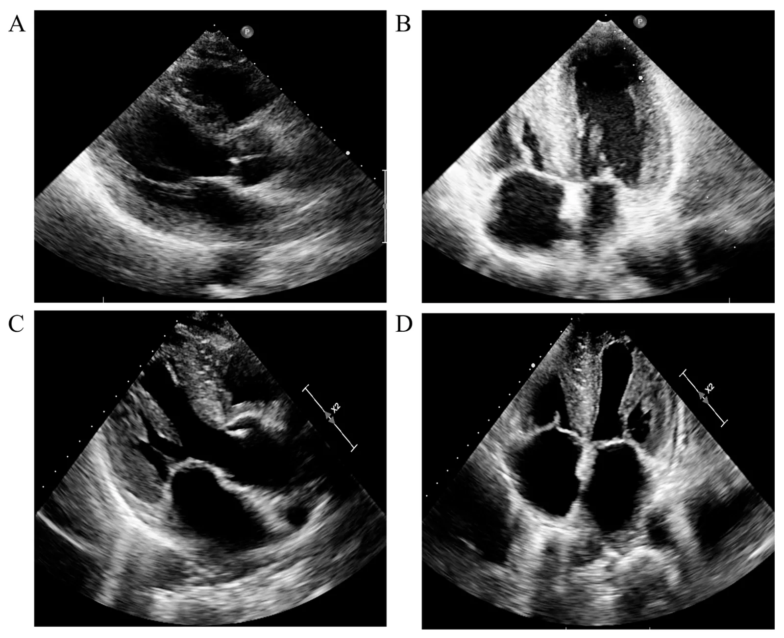
Long description
Four transthoracic echocardiogram panels from a patient with cardiac amyloidosis, a restrictive cardiomyopathy: parasternal long-axis views (A, C) and apical four-chamber views (B, D) show thickened ventricular walls with a granular myocardium and enlarged atria. The apical four-chamber view in panel D shows both atria enlarged relative to the ventricles.
Serum natriuretic peptide, renal and liver function, full blood count, ferritin and transferrin saturation, immunoglobulin light chains in serum and urine, and ECG. The ECG is quick, cheap and often abnormal. Troponin is often mildly and chronically elevated in amyloidosis, which is a prognostic marker rather than an acute infarct.
Echocardiography is the foundation. It demonstrates a non-dilated ventricle with preserved or near-preserved ejection fraction, biatrial enlargement, normal or increased wall thickness (depending on cause), and a restrictive filling pattern: shortened early deceleration time, an elevated E/e′ ratio, and a prominent A wave. Measurement of longitudinal strain characteristically shows reduced basal strain with relative apical sparing in amyloidosis — the so-called relative apical sparing. Tissue Doppler separates amyloid from hypertensive hypertrophy. Right ventricular function, tricuspid regurgitation and pericardial thickness are assessed at the same sitting. Loading conditions change these measurements, so repeated studies are needed before declaring restriction permanent.[1]
CMR is the best test for tissue characterisation. In amyloidosis native T1 values are high, the extracellular volume fraction is increased, and late gadolinium enhancement shows a characteristic diffuse subendocardial or transmural pattern. T2* imaging detects iron overload (T2* values below 20 ms of myocardium indicate significant iron deposition). Parametric mapping also discriminates Fabry disease (low native T1 from lipid). CMR is excellent for distinguishing restriction from constrictive pericarditis, which shows pericardial thickening and enhancement rather than myocardial infiltration.[1] [3]
Myocardial uptake of a bone tracer (99mTc-pyrophosphate, 99mTc-DPD or 99mTc-HMDP, depending on region) strongly suggests ATTR cardiac amyloidosis. Grade 2–3 myocardial uptake carries a sensitivity of more than 99% and a specificity of 86% for ATTR cardiomyopathy, provided there is no monoclonal protein in the serum or urine.[3] A bone scan showing intense myocardial uptake with no monoclonal light chain is sufficient to diagnose ATTR amyloidosis without biopsy in many settings.
Because ATTR cannot be diagnosed on imaging alone in the presence of a monoclonal gammopathy, every patient being considered for this pathway must have a serum free light-chain assay, serum immunofixation electrophoresis and urine immunofixation electrophoresis. An abnormal kappa-to-lambda ratio below 0.26 or above 1.65, in the appropriate clinical setting, is a positive screen for a plasma cell dyscrasia.[3] Interpret the ratio alongside the absolute light-chain concentrations, because renal impairment can elevate both kappa and lambda together and mask a monoclonal excess. Although the scintigraphy result is informative, it is not the last test: all patients undergoing bone scanning in this pathway should also have serum and urine immunofixation and free light-chain measurement.[3]
When imaging is inconclusive or light chains are abnormal, tissue is required. Endomyocardial biopsy has a high diagnostic sensitivity and specificity when performed by experienced operators and is the gold standard for amyloid typing. Abdominal fat pad biopsy is lower risk, with roughly 70 to 80% sensitivity for AL amyloidosis, but it is much less sensitive in ATTR. Cardiac biopsy is therefore commonly preferred when ATTR is suspected and the diagnosis is uncertain.[3] Histology with Congo red (apple-green birefringence) identifies amyloid; immunohistochemistry or mass spectrometry types it. Genetic testing of the TTR gene distinguishes wild-type from variant ATTR and allows family screening; genetic panels are also used in idiopathic restriction and in suspected Fabry, sarcomeric and storage disease.[1] [3]
Cardiac biomarkers (troponin and natriuretic peptide) stage cardiac AL and ATTR amyloidosis and stratify prognosis. These staging systems are applied and interpreted by the amyloid team; the principle is that the degree of cardiac involvement, not the clone or fibril type, is the dominant prognostic driver in AL disease.[3]
Approach to suspected restrictive cardiomyopathy
- Clinical suspicionHeart failure with preserved or mildly reduced EF; biatrial enlargement; a quiet precordium
- ECG, echo with Doppler and strainRestrictive filling, apical sparing in amyloid, pericardium and RV function
- Question: First decision: amyloid, or not?
Red flags for amyloidosis
- Serum and urine FLC, SIFE and UIFERatio <0.26 or >1.65 is abnormal
- Question: Monoclonal protein present?
Yes
- Suspect AL amyloidHaematology review; EMB or fat pad biopsy for typing
No
- Bone scintigraphy (99mTc-PYP/DPD/HMDP)Grade 2–3 uptake diagnoses ATTR-CM without biopsy
Non-amyloid phenotype
- CMR with native T1, ECV and T2*Iron overload, Fabry, sarcoid, fibrosis
- Targeted testsFerritin/transferrin, enzyme assay, genetic panel, EMF work-up
- Biopsy when the diagnosis remains unclearEMB retains gold-standard status
- Define the cause and stage the heartEjection fraction, biomarkers, filling pressures
- Cause-directed treatment plus cautious diuretic and rhythm management
Management rests on three facts about the physiology above. The ventricle is volume sensitive in both directions, so relief of congestion is needed but over-diuresis must be avoided. Stroke volume is essentially fixed, so cardiac output depends on heart rate, which means bradycardia is dangerous and negative chronotropic and inotropic drugs are poorly tolerated. Reverse remodelling is unlikely, so treatment seldom returns the myocardium to normal.[1]
Loop diuretics relieve pulmonary congestion, peripheral oedema and ascites, and are the mainstay of congestion control. Forced diuresis should be avoided, because even mild hypovolaemia can reduce stroke volume and cardiac output. Mineralocorticoid receptor antagonists are generally well tolerated. Beta-blockers have not been shown to improve outcomes in restriction and may be poorly tolerated when cardiac output depends on rate; if they are used for a clear indication such as atrial fibrillation, careful haemodynamic monitoring is needed. Digoxin must be used with extreme caution in cardiac amyloidosis, because it binds to amyloid fibrils, cannot be reliably monitored by serum levels, and is associated with toxicity; in contemporary practice it is usually avoided. Non-dihydropyridine calcium channel blockers (verapamil and diltiazem) and beta-blockers are also poorly tolerated because of their negative chronotropic and inotropic effects.[1] [2] [3]
The use of standard heart-failure therapies in restriction is extrapolated from broader heart failure practice, and the conventional benefits of afterload reduction are less predictable when the ventricle is stiff; refer to the heart failure topic for drug-level detail.
Cardiac amyloidosis — transthyretin. Tafamidis is a transthyretin stabiliser and is the first approved disease-modifying drug. It is given as tafamidis 61 mg (one Vyndamax capsule) orally once daily or tafamidis meglumine 80 mg (four 20-mg Vyndaqel capsules) orally once daily; the two formulations are not interchangeable milligram for milligram.[5] It reduces cardiovascular mortality and hospitalisation and carries a Class I, level of evidence B recommendation for patients with ATTR cardiomyopathy in New York Heart Association classes I and II.[1] Newer ATTR therapies include: acoramidis (a high-affinity transthyretin stabiliser) 712 mg orally twice daily; vutrisiran (a subcutaneous small interfering RNA that suppresses hepatic transthyretin synthesis) 25 mg every three months; and patisiran (an intravenous small interfering RNA) 0.3 mg per kilogram every three weeks (approved for hereditary transthyretin polyneuropathy, with cardiac data from trials).[2] [6] Inotersen, eplontersen and monoclonal antibodies directed at amyloid fibrils, as well as gene-editing approaches, are being evaluated; none replaces specialist involvement. Where approved, stabiliser and gene-silencing therapies should be chosen by the cardiomyopathy and amyloid teams, because head-to-head comparative trial data are limited.[2]
Cardiac amyloidosis — light chain. The immediate aim is a deep and rapid haematologic response. Based on phase III evidence, daratumumab combined with cyclophosphamide, bortezomib and dexamethasone (Dara-CyBorD) is established first-line therapy for most patients and is approved as such. Autologous stem-cell transplantation is offered to selected patients with a good haematologic and cardiac response. Bortezomib should be combined carefully and avoided in regimens that rely on neurotoxic agents in patients with significant neuropathy; melphalan-based regimens are alternatives.[2] Supportive management of cardiac amyloidosis should never delay plasma-cell therapy.
Haemochromatosis. Iron removal — regular phlebotomy where tolerated, with iron chelation (for example deferoxamine or oral chelators) when phlebotomy is not tolerated or secondary iron overload is driven by transfusion dependence — is the disease-modifying treatment, and early removal can reverse cardiac dysfunction.[1] Diabetes, endocrine and liver disease are managed in parallel.
Anderson–Fabry and storage diseases. Enzyme replacement and, where appropriate, substrate-reduction therapies modify the underlying disease. Genetic diagnosis precedes treatment.
Sarcoidosis. Immunosuppression guided by the granulomatous disease, often with corticosteroids as first-line, is the standard approach; cardiac involvement warrants coordinated cardiology and rheumatology care. No dose is stated here, because regimens are individualised.
Eosinophilic and hypereosinophilic disease. The acute inflammatory phase is treated with corticosteroids and, when a parasitic or other trigger is present, targeted therapy against the trigger. Anti-thrombotic therapy is added because endocardial thrombus is frequent. Chronic fibrotic disease is not reversible with drugs and proceeds to valve surgery if regurgitation is severe.
Endomyocardial fibrosis and carcinoid disease. Treatment of the underlying process is limited; management is largely of heart failure, arrhythmia and valve disease. Carcinoid syndrome is managed with somatostatin analogues and hepatic and valvular surgery as indicated.
Atrial fibrillation is very common in amyloid and restriction. Rhythm control with amiodarone is generally well tolerated in cardiac amyloidosis and may be more effective than rate control in this group, although survival data comparing strategies are limited. Beta-blockers and calcium channel blockers are poorly tolerated for rate control and any negative inotrope should be used with care and close monitoring.[2] [3]
Atrial arrhythmias in these patients carry a higher cardioembolic risk than in other heart failure. The 2023 ESC guideline (as endorsed in European practice) recommends prophylactic anticoagulation for atrial fibrillation or flutter in cardiac amyloidosis and hypertrophic cardiomyopathy as a Class I, level B recommendation, and in other restrictive forms as a Class IIa, level B recommendation; the CHA2DS2-VASc score has not been validated in these populations, so anticoagulation is not withheld when the score is low. Direct oral anticoagulants are preferred in the absence of a mechanical valve or moderate-to-severe mitral stenosis, in line with general guidance.[4] Ventricular arrhythmias are treated according to general principles, with the approach to tachyarrhythmias and ventricular arrhythmias as background; implantable cardioverter-defibrillator and cardiac resynchronisation decisions are individualised and evidence in restriction is limited. Conduction system disease may require permanent pacing.[1]
Heart transplantation is a reasonable option in carefully selected patients without the severe systemic disease that would limit survival. Outcomes are similar to transplantation for other heart failure causes, except in cardiac amyloidosis and radiation-induced cardiomyopathy, where results are usually less satisfactory. In recent practice, earlier diagnosis and treatment of amyloidosis have allowed selected patients with ATTR cardiomyopathy to be transplanted with outcomes closer to those of other indications, provided the systemic disease is controlled. Ventricular assist device support is technically challenging because the ventricular cavity is small and the risk of obstruction high.[1]
- Thromboembolism and stroke, especially in cardiac amyloidosis, where atrial infiltration promotes stasis and thrombus formation.[4]
- Worsening right heart failure with recurrent ascites, malnutrition and hepatic congestion.
- Valvular regurgitation secondary to atrial dilatation and ventricular remodelling.
- Conduction disease and sudden death from amyloid or sarcoid infiltration and from the underlying arrhythmia substrate.
- Restrictive physiology that is not reversible once heavy fibrosis or infiltration is established.
Prognosis is driven by the cause rather than by the haemodynamic label. AL amyloidosis carries the worst cardiac prognosis when heart involvement is advanced, and the severity of cardiac involvement is the dominant prognostic factor even with modern haematologic therapy; earlier stage at diagnosis improves survival. ATTR cardiomyopathy has a more indolent course, and stabiliser and gene-silencing therapies now aim to change the natural history. Iron overload cardiomyopathy can be reversed if treated before fibrosis is fixed. Radiation-induced restrictive disease is often severe because of mixed cardiac damage. Endomyocardial fibrosis and idiopathic restriction have variable courses and, in children, may progress to transplant. Overall, disease-modifying treatments are currently available for cardiac amyloidosis and, partially, for iron-overload cardiomyopathy.[1] [3]
Pregnancy. Restriction with a fixed cardiac output and a dependency on heart rate is a high-risk lesion in pregnancy. Care should be coordinated between cardiology, obstetrics and anaesthesia. Tafamidis has animal reproductive toxicity data and is generally avoided. Transmission of hereditary systemic disease (for example, variant ATTR or Fabry disease) requires genetic counselling.
Children. Idiopathic restriction usually presents in childhood and often proves to be genetic on testing. Rhabdomyoma and fibroma are the more common paediatric heart tumours causing obstruction. Palliative and transplant options are considered by the paediatric cardiomyopathy team.
Families. First-degree relatives of a patient with genetic restriction or ATTR variant amyloidosis should be offered cascade testing. In families with amyloidosis, carpal tunnel syndrome or lumbar spinal stenosis in a middle-aged relative can be an early clue.
RCM is under-diagnosed in many resource-limited settings because the diagnosis relies on echocardiography, which is not always available. Useful practical points:
- A normal or near-normal chest radiograph with heart failure signs, elevated jugular venous pressure, hepatomegaly and peripheral oedema should prompt a search for restriction rather than assuming dilated cardiomyopathy.
- The core investigation is a basic echocardiogram with Doppler to look for biatrial enlargement, normal-sized non-dilated ventricles, normal or increased wall thickness, and a restrictive filling pattern. A simple 12-lead ECG supports it.
- Free light-chain assays, immunofixation, scintigraphy and cardiac magnetic resonance imaging are often unavailable. In that case treat on the basis of the best available diagnosis, support with diuretics cautiously, screen for preventable causes (iron deficiency and overload, HIV, tuberculosis, parasites in the case of eosinophilia), and refer.
- Endomyocardial fibrosis is the dominant restrictive disease in equatorial Africa. Late presentation is common, and surgery for fibrosis or valve regurgitation is often the only durable option where available; earlier recognition in the inflammatory eosinophilic phase is the main opportunity to change outcome.
- Tafamidis and gene-silencing therapies for ATTR amyloidosis are not widely available outside high-income settings. Access remains a major equity gap.
Practitioners look up heart failure, ECG interpretation, cardiac physical examination, atrial fibrillation and flutter, supraventricular tachycardias, bradyarrhythmias and AV block and hypertrophic cardiomyopathy alongside this topic. Primary articles on cardiac amyloidosis, iron-overload cardiomyopathy and sarcoid heart disease are cross-referenced as they become available.>
- Restrictive cardiomyopathy is a stiff, non-dilated myocardium with normal or near-normal ejection fraction but restricted filling; stroke volume is essentially fixed and cardiac output depends on heart rate.
- It is a haemodynamic endpoint for many diseases, not a single diagnosis. The four mechanisms are fibrosis, extracellular infiltration, intracellular storage, and endomyocardial fibrosis.
- Cardiac amyloidosis — AL and ATTR — is the commonest identifiable cause in adults in high-income settings; Fabry disease, haemochromatosis, sarcoidosis, sarcomeric mutations, radiation, drugs, carcinoid and idiopathic restriction are the others.
- Clinical presentation is right-sided or biventricular heart failure with a quiet precordium, elevated jugular venous pressure, hepatomegaly, ascites and peripheral oedema; a normal-sized heart on the radiograph and low QRS voltage relative to wall thickness are important clues.
- Echocardiography with Doppler and strain is the first imaging test; CMR characterises tissue; bone scintigraphy diagnoses ATTR in the absence of a monoclonal protein; free light-chain assays and immunofixation exclude AL disease.
- Restriction must be persistent on repeated studies before the label is applied, because congestion can mimic it.
- Constrictive pericarditis is the great imitator: look for pericardial thickening or calcification, septal bounce, enhanced ventricular interdependence and a square-root pressure trace; treatment is pericardiectomy, not drugs.
- Disease-modifying therapy exists for cardiac amyloidosis (tafamidis, acoramidis, vutrisiran and others; AL chemotherapy with Dara-CyBorD) and partially for iron overload (iron removal); otherwise treatment is cautious diuresis, support, arrhythmia and anticoagulation, with transplantation in selected cases.
- Drugs that slow the heart or reduce inotropy — beta-blockers, non-dihydropyridine calcium channel blockers and digoxin — are poorly tolerated in restriction and avoided or used only with extreme caution, particularly in amyloidosis.
- Hereditary causes require genetic testing and cascade screening of relatives. Endomyocardial fibrosis remains a major cause of heart failure in equatorial Africa.
Answer each one in your head first, then open the answer.
A 62-year-old man has exertional breathlessness, ankle swelling and ascites. The electrocardiogram shows low-voltage QRS complexes; echocardiography shows normal ventricular size, mildly increased wall thickness, biatrial enlargement and a restrictive filling pattern. What is the most likely diagnosis group, and what is the single most important first laboratory screen?
Show answerHide answer
The pattern points strongly to cardiac amyloidosis, specifically an infiltrative restrictive cardiomyopathy. The first screen is a serum free light-chain assay with serum and urine immunofixation electrophoresis, to exclude AL amyloidosis before any imaging conclusion is drawn. A negative screen permits a bone scan to diagnose ATTR amyloidosis without biopsy.
Why does the stiff ventricle of restrictive cardiomyopathy tolerate neither over-diuresis nor beta-blockade well?
Show answerHide answer
Because stroke volume is essentially fixed by the stiff myocardium, cardiac output depends chiefly on heart rate and adequate preload. Over-diuresis reduces preload and output; beta-blockers slow the rate and can be negatively inotropic, both of which worsen haemodynamics.
A patient with supposed 'constrictive pericarditis' is found to have marked biatrial enlargement, moderate pulmonary hypertension and no respiratory variation in the Doppler flows. How does this change the diagnosis and the treatment plan?
Show answerHide answer
These features favour restrictive cardiomyopathy rather than constrictive pericarditis, because they show primary myocardial stiffness rather than a tethered pericardium. Treatment shifts from pericardiectomy to a cause-directed plan with cautious diuresis and evaluation for amyloid or another infiltrative disease; transplant may be considered in selected cases.
What are the recommended doses of tafamidis for ATTR cardiomyopathy, and why must serum light-chain studies be done before calling a bone scan 'diagnostic'?
Show answerHide answer
Tafamidis is given as Vyndamax 61 mg once daily or Vyndaqel 80 mg once daily, and the two formulations are not interchangeable milligram for milligram. Light-chain studies must precede the interpretation of scintigraphy because grade 2–3 myocardial bone-tracer uptake is specific for ATTR only when there is no monoclonal protein in serum or urine; otherwise AL amyloidosis cannot be excluded on imaging.
A patient from rural equatorial Africa presents with right heart failure, hepatomegaly and huge biatrial enlargement. Echocardiography shows obliteration of the right and left ventricular apices with fibrous thickening. What is the likely cause, and what does this imply for management?
Show answerHide answer
Endomyocardial fibrosis (tropical EMF), which is a major cause of heart failure in equatorial Africa. Management is largely supportive with cautious diuresis, valve surgery for severe regurgitation where available, and recognition of the eosinophilic inflammatory phase earlier in the disease, when anti-inflammatory therapy may modify outcome.
In cardiac amyloidosis with atrial fibrillation, how do the 2023 cardiomyopathy guideline recommendations differ between cardiac amyloidosis and other restrictive forms, and why is the CHA2DS2-VASc score not the decider?
Show answerHide answer
In cardiac amyloidosis and hypertrophic cardiomyopathy the guideline recommends prophylactic anticoagulation as a Class I, level B recommendation, and in other restrictive forms as a Class IIa, level B recommendation. The CHA2DS2-VASc score has not been validated in these populations, and the cardioembolic risk is increased by atrial infiltration and stasis, so a low score does not protect against thrombus.
Another topic links here.
- Rapezzi C, Aimo A, Barison A, Emdin M, Porcari A, Linhart A, Keren A, Merlo M, Sinagra G. Restrictive cardiomyopathy: definition and diagnosis. European Heart Journal 2022;43(45):4679–4693. doi:10.1093/eurheartj/ehac543. Accessed .
- Alkhatib A, Alashqar R, Abdallah AW, Abu-Nejim H, Hammad A, Khraisat O, Abuzaid AS. A comprehensive review of the management of light-chain (AL) and transthyretin (ATTR) cardiac amyloidosis. Reviews in Cardiovascular Medicine 2025;26(12):42609. doi:10.31083/rcm42609. Accessed .
- Alashqar R, Alkhatib A, Abdallah AW, Odeh M, Al-Taei M, Khraisat O, Al-Hiari M, Taifour H, Hammad A, Abuzaid AS. What cardiologists should know about amyloidosis. Journal of Clinical Medicine 2025;14(18):6668. doi:10.3390/jcm14186668. Accessed .
- Groeneweg JA, van Dalen BM, Cox MPGJ, Heymans S, Braam RL, Michels MP, Asselbergs FW. 2023 European Society of Cardiology guidelines on the management of cardiomyopathies (Dutch endorsement). Netherlands Heart Journal 2025. doi:10.1007/s12471-025-01955-2. Accessed .
- Pfizer Laboratories Div Pfizer Inc. VYNDAQEL and VYNDAMAX (tafamidis / tafamidis meglumine) capsules: full prescribing information. DailyMed, US National Library of Medicine. Updated 12 May 2026. Accessed .
- BridgeBio Pharma Inc. ATTRUBY (acoramidis hydrochloride) tablets: full prescribing information. DailyMed, US National Library of Medicine. Accessed .
Figure credits
Educational content for medical students and doctors. It is not a substitute for clinical judgement or local guidelines.