Aortic dissection
A tear in the aortic wall: recognising it fast, controlling aortic wall stress, and choosing emergency surgery or TEVAR.
- An aortic dissection is a split in the wall of the aorta: blood enters through a tear in the inner layer and travels inside the wall, creating a second channel called the false lumen.
- It is a cardiovascular emergency. A dissection involving the ascending aorta (type A) can kill within hours through rupture, tamponade, coronary occlusion or acute aortic regurgitation.
- The classic story is abrupt, severe chest or back pain in an older person with high blood pressure. The classic trap is a normal ECG, a normal chest radiograph, or pain that sounds musculoskeletal.
- The diagnosis is made by imaging, usually CT angiography. Until imaging is done, the priorities are pain relief, heart-rate control with an intravenous beta-blocker, and then blood-pressure control.
- Type A dissection needs emergency cardiac surgery. Type B dissection is usually managed medically at first, with thoracic endovascular repair (TEVAR) for complications.
By the end of this page you should be able to:
- Define aortic dissection and classify it using the Stanford and DeBakey systems.
- Explain how hypertension, inherited aortopathy, bicuspid aortic valve, pregnancy, cocaine and inflammatory disease lead to a tear.
- Recognise the symptoms and signs, including pulse deficits, blood-pressure differences, aortic regurgitation, tamponade and malperfusion.
- Use the aortic dissection detection risk score (ADD-RS), D-dimer and imaging in a rational sequence.
- Stabilise a patient with impulse control and pain relief, and explain why anticoagulation and thrombolysis are dangerous.
- Decide who needs emergency type A repair, who needs TEVAR, and who can be managed medically.
- Plan surveillance and counsel patients about family screening and pregnancy.
DefinitionAortic dissection
Aortic dissection is a tear in the intima of the aorta that allows blood to track within the media, separating the wall into a true lumen and a false lumen. The false lumen may compress the true lumen, rupture through the adventitia, or extend into branch vessels.
The aorta has three layers: a thin inner intima, a muscular media, and a tough outer adventitia. In dissection the intima tears, and pulsatile blood forces its way into the media. That creates a second channel, the false lumen, alongside the original true lumen. The flap of intima and media that separates them is the intimal flap. Blood can travel along the false lumen for centimetres, forwards or backwards, and the flap can cover the openings of branch arteries.
Aortic dissection is one member of the acute aortic syndrome family, together with intramural haematoma (blood within the wall without a clear tear) and penetrating atherosclerotic ulcer (an ulcer that breaches the inner layer). Intramural haematoma is diagnosed when imaging shows crescentic wall thickening of more than 5 mm without detectable flow.[1] These three conditions overlap and are managed similarly in their acute phase.
The traditional division is simple: the first two weeks are acute, and anything later is chronic.[1] The two-week cut-off reflects the period of highest risk of rupture and death. Some later classification schemes add subacute and chronic stages with different cut-offs, including a four-phase model (hyperacute, acute, subacute, chronic) based on survival data, but the two-week acute threshold is the one that changes immediate management.[1][7]
Figure 1Disekce klasifikace DeBakey a Stanford
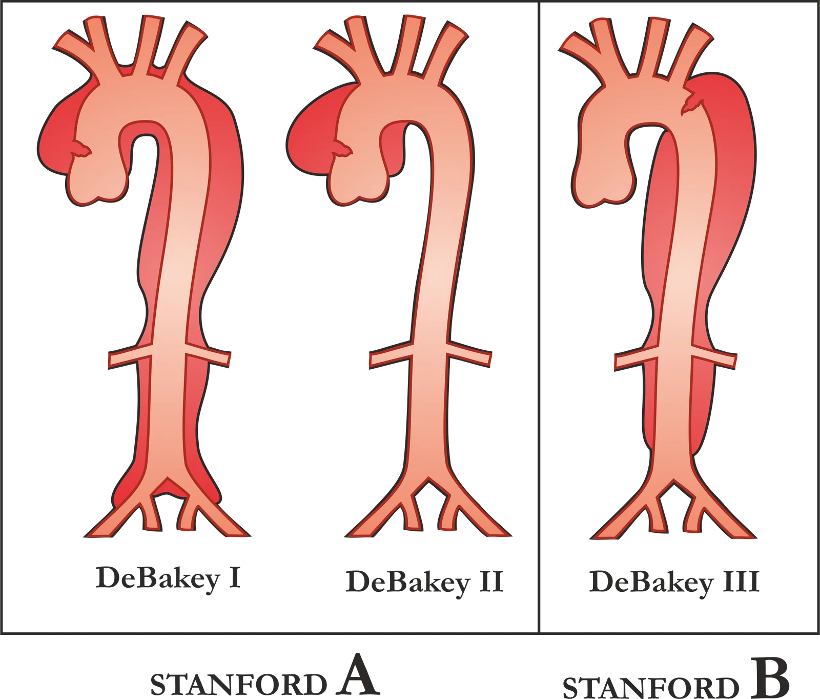
Long description
Classification of the aortic dissection, covering the DeBakey and Stanford systems, from the file description and title.

Two anatomical systems are in everyday use. The Stanford system asks one question: is the ascending aorta involved? The DeBakey system describes where the tear starts and how far the dissection travels.[1]
| System | Category | Meaning |
|---|---|---|
| Stanford | Type A | Any dissection involving the ascending aorta, whatever the site of the tear.[1] |
| Stanford | Type B | Dissection that spares the ascending aorta, including arch dissection that does not reach the ascending aorta.[1] |
| DeBakey | Type I | Tear in the ascending aorta, extending through the arch and often into the descending aorta.[1] |
| DeBakey | Type II | Tear confined to the ascending aorta.[1] |
| DeBakey | Type III | Tear in the descending thoracic aorta; IIIa stays thoracic, IIIb extends below the diaphragm.[1] |
The Stanford classification is deliberately blunt, and that is its strength: type A means the ascending aorta is involved, and the patient usually needs an operation. A tear that begins in the arch but does not involve the ascending aorta is still type B under this scheme.[1]
A newer European classification, TEM, describes the Type of aortic syndrome, the location of the Entry tear, and the presence of Malperfusion. It was introduced because "type A or B" says nothing about which branch vessels are starved of blood, and malperfusion is a major determinant of outcome.[2]
Aortic dissection is uncommon but not rare. The ACC/AHA guideline estimates 5 to 30 cases per million people per year, with men affected more often.[1] Population studies in the United States and Europe give a similar order of magnitude, about 2.6 to 3.5 cases per 100,000 person-years; differences reflect how aggressively cases are found and coded.[7]
- Age. Most patients are between 50 and 70 years old.[1] Type A dissection often presents in the sixth decade.[3]
- Sex. Men are affected more often, and type A dissection is about twice as common in men.[3] Women present later, with a mean age of about 72 years compared with 64 years in men.[7]
- Setting. It is a disease of emergency departments, because the presentation overlaps with myocardial infarction, pulmonary embolism and musculoskeletal pain.
The true burden is higher than hospital series suggest. Up to half of dissections cause sudden death, and almost one in three is misdiagnosed.[8] In some series nearly half of patients die before reaching hospital.[8]
There are important regional differences. In a large international registry, type A accounted for about two-thirds of dissections; in a Chinese registry it accounted for nearer 43%.[8] Access to imaging, referral patterns and the prevalence of hypertension all influence what is counted.
Dissection happens when wall stress exceeds the strength of the aortic wall. Most patients have a combination of a weakened wall and a trigger that raises stress suddenly.
Hypertension is the single most important modifiable factor. In a long-running cohort, 86% of people who later dissected had hypertension.[8] The risk is about four times that of normotensive people, and the population-attributable risk is 54%.[8] About half of dissection events may be preventable with effective blood-pressure treatment.[8]
This is not only a disease of long-standing, severe hypertension. Many patients have untreated or poorly controlled pressure, and the surge during a hypertensive crisis may be the final trigger.
Marfan syndrome is the best-known inherited cause, but it accounts for only about 4 to 5% of dissections.[7] Patients with Marfan syndrome dissect much younger, around 38 years compared with 63 years in non-Marfan patients.[7] Loeys-Dietz syndrome is rarer but more aggressive: almost 98% of affected patients have aortic root aneurysms.[7] Vascular Ehlers-Danlos syndrome is a further inherited cause, and these patients also tend to present at a younger age.[1]
The genetics are broader than these syndromes. Pathogenic variants in 11 genes have been validated as causes of highly penetrant thoracic aortic aneurysm and dissection.[8] Even so, up to 80% of people who dissect have no known family history and no identified variant in a recognised gene.[8] A negative family history is therefore reassuring only up to a point.
A bicuspid aortic valve is present in about 1% of the population, and it is over-represented in dissection cohorts, at about 3.2% in one large registry.[8] The relative risk is roughly eight-fold.[3] The absolute risk for an isolated bicuspid valve is much lower than that figure suggests: about 0.4% over 15 years in one analysis.[8] The valve is a marker of aortopathy, not a guarantee of dissection.
Pregnancy raises the risk about four- to five-fold, through hormonal changes, increased blood volume and the haemodynamic stress of labour.[8][6] It is rare, accounting for 0.2% to 1% of dissections in women, with an incidence of 4 to 12 per million maternities.[8] Around 60% of women with a pregnancy-associated dissection have at least one identifiable risk factor, most often Marfan syndrome, a family history or a bicuspid valve.[8] Type A is about four times more common than type B in pregnancy.[8]
Cocaine and methamphetamine use are recognised triggers, through sudden surges in blood pressure and heart rate. In one series, 37% of patients with dissection had used cocaine within 24 hours of symptoms.[8] Across studies the proportion varies widely, from 0.5% to 28%.[8] The association is important in young patients with no other risk factors.
Blunt trauma, usually from rapid deceleration, can tear the aorta, most often at the isthmus just distal to the left subclavian artery. Iatrogenic dissection follows aortic instrumentation, catheterisation or cardiac surgery.[8] Both are important because the mechanism and treatment differ from spontaneous dissection: traumatic injury is often a contained tear or transection, and iatrogenic dissection may be recognised during the procedure.
Giant cell arteritis typically affects people aged 50 or older and involves the aorta and its main branches in about half of patients.[8] It damages the medial elastic lamina, weakening the wall.[8] In one cohort followed for 52 months, dissection occurred in 5% of patients with giant cell arteritis.[8] Takayasu arteritis, Behçet disease and IgG4-related aortitis can do the same.
Turner syndrome, coarctation, previous aortic dissection, atherosclerosis and weightlifting are also described.[1][8] In practice, the history should ask about all of them, because a young patient with dissection often has an inherited or inflammatory cause that changes long-term care.
The final common pathway is a weak media plus a tear.
Medial degeneration. The media is normally a layered composite of elastic fibres and smooth-muscle cells. In aortopathy, elastic fibres fragment, smooth-muscle cells are lost, and proteoglycans accumulate.[8] The wall becomes less able to bear pulsatile stress, and the layers separate more easily.
The tear. An intimal tear lets blood enter the media. Once inside, the column of blood is exposed to aortic pressure with each beat. The false lumen usually propagates distally with the direction of flow, but it can also track proximally towards the root.
Wall stress. Aortic wall stress rises with pressure, diameter and the rate of pressure rise. That is why beta-blockers are given before vasodilators: slowing the heart and reducing contractility lowers the force of each ejected stroke, while a pure vasodilator can produce reflex tachycardia and a faster pressure rise.
Consequences. The dissection can:
- rupture into the pericardium, causing tamponade;
- rupture into the pleural space or mediastinum, causing haemorrhagic shock;
- distort the aortic valve, causing acute aortic regurgitation and pulmonary oedema;
- involve the coronary ostia, causing myocardial infarction;
- cover or compress branch vessels, causing malperfusion of the brain, spinal cord, kidneys, gut or limbs;
- continue into the abdominal aorta and iliac arteries.
Malperfusion deserves special emphasis. The flap can block a branch dynamically, or the false lumen can thrombose and compress the true lumen statically. The result is end-organ ischaemia even when the patient's blood pressure looks acceptable. A normal blood pressure does not exclude malperfusion.
At operation or autopsy, the aorta contains a longitudinal intimal tear and a false lumen. The false lumen may be filled with blood or thrombus, and the outer wall may be thinned or ruptured. In intramural haematoma, there is blood within the media without a communicating tear.[1]
Microscopically, the characteristic finding is medial degeneration: elastic fibre fragmentation, loss of smooth-muscle cells and accumulation of proteoglycans.[8] Inflammatory aortitis adds a lymphocytic or granulomatous infiltrate and destruction of the elastic lamina.[8]
Pain is the dominant symptom, present in about 90% of patients.[6] It is usually abrupt and severe. The quality helps: sudden onset carries a positive likelihood ratio of 2.6, tearing or ripping pain 10.8, and migratory pain 7.6.[6] But only 30% to 40% of patients actually describe tearing or ripping, and "sharp" is more common.[6]
The pain is often anterior chest pain in type A and interscapular or back pain in type B. Abdominal pain is more common in type B, and can be the presenting feature when the dissection extends to the mesenteric or renal arteries.
Syncope, stroke-like symptoms, limb weakness, abdominal pain or a painless presentation can all occur. Painless dissection is uncommon — reported in under 6% in one review and 6.4% in another — but it is easily missed, especially when the presentation is syncope or stroke.[6][7]
Look for asymmetry and for signs of complications:
- Pulse deficit. A pulse deficit occurs in 19% to 30% of patients and carries a positive likelihood ratio of 5.7.[6] Palpate and compare both radial, brachial and femoral pulses.
- Blood-pressure difference. A difference of more than 20 mmHg between arms is a red flag, although 20% of patients without dissection also have such a difference.[6]
- Aortic regurgitation. A new diastolic murmur is present in up to 40% of patients, but it is often difficult to hear in a noisy emergency department.[6]
- Tamponade. Look for hypotension, raised jugular venous pressure, muffled heart sounds and pulsus paradoxus. Pericardial effusion is found in about one-third of patients, and tamponade complicates about 10%.[6]
- Neurological deficit. Stroke, spinal cord ischaemia and peripheral nerve ischaemia can all occur.
- Malperfusion. Cool, pale or pulseless limbs, oliguria, abdominal tenderness, lactic acidosis or bowel ischaemia.
The clinical picture differs by type. The table summarises the main differences from a large review.[6]
| Feature | Type A | Type B |
|---|---|---|
| Chest pain | 79% | 63% |
| Back pain | 43% | 64% |
| Abdominal pain | 22% | 43% |
| Elevated blood pressure | under 35% | 70% |
| Low blood pressure | 25% | under 5% |
| Syncope | 19% | 3% |
| Stroke-like symptoms | 18% | 4% |
NoteA typical presentation
A man in his sixties with treated hypertension develops sudden severe pain between the shoulder blades while lifting a box. He looks pale and sweaty. The right radial pulse is weaker than the left, and the systolic pressure is much higher in the left arm than in the right. The ECG shows no ST elevation. This is aortic dissection until proved otherwise. The weaker pulse and inter-arm difference are clues, not requirements.
Exam trapWhat the pain does not tell you
A pleuritic or positional component, a normal ECG, or pain that is reproduced by palpation does not exclude dissection. Dissection can cause myocardial infarction, pericardial irritation or musculoskeletal-sounding pain. If the story is abrupt and severe in a patient with risk factors, image the aorta.
The differential is every cause of acute chest, back or abdominal pain. The features below help, but none is decisive, and several conditions can coexist with dissection.
| Condition | Features that point towards it | Why it can mislead |
|---|---|---|
| Acute coronary syndrome | Pressure-like chest pain, ST changes, raised troponin, response to nitrates | Dissection can involve a coronary ostium and cause true infarction; giving thrombolysis would be disastrous. |
| Pulmonary embolism | Pleuritic pain, breathlessness, hypoxaemia, right ventricular strain | D-dimer is raised in both; a positive D-dimer cannot separate them. |
| Pericarditis | Positional pain, pericardial rub, diffuse ST elevation, small effusion | Dissection can cause a sympathetic effusion and pericarditic pain. |
| Musculoskeletal or rib pain | Focal tenderness, pain on movement or breathing, no haemodynamic upset | Tenderness is common in dissection too; it does not exclude it. |
| Oesophageal or biliary pain | Burning or colicky pain, relation to food, epigastric tenderness | Abdominal dissection can present with epigastric pain and vomiting. |
| Renal or ureteric colic | Loin-to-groin pain, haematuria, obstructed urine | Renal malperfusion can cause flank pain and haematuria. |
| Stroke of another cause | Focal deficit, atrial fibrillation, carotid disease | Dissection can cause stroke by carotid or vertebral malperfusion. |
These are immediate, widely available and never sufficient. The ECG is normal in about 30% of patients with dissection; 19% have ischaemic ST or T-wave changes, and 7% have a myocardial infarction.[6] A normal ECG is therefore not reassuring.
The chest radiograph is abnormal in many patients but normal in others. A widened mediastinum is seen in nearly 60%, and a width above 6 to 8 cm is generally considered abnormal.[6] A completely normal chest radiograph is reported in about 28% of patients.[7] Neither test can exclude the diagnosis.
D-dimer is a breakdown product of cross-linked fibrin. Aortic dissection activates coagulation within the false lumen, so D-dimer is usually raised. A level above 500 ng/mL is the usual threshold for a positive result.[3][6][9]
The aortic dissection detection risk score (ADD-RS) structures the pre-test assessment. It gives one point for each of three domains: a high-risk condition, a high-risk pain feature, and a high-risk examination finding, for a total of 0 to 3.[4]
- High-risk conditions include connective tissue disease, aortic valve disease, recent aortic manipulation, family history of aortic disease, and known aortic aneurysm.[9]
- High-risk pain features are abrupt or thunderclap onset, severe or worst-ever pain, tearing or ripping pain, and migrating or radiating pain.[9]
- High-risk examination findings are a pulse deficit, neurological deficit, aortic insufficiency, and hypotension or pericardial effusion.[9]
The score performs differently at different cut-offs. In a meta-analysis, an ADD-RS above 0 had a sensitivity of 94.6% and a specificity of 34.7%; an ADD-RS above 1 had a sensitivity of 43.4% and a specificity of 89.3%.[4] Combining the score with D-dimer improves the trade-off. An ADD-RS above 0 or a positive D-dimer gave a sensitivity of 99.8% and a specificity of 21.8%; an ADD-RS above 1 or a positive D-dimer gave 98.3% and 51.4%.[4] A Canadian-style strategy, using D-dimer when the score is 1 and imaging when it is above 1, gave 93.1% sensitivity and 67.1% specificity.[4]
The safest rule is the one for the lowest-risk group: an ADD-RS of 0 with a D-dimer below 500 ng/mL had a sensitivity of 99.9% in one analysis, although specificity was variable.[4] That combination can support discharge without imaging only when the history is genuinely low risk and the D-dimer is interpreted with its limitations.
Exam trapWhen D-dimer lets you down
D-dimer can be falsely negative after 24 hours of symptoms and in intramural haematoma.[9] It is less useful in pregnancy, after recent surgery, and in hospitalised patients, because it is often raised for other reasons.[9] A negative D-dimer does not overrule a high clinical suspicion or an abnormal chest radiograph.
Contrast-enhanced CT angiography (CTA) is the first-line imaging test in a stable patient.[5] The European guideline advises ECG-gated contrast CT from the neck to the pelvis.[2] Gating reduces motion artefact at the aortic root, where a flap is easiest to miss. CTA shows the intimal flap, the true and false lumens, the entry tear, branch-vessel involvement, pericardial effusion and extent of the dissection.
CTA is accurate. A systematic review of 82 studies including 57,311 patients reported a sensitivity of 100% when contrast timing was correct,[6] while contemporary reviews describe sensitivity in the high 90% range.[3] It is not infallible: a non-gated study, poor contrast timing, or a subtle intramural haematoma can mislead.
Transoesophageal echocardiography (TOE) places the probe in the oesophagus, immediately behind the heart and beside the descending aorta, giving high-resolution images of the ascending aorta, arch and descending aorta. It is useful when the patient is too unstable to leave the resuscitation room, and it is recommended to guide perioperative management and detect early complications.[2][5] It also shows aortic regurgitation, pericardial effusion and ventricular function.
TOE is operator-dependent and needs a trained team. A normal transthoracic echocardiogram does not exclude dissection; echo can be completely normal in about 39% of cases.[7] A bedside transthoracic study is still valuable for showing a pericardial effusion or a dilated root in an unstable patient.
MRI and magnetic resonance angiography avoid ionising radiation and give excellent tissue contrast. They are preferred for subacute and chronic dissection, especially in younger patients, and are useful in stable patients with renal insufficiency because they avoid iodinated contrast.[7][5] They are less practical in the acute unstable patient because of scanning time and monitoring needs.
Suspected acute aortic syndrome: a diagnostic pathway
- Suspect aortic syndromeAbrupt severe chest, back or abdominal pain; syncope; stroke; pulse or BP asymmetry; malperfusion
- Resuscitate and monitorAirway, breathing, two large cannulae, arterial line, ECG, chest radiograph, troponin
- Question: Haemodynamically unstable?
Yes
- Bedside echo firstTOE or transthoracic echo for flap, effusion, tamponade, aortic regurgitation
- Question: Aortic syndrome confirmed or likely?
Yes
- Treat and call cardiac surgeryDo not delay for CT if unstable; image when safe
No
- Look for another causeECG, troponin, ultrasound, focused assessment
No
- Question: Low clinical probability and D-dimer available?
Yes
- Calculate ADD-RS and send D-dimerIf ADD-RS 0 and D-dimer below 500 ng/mL, consider another diagnosis with close review
No or high probability
- Go straight to ECG-gated CTANeck to pelvis; include non-contrast phase for intramural haematoma
- Question: Does CT show ascending aortic involvement?
Yes: type A
- Emergency surgical repairImpulse control, pain relief, cross-match, transfer to aortic centre if stable
No: type B
- Assess for complicationsMalperfusion, rupture, refractory hypertension, hypotension
There is no blood test that diagnoses dissection. The diagnosis is made by imaging: an intimal flap separating two lumens on CTA, TOE or MRI, or crescentic mural thickening without flow in intramural haematoma.[1]
Once the diagnosis is made, the questions that determine treatment are:
- Is the ascending aorta involved? If yes, it is type A and needs emergency surgery.
- Is there malperfusion or rupture? This makes a type B complicated and pushes towards intervention.
- Is the patient stable? Shock, tamponade, stroke and visceral ischaemia change the order of priorities.
- What is the anatomy? TEVAR needs a suitable landing zone and access vessels.
About a quarter of type B dissections present with high-risk features such as imminent rupture, organ or limb ischaemia, refractory hypertension, hypotension with a systolic pressure below 90 mmHg, or cardiogenic shock.[7]
Dissection is a haemodynamic emergency before it is a surgical one. Start with:
- Airway and breathing. Give oxygen if hypoxaemic. Intubate if the patient is unconscious or in respiratory failure.
- Circulation. Insert two large-bore cannulae, take blood for cross-match, full blood count, coagulation, renal function, troponin and blood gas, and place an arterial line.
- Monitoring. Continuous ECG, arterial pressure and urine output. Admit to a high-dependency or intensive care area.
- Analgesia. Pain drives sympathetic tone and blood pressure. Give intravenous opioid, titrated to effect.[1][3]
- Urgent senior help. Involve cardiology, cardiac surgery and, where available, vascular surgery immediately.
The aim is to reduce the force of each pulse against the torn wall. The ACC/AHA guideline recommends anti-impulse therapy with invasive blood-pressure monitoring, targeting a systolic pressure below 120 mmHg or the lowest pressure that preserves organ perfusion, and a heart rate of 60 to 80 beats per minute.[1] A review of emergency protocols uses a stricter target, with heart rate below 60/min (or below 80/min) and systolic pressure of 100 to 120 mmHg, reached within 20 to 30 minutes.[5] The exact numbers differ between protocols; the principle is the same.
Beta-blocker first. Give an intravenous beta-blocker before any vasodilator. Esmolol is short-acting and easy to titrate: a loading dose of 250 to 500 micrograms/kg over one minute in published emergency protocols (the product label specifies 500 micrograms/kg over one minute), then an infusion of 25 to 50 micrograms/kg/min, up to a maximum of 300 micrograms/kg/min.[5] Labetalol and metoprolol are alternatives.[3] In asthma, severe heart failure or high-grade atrioventricular block, a non-dihydropyridine calcium-channel blocker may be needed instead.
Then a vasodilator. If blood pressure remains high after the heart rate is controlled, add sodium nitroprusside at 0.25 to 0.50 micrograms/kg/min in published emergency protocols (the product label recommends starting at 0.3 micrograms/kg/min), up to a maximum of 10 micrograms/kg/min, with close monitoring for hypotension and cyanide toxicity.[5] Never start a vasodilator alone: reflex tachycardia increases the rate of pressure rise and can extend the dissection.[5]
| Drug | Typical dose | Role and cautions |
|---|---|---|
| Esmolol | 250 to 500 micrograms/kg IV over 1 minute (product label 500 micrograms/kg over 1 minute), then 25 to 50 micrograms/kg/min; maximum 300 micrograms/kg/min.[5] | First-line beta-blocker. Short half-life, easy to titrate; caution in asthma, bradycardia and heart failure. |
| Sodium nitroprusside | 0.25 to 0.50 micrograms/kg/min in published protocols (product label 0.3 micrograms/kg/min to start); maximum 10 micrograms/kg/min.[5] | Added after beta-blockade if pressure remains high. Monitor for hypotension and cyanide toxicity; caution in renal impairment. |
| Morphine | IV, titrated to pain.[5][3] | Analgesia and venodilation; monitor respiration and pressure. |
| Fentanyl | IV, titrated to pain.[6] | Useful when a rapid-onset opioid is preferred; monitor respiration. |
Red flagDo not treat dissection with clot-busting drugs
Thrombolysis is designed to dissolve thrombus. It cannot repair a torn aortic wall, and it can turn a contained dissection into fatal haemorrhage. If dissection is a realistic possibility, exclude it before giving fibrinolytic therapy for a presumed myocardial infarction. When pulmonary embolism is suspected and the patient has high-risk features for aortic syndrome, the Canadian guideline advises holding anticoagulation until the diagnosis is confirmed.[9]
Anticoagulation is not a treatment for dissection. It may be needed later for another indication, such as atrial fibrillation or a mechanical valve, but in the acute phase it increases the risk of bleeding into the false lumen, mediastinum or pericardium. The exception is a patient with a separate, compelling indication, where the decision is made by the treating team after weighing the bleeding risk.
Type A dissection is a surgical emergency. The ACC/AHA guideline recommends immediate surgical consultation and intervention because of the high risk of rupture, tamponade, coronary malperfusion and acute aortic regurgitation.[1] Medical management alone has a mortality two to three times that of surgery.[1]
The operation usually replaces the ascending aorta, often with a composite graft or a valve-sparing root procedure. The repair should include an open distal anastomosis rather than a simple supracoronary graft, because it addresses the arch and reduces the risk of a residual false lumen.[1] If the patient is stable, transfer to a high-volume aortic centre is associated with better survival.[1]
Timing matters. Repair within the first 48 hours is ideal.[3] In the International Registry of Acute Aortic Dissection, medically treated patients had an early mortality of 23.7% (0.5% per hour), compared with 4.4% (0.09% per hour) in those who underwent surgery.[3] Registry comparisons of this kind are confounded by selection. Operative mortality still varies widely, from about 5% to 75% depending on the patient and centre.[3]
For an uncomplicated type B dissection, medical therapy is the initial strategy.[1] That means impulse control, pain relief, bed rest and close surveillance. The early results are good: 30-day mortality is about 10%.[1] But the longer-term course is not benign. Over five years, morbidity and mortality can reach 30% and 42%, respectively, with medical management alone.[7]
A type B dissection is complicated when it causes malperfusion, rupture or refractory hypertension, or when the patient is hypotensive or in shock.[1][7] For complicated type B dissection with anatomy suitable for a stent graft, the European guideline recommends TEVAR, with open surgery as the alternative.[2] TEVAR covers the entry tear, redirects flow into the true lumen and expands the true lumen, improving perfusion.
The management of uncomplicated type B dissection is where guidelines diverge. The ACC/AHA guideline notes an increasing role for TEVAR but still recommends initial medical therapy.[1] The 2024 European guideline goes further: for uncomplicated type B dissection with favourable TEVAR anatomy, it recommends endovascular repair in the subacute phase (IIa/B).[2] The disagreement is real. It reflects uncertainty about whether early TEVAR prevents late aortic events in every patient, or only in those with high-risk features. The decision should be individualised, taking anatomy, age, comorbidity and patient preference into account.
Management after the diagnosis is confirmed
- Confirmed aortic dissectionType, entry tear, malperfusion, haemodynamic state
- Question: Ascending aorta involved?
Yes: type A
- Emergency surgeryImpulse control and analgesia while arranging theatre; open distal anastomosis
- Transfer if stableHigh-volume aortic centre; do not delay for non-essential tests
No: type B
- Question: Complicated?
Yes: malperfusion, rupture, refractory hypertension, shock
- TEVAR if anatomy suitableOpen surgery is the alternative; treat malperfusion urgently
No: uncomplicated
- Medical therapy firstImpulse control, analgesia, surveillance imaging
- Consider subacute TEVARIf favourable anatomy; ESC IIa/B, ACC/AHA more cautious
Dissection is a chronic disease even after successful treatment. The false lumen can thrombose and shrink, or it can remain patent and dilate into an aneurysm. Regular imaging is essential.
After TEVAR, CT surveillance is recommended at one month and twelve months, then annually if stable.[1] After medical management of type B dissection, delayed aortic expansion occurs in 20% to 50% of patients over four years.[1] Imaging is closer in the first two years after repair, when procedure-related complications such as endoleak and suture aneurysm are most likely, and can then be spaced out.[7] Surveillance continues for life.[7]
Blood pressure should be controlled long term, usually with a beta-blocker as part of the regimen. Treat other vascular risk factors. Avoid strenuous straining and cocaine. Discuss pregnancy before conception in women with aortic disease.
Refer immediately to cardiac surgery for any type A dissection, any dissection with tamponade, rupture or coronary involvement, and any patient with a stroke or malperfusion. Refer to a vascular or aortic team for complicated type B dissection, for TEVAR assessment, and for elective repair of a dilated residual aorta. Refer stable patients with connective tissue disease or a family history to a specialist aortic clinic, even when the diameter is not yet at a surgical threshold.
- Rupture and tamponade. The most feared early complication; sudden hypotension, distended neck veins and pulseless electrical activity.
- Acute aortic regurgitation. The flap distorts the valve, causing pulmonary oedema and shock.
- Myocardial infarction. Coronary ostial involvement, usually the right coronary artery.
- Stroke and spinal cord ischaemia. Carotid, vertebral or intercostal malperfusion.
- Visceral and limb malperfusion. Renal failure, mesenteric ischaemia, limb ischaemia and lactic acidosis.
- Late aneurysm and reoperation. The residual false lumen can dilate over years.
- Chronic dissection and false-lumen thrombosis. Some patients develop a chronic dissection that needs later intervention.
- Postoperative complications. Neurological complications affect about 18% of patients after type A repair.[3]
Untreated type A dissection is rapidly lethal, with an early mortality of 1% to 2% per hour after symptoms begin.[1] Up to half of all dissections cause sudden death, and nearly half of patients may die before reaching hospital.[8] With surgery, outcomes are much better but still serious: operative mortality ranges from about 5% to 75%, and long-term survival is limited by comorbidity and residual aortic disease.[3]
For type B dissection managed medically, 30-day mortality is about 10%, but five-year morbidity and mortality are substantial.[1][7] Across all types, reported ten-year survival is about 30% to 60%.[7] These figures are old and heterogeneous, and they may improve with earlier diagnosis, centralised care and better blood-pressure control.
Control blood pressure. This is the single most effective preventive measure. About half of dissection events may be preventable with effective antihypertensive treatment, and the population-attributable risk of hypertension is 54%.[8]
Find the inherited cases. The ACC/AHA guideline recommends screening first-degree relatives of patients with aortic root or ascending aortic aneurysm, or aortic dissection, using aortic imaging.[1] Take a three-generation family history. Refer for genetic counselling when there is a young patient, a family history, features of Marfan or Loeys-Dietz syndrome, or a bicuspid valve with aortic dilation. Pathogenic variants in 11 genes are validated causes, and cascade testing can identify relatives at risk.[8]
Repair before it dissects. Elective surgery is considered for an ascending aorta of 55 mm or more, and for a bicuspid valve with a root phenotype above 50 mm.[2] Selected low-risk patients with a tricuspid valve may be offered surgery when the ascending aorta is above 52 mm.[2] These decisions belong to an aortic team, because the risks of surgery and the patient's anatomy matter as much as the number.
Counsel women with aortopathy. Discuss pregnancy before conception. In Marfan syndrome, vaginal delivery is generally recommended when the aortic root is below 4 cm, and caesarean delivery when it is above 4.5 cm; the range in between is a specialist decision.[8]
Avoid triggers. Stop cocaine and amphetamine use. Treat severe hypertension promptly. Avoid heavy straining and weightlifting in people with a dilated aorta.
Pregnancy. Pregnancy raises the risk about four- to five-fold.[8][6] Most pregnancy-associated dissections occur in the third trimester or within 12 weeks of delivery.[1] Type A is about four times more common than type B.[8] Around 60% of women with a pregnancy-associated dissection have an identifiable risk factor, so a woman with Marfan syndrome, a bicuspid valve or a family history needs pre-pregnancy counselling and surveillance.[8] Management requires a multidisciplinary team of obstetrics, cardiology, anaesthesia and cardiac surgery.
Older adults. Women present later than men and may have atypical symptoms, which contributes to diagnostic delay.[7] A normal ECG and a normal chest radiograph are common, so age should lower the threshold for imaging, not raise it.
Connective tissue disease. Patients with Marfan syndrome dissect at a much younger age, around 38 years versus 63 years in non-Marfan patients.[7] Loeys-Dietz syndrome carries a very high risk of root aneurysm, with almost 98% affected.[7] These patients need lifelong aortic surveillance and a low threshold for elective repair.
Bicuspid aortic valve. A bicuspid valve is present in about 1% of the population and is associated with an eight-fold relative risk of dissection, but the absolute risk in an isolated valve is low.[3][8] Surveillance should focus on aortic diameter and growth, not on the valve alone.
East Africa
Aortic dissection is often diagnosed late where CT angiography is not immediately available. A normal chest radiograph or ECG does not exclude it. If CT is unavailable, look for a widened mediastinum, a pleural effusion, a pericardial effusion on bedside ultrasound, and asymmetry of pulses or blood pressure. Start impulse control with an intravenous beta-blocker (whichever is available) and an opioid, and arrange urgent transfer to a centre with cardiac surgery. Do not give thrombolysis for a suspected myocardial infarction unless dissection has been excluded. If surgery is not available locally, transfer is the treatment.
Even without advanced imaging, several steps save lives:
- Treat the patient, not the test: control heart rate and pain before sending them on a long journey.
- Use bedside ultrasound to look for a pericardial effusion, a dilated aortic root or an intimal flap; a normal study does not exclude dissection.
- Record blood pressure in both arms and palpate all peripheral pulses.
- If CT is available but cardiac surgery is not, image first only if the result will change transfer or treatment; do not delay transfer in an unstable patient.
- Teach the referral pathway before the emergency happens, so that the nearest surgical centre can accept the patient quickly.
- Aortic dissection is a tear in the intima that lets blood track within the media, creating a true and a false lumen.
- The first two weeks are the acute phase; later disease may be classified as subacute or chronic depending on the scheme. Stanford type A involves the ascending aorta and is a surgical emergency; type B spares it.
- Hypertension is the dominant risk factor, present in 86% of patients in one cohort and associated with a four-fold increase in risk.
- Marfan syndrome, Loeys-Dietz syndrome, vascular Ehlers-Danlos syndrome, bicuspid aortic valve, pregnancy, cocaine, trauma and giant cell arteritis are important causes.
- The classic presentation is abrupt severe chest or back pain, often with a pulse deficit, inter-arm blood-pressure difference, aortic regurgitation murmur or neurological deficit.
- A normal ECG and a normal chest radiograph do not exclude dissection.
- The ADD-RS structures pre-test probability; a score of 0 with a D-dimer below 500 ng/mL identifies a very low-risk group, but D-dimer has important false negatives.
- ECG-gated CT angiography is the first-line imaging test in stable patients; TOE is useful in unstable patients and perioperatively; MRI is preferred for subacute and chronic disease.
- Immediate treatment is pain relief, intravenous beta-blockade, then a vasodilator if needed. Never give a vasodilator alone.
- Type A dissection needs emergency surgery, ideally within 48 hours; stable patients should be transferred to a high-volume aortic centre.
- Uncomplicated type B dissection is treated medically at first. Complicated type B dissection is treated with TEVAR when the anatomy is suitable.
- Guidelines differ on early TEVAR for uncomplicated type B dissection: the 2024 European guideline recommends it in the subacute phase for favourable anatomy, while ACC/AHA 2022 remains more cautious.
- Lifelong surveillance is required because the residual aorta can dilate or rupture years later.
Answer each one in your head first, then open the answer.
A 58-year-old man with hypertension has sudden severe interscapular pain. The systolic pressure is clearly higher in the left arm than in the right. The ECG is normal. What is the next step, and why is the normal ECG misleading?
Show answerHide answer
Treat him as a possible aortic dissection. The inter-arm difference is a red flag, and a normal ECG is common: about 30% of patients with dissection have a normal ECG. Give analgesia and start intravenous beta-blockade, then arrange urgent ECG-gated CT angiography. Do not send him home because the ECG is normal.
Why is a beta-blocker given before sodium nitroprusside in acute dissection?
Show answerHide answer
A vasodilator lowers blood pressure but can cause reflex tachycardia, which increases the rate of pressure rise against the aortic wall and may extend the dissection. A beta-blocker slows the heart and reduces contractility first, lowering the force of each pulse. The vasodilator is then added if pressure remains high.
A 70-year-old woman has a type B dissection with severe abdominal pain, oliguria and a raised lactate. Is this uncomplicated or complicated, and what treatment does she need?
Show answerHide answer
This is a complicated type B dissection with malperfusion. She needs urgent assessment by a vascular or aortic team for TEVAR if the anatomy is suitable, with open surgery as the alternative. She also needs impulse control, analgesia and supportive care while the team is assembled.
A 34-year-old woman with Marfan syndrome has a type B dissection and wants to know whether she can have a vaginal delivery in a future pregnancy. What information matters?
Show answerHide answer
The aortic root diameter is the key factor. In Marfan syndrome, vaginal delivery is generally recommended when the root is below 4 cm, and caesarean delivery when it is above 4.5 cm; between those values the decision is individualised. She needs pre-pregnancy counselling with cardiology, obstetrics and anaesthesia, plus surveillance imaging and optimisation of blood pressure and beta-blockade.
A patient with suspected dissection has an ADD-RS of 0 and a D-dimer below the threshold. Can dissection be excluded?
Show answerHide answer
The combination is associated with a very high sensitivity, about 99.9% in one analysis, so it supports a low probability. But it does not replace clinical judgement: D-dimer can be falsely negative after 24 hours and in intramural haematoma, and it is less reliable in pregnancy and after surgery. If the history remains compelling or the chest radiograph is abnormal, image the aorta.
Why is thrombolysis dangerous when a patient with dissection is misdiagnosed as having a myocardial infarction?
Show answerHide answer
Thrombolysis dissolves clot but cannot repair the torn aortic wall. It can cause bleeding into the false lumen, mediastinum or pericardium, and can precipitate rupture or tamponade. Dissection must be excluded before fibrinolytic therapy is given for a suspected infarction, especially when the pain is abrupt or the patient has pulse or blood-pressure asymmetry.
What surveillance does a patient need after TEVAR for a type B dissection?
Show answerHide answer
CT imaging at one month and twelve months, then annually if stable. Surveillance is lifelong, because the residual false lumen can dilate, and endoleaks or graft complications can appear later. Blood pressure and heart rate should remain controlled.
- Cardiac physical examination — how to assess pulses, blood-pressure differences, murmurs and tamponade.
- ECG interpretation — why a normal ECG cannot exclude aortic dissection, and how to recognise myocardial ischaemia caused by coronary malperfusion.
- Heart failure — acute aortic regurgitation and the haemodynamic consequences of a damaged aortic valve.
- Isselbacher EM, Preventza O, Hamilton Black J 3rd, et al. 2022 ACC/AHA Guideline for the Diagnosis and Management of Aortic Disease. Circulation. 2022;146(24):e334-e482. doi:10.1161/CIR.0000000000001106. Accessed .
- Ferrante G, Bacallao BA, Gioia F, Del Monaco G, Amata F. Commentary on the 2024 European Society of Cardiology Guidelines on Peripheral Arterial and Aortic Diseases. European Cardiology Review. 2024;19:e24. doi:10.15420/ecr.2024.45. Accessed .
- Loguercio M, Romeo MG, Akinci B, et al. Type A Aortic Dissection: From Diagnosis to Cardiac Rehabilitation. Journal of Clinical Medicine. 2026;15(7):2749. doi:10.3390/jcm15072749. Accessed .
- Ren S, Essat M, Pandor A, et al. Diagnostic accuracy of the aortic dissection detection risk score alone or with D-dimer for acute aortic syndromes: systematic review and meta-analysis. PLoS One. 2024;19(6):e0304401. doi:10.1371/journal.pone.0304401. Accessed .
- Hu H, Wu Z. An Evidenced-Based Review of Emergency Target Blood Pressure Management for Acute Aortic Dissection. Emergency Medicine International. 2023;2023:8392732. doi:10.1155/2023/8392732. Accessed .
- Briggs B, Cline D. Diagnosing aortic dissection: A review of this elusive, lethal diagnosis. Journal of the American College of Emergency Physicians Open. 2024;5(4):e13225. doi:10.1002/emp2.13225. Accessed .
- Yuan X, Mitsis A, Nienaber CA. Current Understanding of Aortic Dissection. Life (Basel). 2022;12(10):1606. doi:10.3390/life12101606. Accessed .
- Zhou Z, Cecchi AC, Prakash SK, Milewicz DM. Risk Factors for Thoracic Aortic Dissection. Genes (Basel). 2022;13(10):1814. doi:10.3390/genes13101814. Accessed .
- Ohle R, Yan JW, Yadav K, et al. Diagnosing acute aortic syndrome: a Canadian clinical practice guideline. CMAJ. 2020;192(29):E832-E843. doi:10.1503/cmaj.200021. Accessed .
Figure credits
Educational content for medical students and doctors. It is not a substitute for clinical judgement or local guidelines.