Aortic aneurysm
How aortic aneurysms form, who to screen, when to repair, and how to recognise and manage rupture.
- An aortic aneurysm is a permanent, focal ballooning of the aorta. It is dangerous because the wall can tear (dissection) or burst (rupture), and rupture is often fatal before the patient reaches hospital.
- Abdominal aortic aneurysms (AAAs) are common in older men who smoke. Most cause no symptoms and are found by ultrasound screening or by imaging done for another reason.
- The strongest modifiable risk factor is smoking. Age, male sex, high blood pressure and a family history also matter; Marfan syndrome, Loeys-Dietz syndrome, vascular Ehlers-Danlos syndrome and a bicuspid aortic valve are important inherited causes.
- Small aneurysms are watched with ultrasound. Repair is offered when the risk of rupture outweighs the risk of surgery, usually at 5.5 cm in men and 5.0 cm in women for an AAA, or at a cause-specific threshold in the chest.[1]
- A ruptured AAA is a race against time: recognise it, call the vascular team, resuscitate with restrictive fluids, and move to immediate repair. Endovascular repair (EVAR) is preferred when the anatomy is suitable.
By the end of this page you should be able to:
- Define a true and a false aneurysm, and classify aneurysms by segment, shape and cause.
- Explain how medial degeneration, wall stress and inflammation lead to enlargement and rupture.
- Recognise the asymptomatic patient, the patient with compressive symptoms, and the patient with a ruptured or symptomatic aneurysm.
- Choose and interpret ultrasound, CT angiography, MRI and echocardiography, and measure the aorta consistently.
- Apply the main surveillance intervals and repair thresholds, and explain why guidelines differ.
- Outline medical therapy, open repair and EVAR, including the trade-off between early safety and late durability.
- Manage a suspected rupture and plan follow-up after repair.
The aorta is the body's main artery. It leaves the left ventricle as the aortic root, turns upward as the ascending aorta, arches over the pulmonary artery, then descends through the chest and abdomen before splitting into the iliac arteries. Each segment has a different normal diameter, a different pattern of disease and a different repair.
An aneurysm is not simply a widened artery that can be ignored. Once the wall has dilated, it is permanently weakened. The wall tension needed to contain the same blood pressure rises as the radius increases, so enlargement feeds further enlargement. That is why an aneurysm tends to grow, and why the risk of rupture rises steeply with size.
This page covers aneurysms of the thoracic aorta, the thoracoabdominal aorta and the abdominal aorta. It does not cover aortic dissection in detail, although dissection and aneurysm are closely related: a dilated aorta is more likely to dissect, and a chronic dissection can become an aneurysm. The approach to the patient with chest pain and a possible acute aortic syndrome belongs in the acute aortic syndrome topic.
NoteWhy the distinction matters
An aneurysm is a chronic structural disease. A rupture is an acute emergency. The same patient can move from one to the other within minutes, so every encounter with a known aneurysm is also an opportunity to check size, symptoms, blood pressure and follow-up.
For the abdominal aorta, the practical definition is a maximum anteroposterior or transverse diameter greater than 3.0 cm, measured outer-edge to outer-edge in the 2022 ACC/AHA guideline.[1] The American College of Radiology uses the general rule of a focal dilatation at least 50% greater than normal.[12] For the ascending aorta, the 2022 ACC/AHA guideline calls 4.0 to 4.4 cm "dilated" and 4.5 cm or more "aneurysmal", because the risk of dissection rises sharply at that point.[1] These are not interchangeable definitions: a number means little without knowing the segment, the patient's body size and the measurement method.
A true aneurysm involves all three layers of the aortic wall: the intima, the media and the adventitia. The wall stretches and thins but remains continuous. Most degenerative, genetic and inflammatory aneurysms are true aneurysms.
A false aneurysm, or pseudoaneurysm, is a contained breach of the wall. Blood escapes through a defect and is held back by the adventitia, surrounding tissue or a haematoma. Trauma, infection, a previous anastomosis and a penetrating ulcer can all produce one. A false aneurysm has little normal wall to hold it together and can rupture at a smaller size than a true aneurysm.
- Fusiform aneurysms involve the whole circumference and taper at both ends. This is the usual shape of a degenerative AAA.
- Saccular aneurysms involve only part of the circumference and form a pouch. Intervention may be reasonable for a saccular aneurysm, so these are discussed early with a vascular team.[1]
- Thoracic aneurysms are named by segment: root, ascending, arch or descending.
- Thoracoabdominal aneurysms cross the diaphragm. The Crawford system (extent I to IV) and the Safi modification (extent V) describe how much of the aorta is involved and predict the difficulty of repair.[1]
- Abdominal aneurysms are described as suprarenal, juxtarenal or infrarenal according to their relation to the renal arteries. Most are infrarenal.
| Category | What it means | Typical examples and clinical point |
|---|---|---|
| True aneurysm | All three wall layers are continuous but stretched | Degenerative AAA, Marfan root aneurysm, inflammatory aortitis; risk rises with diameter |
| False aneurysm | A contained breach of the wall | Trauma, infection, anastomotic leak, penetrating ulcer; can rupture at a smaller size |
| Fusiform | Circumferential dilatation | Common degenerative AAA; surveillance by maximum diameter |
| Saccular | Pouching of part of the circumference | Intervention may be reasonable for a saccular AAA[1] |
| Thoracic | Root, ascending, arch or descending | Compression symptoms, aortic regurgitation, dissection; cause-specific thresholds |
| Thoracoabdominal | Thoracic aneurysm extending into the abdomen | Crawford I-IV and Safi V; repair is major surgery[1] |
| Abdominal | Suprarenal, juxtarenal or infrarenal | Screening, ultrasound surveillance and EVAR are best established here |
Causes fall into four groups: degenerative and atherosclerotic, genetic and developmental, inflammatory, and infectious or traumatic. The group predicts the patient's age, the segment involved and the repair threshold, and each is covered below.
Abdominal aortic aneurysm is a global disease, but its frequency varies widely with age, sex, ancestry, smoking and how hard you look for it. A 2023 systematic review and modelling study estimated that 0.92% of people aged 30 to 79 years worldwide had an AAA in 2019, about 35.12 million people.[7] Prevalence was 1.46% in men and 0.39% in women, and about 79% of affected people were men.[7] The Western Pacific region had the highest estimated prevalence at 1.31%, and the African region the lowest at 0.33%.[7] These figures should be read with caution: many studies relied on ultrasound in selected groups, and access to diagnosis differs between countries.
Thoracic aortic aneurysms are less common than AAAs. The 2022 ACC/AHA guideline reports an incidence of 5 to 10 per 100,000 person-years.[1] Of all thoracic aneurysms, about 60% involve the root or ascending aorta, about 30% the descending aorta and fewer than 10% the arch.[1] About 20% are related to a genetic or heritable condition, and up to 20% of people with a thoracic aneurysm or dissection have an affected first-degree relative.[1]
The risk factors for AAA are well established. In a large systematic review, the relative risks were approximately 5.93 for male sex, 4.87 for current smoking and 2.10 for former smoking, 3.80 for a family history of aortic aneurysm and 1.66 for hypertension.[6] Diabetes was associated with a lower risk (relative risk 0.58).[6] A family history raises risk about two- to four-fold, and twin studies suggest that heritability is high, between 70% and 77%.[6]
- Age. Risk rises with age, which is why screening targets older adults. Screening programmes therefore target the 65-to-75 age group.
- Sex. Men are affected far more often, but women have a higher risk of rupture at a given size, and NICE advises that AAAs are more likely to rupture in women.[2][6]
- Smoking. The strongest modifiable risk factor, with a relative risk of about 4.9 for current smoking.[6] It also increases perioperative risk.
- Hypertension. A modest but consistent risk factor, and a target for treatment.
- Family history and genetics. A first-degree relative with an AAA should prompt imaging, and a family history of thoracic aortic aneurysm or dissection should be discussed with an aortic team.[1]
- Atherosclerosis. Coronary, cerebrovascular and peripheral artery disease commonly coexist, and they share risk factors, but the mechanisms are not identical.
- Diabetes. Associated with a lower risk of AAA in observational studies, for reasons that are not fully understood.[6]
An AAA is usually silent until it ruptures. Rupture is one of the most lethal cardiovascular emergencies. A meta-analysis of 24 cohort studies found an overall fatality of 81% after ruptured AAA, with about one third of patients dying before reaching hospital.[7] The Global Burden of Disease collaborators attributed about 167,200 deaths and 3 million disability-adjusted life years to AAA in 2017.[6] These figures understate the problem in places where post-mortem examination and emergency care are limited.
Most AAAs and many descending thoracic aneurysms are degenerative. The media loses elastic fibres and smooth muscle cells, the remaining matrix is remodelled, and the wall becomes thinner and stiffer. Smoking and hypertension accelerate this process. Atherosclerosis usually coexists, but the aneurysm is a disease of the whole wall, not just an atherosclerotic plaque.
A genetic cause should be suspected when the patient is young, has a family history, has disease in the root or ascending aorta, has multiple aneurysms, or has features of a syndrome.
- Marfan syndrome. Autosomal dominant, caused by FBN1 variants, affecting about 1 in 5,000 people.[1] It causes aortic root aneurysm, long limbs, lens dislocation, pectus deformity and other features. The root is the main danger.
- Loeys-Dietz syndrome. Caused by variants in TGFBR1, TGFBR2, SMAD3, TGFB2 or TGFB3. It causes aortic and branch-vessel aneurysms, arterial tortuosity and craniofacial features. Repair thresholds are lower for TGFBR1, TGFBR2 and SMAD3 variants than for other forms of thoracic aortic disease.[1]
- Vascular Ehlers-Danlos syndrome. Caused by COL3A1 variants and affects about 1 in 50,000 to 100,000 people.[1] The aorta and branches can rupture or dissect without significant dilatation, and invasive procedures are hazardous.[1]
- Bicuspid aortic valve. Associated with ascending aortic dilatation and a higher risk of dissection, although the risk is concentrated in those with marked enlargement.
- Turner syndrome. Associated with bicuspid valve, coarctation and aortic dilatation, so the aorta should be assessed at diagnosis.[1]
- Familial thoracic aortic aneurysm. A family history of aortic aneurysm or dissection, or an unexplained sudden death at a young age, should be taken seriously and discussed with an aortic team.[1]
Inflammatory aortitis is uncommon but important because it affects younger patients and requires immunosuppression rather than simple surveillance. Giant cell arteritis typically affects older adults and the branches of the arch; Takayasu arteritis affects younger patients, often women, and the aorta and its main branches.
Infectious aortitis, sometimes called a mycotic aneurysm, is usually bacterial rather than fungal. The commonest organisms are Staphylococcus aureus, pneumococcus, Escherichia coli and Salmonella.[1] Infection seeds a damaged area of intima, multiplies in the wall and produces rapid destruction. Syphilitic aortitis classically appears 10 to 25 years after infection and is now rare.[1]
Clinical pearlThink infection when the aneurysm behaves badly
Fever, back pain, a rapidly growing aneurysm, a saccular false aneurysm or positive blood cultures should make you consider infectious aortitis. Do not treat it as ordinary degenerative disease: it needs blood cultures, prolonged antibiotics and usually surgical debridement.
Fluoroquinolones are associated with a small increase in the risk of aortic aneurysm and dissection, particularly in people with existing aortic disease or other risk factors.[5] The MHRA advises using them only after a careful benefit-risk assessment in patients at risk, and after considering other antibiotics.[5] The absolute excess is small, but the warning matters because these antibiotics are common.
Trauma can produce a false aneurysm, most often at the aortic isthmus after blunt deceleration. Previous aortic surgery can produce an anastomotic false aneurysm. Pregnancy and childbirth increase the risk of dissection in women with a predisposing aortopathy.
The aortic wall has three layers. The intima is the smooth inner lining. The media contains elastic lamellae and smooth muscle cells and gives the aorta its stretch and recoil. The adventitia is a collagen-rich outer coat with the vasa vasorum, the small vessels that supply the outer wall.
Aneurysm formation is a failure of the media. Elastic fibres fragment, smooth muscle cells are lost, and matrix metalloproteinases and other proteases break down collagen and elastin. Inflammatory cells, including macrophages and lymphocytes, enter the wall and release cytokines. The vasa vasorum may become inadequate, producing chronic hypoxia. The result is a thin, stiff wall that cannot bear normal pressure.
For a thin-walled cylinder, wall tension is proportional to pressure multiplied by radius, divided by wall thickness. For a sphere, it is proportional to pressure multiplied by radius, divided by twice the wall thickness. The exact formula matters less than the relationship: at the same blood pressure, a larger radius means more tension on each remaining fibre, and a thinner wall means more stress per fibre.
This explains several clinical facts. Aneurysm growth accelerates as the aorta enlarges. High blood pressure increases the risk of rupture. A small aorta in a small person may rupture at a diameter that would be safe in a larger person. And the wall does not need to be inflamed on the day of rupture; it may simply have reached the limit of its residual strength.
Most large aneurysms contain laminated intraluminal thrombus. The thrombus is not inert: it contains inflammatory cells, proteolytic enzymes and cytokines, and its volume correlates with growth.[6] It may also produce a hypoxic environment in the underlying wall. This is one reason why an aneurysm can continue to expand even when the lumen is partly filled with clot.
Marfan syndrome involves fibrillin-1, a component of the extracellular matrix that also regulates TGF-beta signalling. Loeys-Dietz syndrome involves components of the TGF-beta pathway. Vascular Ehlers-Danlos syndrome involves type III collagen, which is essential for the strength of blood vessels and hollow organs. These are not merely "weak connective tissue" diseases: they alter signalling, matrix repair and the response to mechanical stress.
Rupture usually begins with a small tear in the wall, often at a point of maximum stress. Blood escapes into the retroperitoneum or peritoneal cavity. A contained rupture may tamponade itself temporarily, producing back or flank pain and a falling haemoglobin. A free rupture produces sudden hypotension, abdominal distension and cardiac arrest.
Thoracic rupture behaves differently. Blood can enter the pleural space, causing haemothorax and shock, or the pericardium, causing tamponade. An aortoesophageal fistula can present with a herald bleed and then catastrophic haemorrhage.
Grossly, a degenerative aneurysm is a dilated segment with a thin, whitish wall. Atherosclerotic plaque, calcification and mural thrombus are common. The aneurysm may be fusiform or saccular. Inflammatory aneurysms have a thickened, fibrotic wall that may adhere to surrounding structures. Infectious aneurysms are often saccular, friable and surrounded by inflammation.
Microscopically, the characteristic change is cystic medial degeneration: pools of mucoid material, fragmentation and loss of elastic fibres, loss of smooth muscle cells, and replacement by collagen. The term "cystic" is descriptive; these are not true cysts. Inflammatory aortitis shows a lymphoplasmacytic infiltrate, sometimes with giant cells. Infectious aortitis shows neutrophils, necrosis and organisms.
The pathology explains the imaging. Calcification is bright on CT. Thrombus is visible as a crescent of lower attenuation. Inflammatory wall thickening enhances with contrast. Infection may show a saccular outpouching, periaortic gas or a soft-tissue mass.
Most aortic aneurysms are asymptomatic. An AAA may be found during ultrasound screening, during imaging for another condition, or when a clinician notices a pulsatile abdominal mass. A thoracic aneurysm may be found on a chest radiograph, CT or echocardiogram performed for an unrelated reason.
Because symptoms are absent, the diagnosis depends on looking for the disease in the right people. That is the rationale for screening and for taking a family history.
Examination. A large AAA may produce a pulsatile, expansile abdominal mass. Palpation is neither sensitive nor specific enough to be a screening test: physical examination has a sensitivity of 39% to 68% and a specificity of 75% for AAA.[4] Obesity, abdominal distension and a small aneurysm all reduce the chance of finding it. A normal examination therefore does not exclude an aneurysm.
Symptoms. Most patients have none. When symptoms occur, they may be due to expansion, compression or rupture:
- Abdominal, back or flank pain is the commonest symptom of an expanding or leaking aneurysm. In a 2024 cohort of ruptured AAAs, 93.3% of patients presented with abdominal or back pain.[9]
- Pulsatile mass may be visible or palpable, especially in a thin patient.
- Hypotension, syncope or cardiac arrest suggests free rupture or a large contained bleed.
- Distal ischaemia can occur if thrombus or atheroma embolises, or if a dissection extends into the iliac arteries.
- Aortoenteric fistula can cause a herald gastrointestinal bleed, often followed by massive bleeding.
- Aortocaval fistula can cause high-output heart failure, a pulsatile mass and a continuous bruit.
Rupture. The classic picture is sudden severe abdominal or back pain, collapse and a pulsatile mass, but the presentation can be subtler. NICE advises thinking of rupture in anyone with new abdominal or back pain, cardiovascular collapse or loss of consciousness.[2] Women are more likely to rupture at a given size.[2]
Red flagDo not wait for the classical triad
The full triad of pain, hypotension and a pulsatile mass is present in only a minority of ruptured AAAs. A normal blood pressure does not exclude a contained rupture, and a patient who initially looks stable can deteriorate within minutes. If the story fits, get help and image or transfer immediately.
Thoracic aneurysms are often silent until they compress a neighbouring structure or rupture. Because the arch lies next to the trachea, oesophagus, recurrent laryngeal nerve and superior vena cava, mass effect can produce:
- Hoarseness from stretching of the left recurrent laryngeal nerve (Ortner's syndrome).[1]
- Dysphagia from oesophageal compression (dysphagia aortica).[1]
- Dyspnoea from tracheal compression.[1]
- Superior vena cava syndrome from compression of the innominate vein or superior vena cava.[1]
- Chest, neck, jaw or back pain, which may be nonspecific or may signal rapid growth.[1]
A root or ascending aneurysm can distort the aortic valve and cause aortic regurgitation. The patient may present with exertional breathlessness, orthopnoea or heart failure; see heart failure. A large ascending aneurysm can also cause chest pain or sudden tamponade if it ruptures into the pericardium.
These often present with pain, fever, malaise and weight loss rather than with a pulsatile mass. The aneurysm may grow rapidly. Inflammatory aortitis may be accompanied by a raised erythrocyte sedimentation rate or C-reactive protein, and infectious aortitis by positive blood cultures. A high index of suspicion is needed because the treatment is different from that of degenerative disease.
NoteA vignette
A 74-year-old man who has smoked for 50 years collapses at home with severe lower back pain. He is pale and sweaty. Blood pressure is 86/54 mmHg, pulse 118, and there is a tender, pulsatile abdominal mass. He has a known 5.8 cm AAA that was due for review. This is a ruptured AAA until proved otherwise. He needs immediate vascular review, restrictive fluid resuscitation, blood products and transfer to the operating theatre or angiography suite, not a period of observation.
Use the general approach in cardiac physical examination, then focus on the aorta.
- General observation. Pallor, sweating, confusion, cool peripheries and a low urine output suggest shock.
- Abdomen. Inspect for distension and visible pulsation. Palpate gently for an expansile mass; do not press hard on a suspected rupture. Listen for a bruit, although a bruit is nonspecific.
- Vascular status. Check femoral, popliteal and foot pulses, and look for signs of limb ischaemia.
- Chest. Look for tracheal deviation, distended neck veins, unequal arm pressures and a widened mediastinum on the chest radiograph. Listen for a diastolic murmur of aortic regurgitation.
- Signs of a syndrome. Tall stature, long fingers, lens dislocation, pectus deformity, joint hypermobility, translucent skin, easy bruising and craniofacial features.
The differential depends on the presentation. A stable patient with abdominal pain may have renal colic, pancreatitis, diverticulitis, biliary disease or mesenteric ischaemia. A shocked patient may have a ruptured AAA, a myocardial infarction, a pulmonary embolism, sepsis or a gastrointestinal bleed. A patient with chest or back pain may have an acute aortic syndrome, musculoskeletal pain, pericarditis or pneumonia.
The safest rule is simple: in a patient with a known or suspected aneurysm and new severe pain, image the aorta and involve the vascular team early. Do not attribute pain to "musculoskeletal" until the aorta has been assessed.
| Condition | Features that point towards it | Feature that separates it from a leaking aneurysm |
|---|---|---|
| Renal colic | Loin-to-groin pain, restlessness, haematuria | No pulsatile mass; CT shows a stone; pain comes in waves |
| Pancreatitis | Epigastric pain radiating to the back, vomiting, raised lipase | No pulsatile mass; pancreatic inflammation on CT |
| Diverticulitis | Left lower quadrant pain, fever, altered bowel habit | Tenderness and inflammation in the colon, not the aorta |
| Mesenteric ischaemia | Pain out of proportion to examination, atrial fibrillation, metabolic acidosis | Bowel ischaemia on CT angiography; aorta may be normal |
| Myocardial infarction | Chest pain, diaphoresis, ECG changes, raised troponin | Troponin and ECG; no aortic dilatation on CT |
| Aortic dissection | Tearing chest or back pain, pulse deficit, new aortic regurgitation | Dissection flap on CT; aneurysm may also be present |
| Pulmonary embolism | Pleuritic pain, breathlessness, tachycardia, risk factors | CT pulmonary angiography; aorta normal |
| Musculoskeletal or pericardial pain | Positional or pleuritic pain, reproducible tenderness | Normal aorta on imaging; no shock |
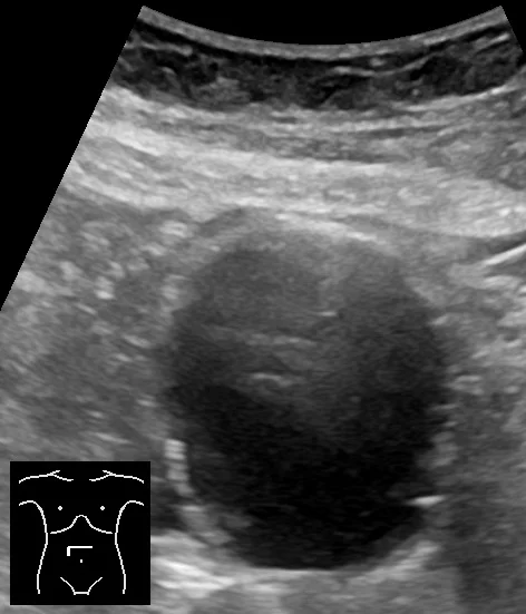
Figure 1Ultrasonography of abdominal aortic aneurysm in axial plane
Long description
Abdominal ultrasonography of a 78-year-old woman in the axial plane, showing an abdominal aortic aneurysm.

Ultrasound is the first test for a suspected AAA and the standard method for surveillance.[12] It is quick, portable, avoids radiation and has high accuracy: a systematic review found sensitivity of 94% to 100% and specificity of 98% to 100% for detecting AAA.[4] Obesity and bowel gas can limit the view, so a negative scan in a patient with a high clinical suspicion still needs review.
Two measurement conventions are in use. The ACC/AHA guideline measures outer-edge to outer-edge.[1] NICE and the NHS screening programme measure inner-to-inner.[2] The difference can be several millimetres, which matters near a repair threshold. Always record which method was used and compare like with like.
Exam trapA change in method can look like a change in size
If a patient's aneurysm is measured inner-to-inner at one visit and outer-to-outer at the next, the diameter may appear to jump without any real growth. Check the method before acting on a single measurement, especially when the result is close to a threshold.
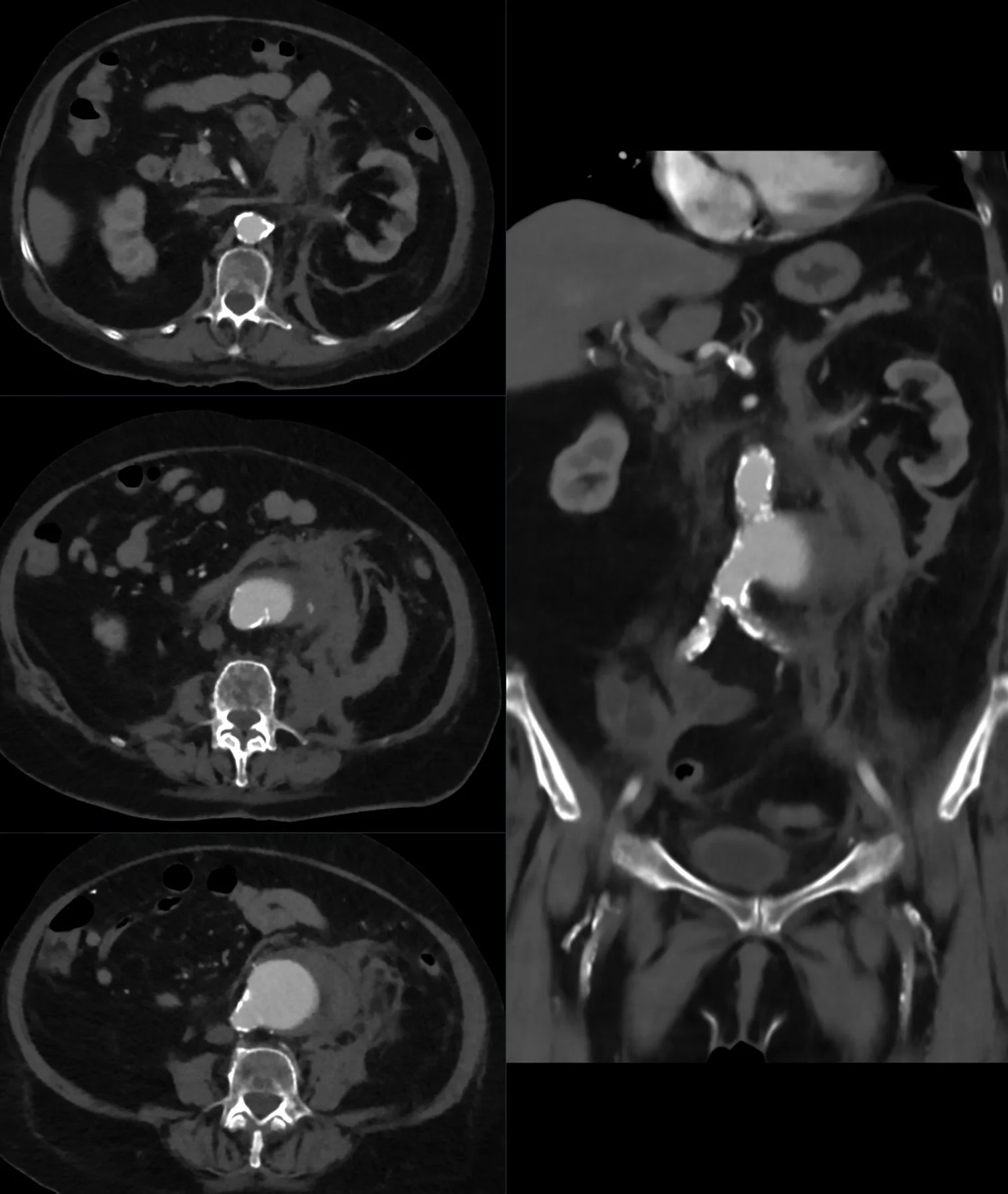
Figure 2Rupturierendes Bauchaortenaneurysma 86W - CT axial und koronar - 001
Long description
A ruptured abdominal aortic aneurysm shown on axial and coronal CT images.

CT angiography (CTA) is the best test for planning AAA repair.[12] It shows the extent of the aneurysm, the diameter at each level, the relation to branch vessels, the presence of thrombus and calcification, and the anatomy of the iliac access vessels. It is also the main test for assessing the thoracic aorta, and it detects rupture, dissection, penetrating ulcer and a false aneurysm.
CTA is the standard for planning elective repair, because it defines the anatomy, branch vessels and access route.[1] In an unstable patient, imaging must not delay transfer to the operating theatre. The 2022 ACC/AHA guideline recommends CTA in stable patients with a suspected ruptured AAA to see whether the anatomy is suitable for EVAR.[1]
MRI and magnetic resonance angiography avoid ionising radiation and are useful in younger patients, in genetic aortopathies and in repeated imaging. They are also an alternative when iodinated contrast is contraindicated. Gadolinium should be used cautiously in severe renal impairment. MRI is less practical in an unstable patient and is not the first test in a suspected rupture.
Transthoracic echocardiography (TTE) is well suited to the aortic root and ascending aorta. It measures the root, detects aortic regurgitation and can show a dissection flap or pericardial effusion. Transoesophageal echocardiography (TEE) gives better views of the ascending aorta and arch and is useful when TTE is inadequate or when a dissection is suspected. Echocardiography is also used during cardiac surgery to check the valve and the repair.
- ECG. Not diagnostic of an aneurysm, but essential in chest or abdominal pain to look for myocardial ischaemia and arrhythmia; see ECG interpretation.
- Chest radiograph. May show a widened mediastinum, an abnormal aortic contour or a pleural effusion, but it is insensitive and cannot exclude a thoracic aneurysm.
- Blood tests. Full blood count, renal function, electrolytes, coagulation, blood group and crossmatch, and troponin when cardiac ischaemia is possible. There is no blood test that diagnoses an aneurysm.
- Blood cultures. Take them before antibiotics if infectious aortitis is suspected.
- Genetic testing. Consider for young patients, familial disease, syndromic features or a bicuspid valve with aortic disease. Refer to clinical genetics.
The diagnosis rests on imaging plus the clinical context. There is no formal staging system for aneurysms as there is for cancer. Severity is described by diameter, rate of growth, symptoms, segment, cause and the presence of complications. For thoracoabdominal aneurysms, the Crawford extent is the standard anatomical description.[1]
The table below summarises the main surveillance and repair thresholds. It is deliberately explicit about which guideline says what, because the recommendations are not identical.
| Situation | ACC/AHA 2022 | NICE/NHS | Practical comment |
|---|---|---|---|
| AAA 3.0-3.9 cm | Ultrasound every 3 years[1] | NHS: annual scan for 3.0-4.4 cm[3] | The NHS band is wider and more frequent |
| AAA 4.0-4.9 cm in men | Annual ultrasound[1] | NHS: annual for 3.0-4.4 cm; every 3 months for 4.5-5.4 cm[3] | A 4.6 cm AAA is annual under ACC/AHA but 3-monthly under NHS |
| AAA 4.0-4.4 cm in women | Annual ultrasound[1] | NHS: annual for 3.0-4.4 cm[3] | Similar interval, but the UK programme does not usually scan women |
| AAA 5.0-5.4 cm in men | Ultrasound every 6 months[1] | NHS: every 3 months for 4.5-5.4 cm[3] | NICE is more frequent |
| AAA 4.5-4.9 cm in women | Ultrasound every 6 months[1] | NHS: every 3 months for 4.5-5.4 cm[3] | NICE is more frequent; the UK programme does not usually scan women |
| AAA repair threshold | Men at least 5.5 cm; women at least 5.0 cm[1] | Symptomatic; larger than 4.0 cm and grown by more than 1 cm in a year; or at least 5.5 cm[2] | The main international disagreement; see below |
| Ascending/root sporadic | Surgery at 5.5 cm; 5.0 cm reasonable at experienced centres[1] | Not covered | Individualise by size, growth, symptoms and body size |
| Descending thoracic | Repair at 5.5 cm; consider earlier with high-risk features[1] | Not covered | Open or endovascular depending on anatomy and fitness |
| Thoracoabdominal | Repair at 6.0 cm; 5.5 cm at experienced centres[1] | Not covered | Crawford extent predicts operative risk |
| Bicuspid aortic valve | 5.5 cm; 5.0-5.4 cm with an additional risk factor; area/height at least 10 cm2/m[1] | Not covered | Assess valve and root together |
| Marfan syndrome | 5.0 cm; 4.5 cm with high-risk features[1] | Not covered | Root replacement, often valve-sparing |
| Loeys-Dietz syndrome | Gene-specific: 4.0-5.0 cm (see text)[1] | Not covered | Earlier than Marfan in high-risk genotypes |
| Vascular Ehlers-Danlos | No diameter threshold; individualise[1] | Not covered | Rupture can occur without dilatation |
NoteThe guidelines disagree, and that is not a typo
The 2022 ACC/AHA guideline repairs AAAs at 5.5 cm in men and 5.0 cm in women.[1] NICE uses a 5.5 cm threshold for everyone, alongside its growth and symptom criteria, and measures inner-to-inner.[2] The 2024 ESVS guideline is more conservative still: a 2024 analysis reports that it issued a strong recommendation against elective repair below 5.5 cm and downgraded the 5.5 cm recommendation in men to Class IIa.[9][10] The difference reflects different evidence syntheses, different health systems and, in part, the absence of strong trial evidence for the 5.0 cm threshold in women.[6] Whichever protocol you follow, apply it consistently and document the measurement method.
Screening finds AAAs before they rupture. All the population-based randomised trials of AAA screening used a one-time scan, and the benefit depends on reliable surveillance and timely repair.[4] Screening is only useful if the health system can offer surveillance and timely repair to the people it finds.
Recommendations differ by country and by sex.
- UK (NHS). All men are invited in the year they turn 65 for a one-off ultrasound scan.[3] Men are invited because they are much more likely to have an AAA. The programme does not usually screen women or men under 65.[3]
- United States (USPSTF). One-time ultrasound screening is recommended for men aged 65 to 75 who have ever smoked (Grade B).[4] For men who have never smoked, selective screening is recommended rather than routine screening (Grade C).[4] Women who have never smoked and have no family history should not be screened (Grade D).[4] For women aged 65 to 75 who have ever smoked or have a family history, the evidence is insufficient to recommend for or against screening (I statement).[4]
- ACC/AHA. Ultrasound screening is recommended for men aged 65 or older who have ever smoked, and for men or women aged 65 or older with a first-degree relative with AAA.[1] Screening is reasonable for women aged 65 or older who have ever smoked.[1] For people under 65 with multiple risk factors or a family history, screening may be considered.[1] Repeat screening after a negative scan is not recommended for asymptomatic people over 75.[1]
United Kingdom (NICE)
In the UK, the NHS AAA screening programme invites men in the year they turn 65 and uses a one-off ultrasound scan. A small aneurysm (3.0-4.4 cm) is scanned annually, a medium one (4.5-5.4 cm) every 3 months, and a large one (5.5 cm or more) is referred to a vascular surgeon. Measurement is inner-to-inner.[2][3]
United States
In the United States, the USPSTF recommends one-time ultrasound screening for men aged 65 to 75 who have ever smoked, selective screening for men who have never smoked, and no screening for women who have never smoked and have no family history. The ACC/AHA guideline also recommends screening first-degree relatives of patients with AAA.[1][4]
There is no population screening programme for thoracic aortic aneurysm. Case finding is used instead. Up to 20% of people with a thoracic aneurysm or dissection have an affected first-degree relative, so take a family history and discuss relatives with the aortic team.[1] People with Marfan syndrome, Loeys-Dietz syndrome, vascular Ehlers-Danlos syndrome, Turner syndrome or a bicuspid aortic valve need surveillance according to their condition.
The same measures that reduce cardiovascular risk also reduce the risk of aneurysm growth and rupture:
- Do not start smoking; if you smoke, stop. Offer referral to a stop-smoking service.[2]
- Treat hypertension. The ACC/AHA target for patients with AAA is below 130/80 mmHg; for thoracic aortic aneurysm the target is 130/80 mmHg or lower.[1]
- Treat dyslipidaemia, especially in people with atherosclerosis.
- Maintain a healthy weight and exercise, although exercise should be individualised in people with large or symptomatic aneurysms.
- Avoid fluoroquinolones when another antibiotic will do, particularly in people with existing aortic disease.[5]
The management of an aortic aneurysm has three parts: reduce the risk of growth and rupture, decide when to repair, and choose the right repair for the patient. These decisions are shared with the patient and, for complex disease, with a multidisciplinary aortic team.
Blood pressure. In patients with AAA and an average systolic pressure of 130 mmHg or more, or diastolic pressure of 80 mmHg or more, antihypertensive treatment is recommended to reduce cardiovascular events.[1] The ACC/AHA guideline sets a target below 130/80 mmHg for AAA and 130/80 mmHg or lower for thoracic aortic aneurysm.[1] In thoracic aortic disease, beta blockers and angiotensin receptor blockers (ARBs) are reasonable to achieve the target.[1] In AAA, no specific antihypertensive drug has been shown to slow growth in a randomised trial.[1][6]
Statins. In patients with AAA and evidence of aortic atherosclerosis, moderate- or high-intensity statin therapy is recommended.[1] For thoracic aortic disease, the decision follows the patient's overall atherosclerotic risk, because statins treat the atherosclerosis rather than the aneurysm itself.
Smoking cessation. This is recommended for everyone with an aortic aneurysm who smokes.[1] It is probably the single most useful lifestyle intervention, because smoking affects development, growth, rupture and operative risk.
Antiplatelet therapy. Low-dose aspirin (75-162 mg/day) is reasonable in patients with atherosclerotic thoracic aortic aneurysm and concomitant atheroma or penetrating ulcer.[1] It is not routinely indicated for every AAA, and the decision should be individualised according to cardiovascular risk and bleeding risk.
What does not work. No drug has been shown convincingly to slow AAA growth in randomised controlled trials.[6] This is important counselling: surveillance is not a failure of treatment, and patients should not be given false reassurance that a statin or beta blocker will make repair unnecessary.
Exam trapDo not promise that a drug will shrink the aneurysm
Statins and antihypertensives reduce cardiovascular events, but they do not reliably shrink an aneurysm or remove the need for surveillance. The decision to repair remains based on size, growth, symptoms and the balance of operative risk.
Abdominal aortic aneurysm. The ACC/AHA guideline recommends repair for an asymptomatic AAA at a maximum diameter of 5.5 cm in men or 5.0 cm in women.[1] Repair is also recommended for symptoms attributable to the aneurysm, and may be reasonable for a saccular aneurysm or for growth of 0.5 cm or more in 6 months.[1] NICE considers repair for symptoms, for an asymptomatic AAA larger than 4.0 cm that has grown by more than 1 cm in a year, or for an asymptomatic AAA of 5.5 cm or more.[2]
Thoracic aortic aneurysm. The threshold depends on the segment and the cause:
- Root and ascending aorta, sporadic: surgery at 5.5 cm; 5.0 cm is reasonable at experienced centres.[1] Operate for symptoms or rapid growth (0.3 cm per year over 2 consecutive years, or 0.5 cm in 1 year).[1]
- Bicuspid aortic valve: 5.5 cm; 5.0 to 5.4 cm with an additional risk factor; or an aortic root/ascending area-to-height ratio of 10 cm2/m or more.[1]
- Marfan syndrome: 5.0 cm; 4.5 cm with high-risk features such as a family history of dissection or rapid growth.[1]
- Loeys-Dietz syndrome: gene-specific thresholds. For TGFBR1 or TGFBR2 without high-risk features, 4.5 cm; with high-risk features, 4.0 cm. For SMAD3 or TGFB2, 4.5 cm; for TGFB3, 5.0 cm.[1] Arch, descending or abdominal disease in TGFBR1, TGFBR2 or SMAD3 may be considered for repair at 4.5 cm.[1]
- Vascular Ehlers-Danlos syndrome: no diameter threshold can be recommended; decisions are individualised because the aorta can rupture without dilatation.[1]
- Descending thoracic aorta: 5.5 cm, with earlier consideration for rapid growth or symptoms.[1]
- Thoracoabdominal aorta: 6.0 cm, or 5.5 cm at experienced centres.[1]
Symptoms matter more than size. A symptomatic aneurysm is repaired urgently, whatever its diameter, because pain or compression suggests impending rupture. The same applies to a documented rapid growth rate.
Before elective repair, the patient needs a clear anatomical plan and an honest assessment of operative risk.
- Imaging. CTA of the whole aorta and iliac arteries is the standard.[1] It defines the proximal and distal landing zones, the branch vessels, the access vessels and the presence of thrombus or calcification.
- Cardiac and pulmonary assessment. Look for coronary disease, heart failure, valvular disease and chronic lung disease. The result informs the choice of open or endovascular repair and the discussion of operative risk.
- Renal function. Contrast and cross-clamping both threaten the kidneys. Optimise hydration and avoid nephrotoxins.
- Frailty and goals of care. A repair that is technically possible may not be in the patient's interest if life expectancy is short or the recovery burden is unacceptable.
- Medication review. NICE advises against routinely offering preoperative beta blockers solely for AAA repair.[2] Continue beta blockers for another indication, such as atrial fibrillation.
- Antibiotic prophylaxis. Patients with an aortic prosthesis should receive prophylaxis before dental procedures that manipulate the gingiva or periapical tissues, according to the SVS guideline.[11]
Open repair replaces the diseased segment with a synthetic graft through a laparotomy (for AAA) or thoracotomy (for thoracic disease). It is durable, does not usually require lifelong aortic imaging, and is often preferred in younger, fitter patients. The cost is a major operation with a period of aortic clamping, a systemic inflammatory response, and risks of bleeding, renal injury, myocardial infarction and bowel ischaemia.
Endovascular aneurysm repair (EVAR) places a stent graft inside the aorta through the femoral arteries. It avoids a large abdominal incision, reduces early morbidity and has a lower 30-day mortality. In a pooled analysis of four randomised trials, 30-day mortality was lower with EVAR (odds ratio 0.40, 95% CI 0.22 to 0.74).[8] By 5 years, survival was similar in both groups at 73.6%.[8] Beyond 3 years, aneurysm-related mortality was higher in the EVAR group (hazard ratio 5.16), and secondary rupture occurred in 2.4% of EVAR patients versus 0.3% of open-repair patients, with a median time to rupture of 3.5 years.[8]
EVAR depends on anatomy: an adequate proximal neck, suitable iliac access and no major branch vessel that cannot be covered. Fenestrated and branched endografts extend EVAR to juxtarenal and thoracoabdominal aneurysms, but they are complex, require specialised centres and need lifelong surveillance.
| Feature | Open repair | EVAR |
|---|---|---|
| Approach | Laparotomy, aortic clamp, synthetic graft | Femoral artery access, stent graft |
| Anaesthesia | General or regional | General, regional or local |
| Early mortality | Higher than EVAR in pooled trials[8] | Lower 30-day mortality (odds ratio 0.40)[8] |
| Late durability | Durable; fewer late aneurysm-related deaths[8] | Higher late aneurysm-related mortality and reintervention[8] |
| Surveillance | Usually limited | Lifelong imaging for endoleak and sac growth[1] |
| Anatomy | Less dependent on neck and iliac anatomy | Requires suitable landing zones and access |
| Best fit | Younger, fitter patients; unsuitable EVAR anatomy | Older or higher-risk patients; suitable anatomy; ruptured AAA |
The guidelines differ on the default operation. NICE offers open repair first-line for most people with an unruptured AAA, and considers EVAR when open repair is contraindicated by abdominal copathology such as a hostile abdomen, horseshoe kidney or stoma, or by anaesthetic and medical risk.[2] The ACC/AHA guideline weighs EVAR's lower perioperative mortality against its higher late aneurysm-related mortality, reintervention and secondary rupture, and leaves the choice to anatomy, fitness and patient preference.[1] Both agree that the patient's anatomy, fitness and preferences must decide the individual case.
NoteEarly safety versus late durability
EVAR wins at 30 days; open repair catches up over years. The trade-off is not just mortality: EVAR commits the patient to lifelong imaging and a small but real risk of late rupture, while open repair commits them to a larger operation now. That is why the "best" operation depends on the patient, not only on the aneurysm.
A ruptured AAA is a surgical emergency. The patient may be pale, sweaty, confused or hypotensive, and the blood pressure can be deceptively normal at first. The priorities are to recognise the problem, call for help, resuscitate while arranging definitive control, and avoid unnecessary delay.
- Call the vascular team immediately. Do not wait for confirmatory imaging if the patient is unstable.
- Resuscitate. Secure large-bore intravenous access, take blood for crossmatch, full blood count, renal function and coagulation, and activate the massive transfusion protocol if needed. Use a restrictive approach to fluids (permissive hypotension) while awaiting haemorrhage control.[1][2] The aim is to avoid dislodging clot with a high blood pressure; follow the local protocol.
- Image if stable. Bedside ultrasound can confirm an AAA. In a stable patient, CTA can show whether the anatomy is suitable for EVAR.[1] In an unstable patient, transfer to the operating theatre or angiography suite takes priority.
- Repair. EVAR is preferred when the anatomy is suitable, because it reduces morbidity and mortality compared with open repair.[1] Local anaesthesia is preferred for EVAR in this setting.[1] Open repair is used when EVAR is unsuitable or unavailable. An aortic occlusion balloon can be used to gain proximal control in selected patients.[10]
- Treat complications. Abdominal compartment syndrome and colonic ischaemia are recognised after ruptured AAA repair and require vigilant monitoring.[10]
NICE adds an age and sex nuance: EVAR provides more benefit than open repair for most people, especially men over 70 and women of any age, while open repair may offer a better balance in men under 70.[2] The SVS recommends a door-to-intervention time of less than 90 minutes for ruptured AAA.[11]
Suspected ruptured abdominal aortic aneurysm
- Suspected ruptureNew abdominal or back pain, collapse, hypotension or a known AAA
- Question: Is the patient unstable or arresting?
Yes
- Resuscitate and call the vascular teamLarge-bore access, crossmatch, restrictive fluids, massive transfusion if needed
- Move to immediate haemorrhage controlBedside ultrasound if it does not delay; theatre or angiography suite; EVAR if anatomy suitable
No
- Urgent CTAConfirm rupture, assess anatomy for EVAR, plan access
- RepairEVAR preferred if suitable; open repair if not
After open repair, the graft is durable, but patients still need cardiovascular risk management and clinical review. After EVAR, lifelong surveillance is required because the endograft can leak, migrate, fracture or separate, and the aneurysm sac can continue to expand.[1] NICE recommends enrolling every patient who has had EVAR in a surveillance programme and basing the frequency on the risk of complications.[2]
An endoleak is blood flow outside the stent graft but within the aneurysm sac. There are five types:[1]
- Type I: leak at the proximal (Ia) or distal (Ib) attachment site.
- Type II: backflow into the sac from a branch vessel.
- Type III: a defect in the graft fabric or a misalignment between components.
- Type IV: leakage through the graft wall due to porosity.
- Type V: endotension, with sac expansion but no visible leak.
Type I and type III endoleaks are usually treated because they expose the sac to systemic pressure. Type II endoleaks are treated if the sac is expanding; if the sac is stable, they can be observed.[2][11]
Clinical pearlA growing sac after EVAR is an endoleak until proved otherwise
A growing sac suggests an endoleak or graft failure. Any sac expansion or new endoleak deserves CT angiography, or contrast-enhanced ultrasound when CT is contraindicated. Do not exclude an endoleak on colour duplex alone.[2]
- AAA of 5.5 cm or more: refer to a regional vascular service to be seen within 2 weeks.[2]
- AAA of 3.0 to 5.4 cm: refer to a regional vascular service to be seen within 12 weeks.[2]
- Symptomatic or suspected ruptured AAA: immediate vascular discussion and emergency transfer.[2]
- Thoracic or thoracoabdominal aneurysm: refer to a multidisciplinary aortic team, especially if the diameter is near a threshold, growth is rapid, symptoms are present, or the patient has a genetic syndrome.
- Genetic aortopathy or family history: refer to cardiology, clinical genetics and an aortic team before pregnancy or before elective surgery.
- Rupture. The most important complication. It may be contained (retroperitoneal) or free (intraperitoneal), and it can be fatal within minutes.
- Dissection. A dilated aorta is more likely to tear. Dissection can cause malperfusion of the brain, limbs, gut or kidneys.
- Thromboembolism. Mural thrombus can embolise to the legs, gut, kidneys or brain.
- Compression. A thoracic aneurysm can compress the trachea, oesophagus, recurrent laryngeal nerve or superior vena cava. An abdominal aneurysm can compress the ureter or cause abdominal pain.
- Fistula. An aortoenteric fistula can cause gastrointestinal bleeding; an aortocaval fistula can cause high-output heart failure.
- Infection. Infectious aortitis can destroy the wall and cause a false aneurysm.
- Bleeding and transfusion. Open repair has larger blood loss; EVAR can cause access-site bleeding or retroperitoneal haemorrhage.
- Myocardial infarction, stroke and arrhythmia. Patients usually have widespread atherosclerosis; see atrial fibrillation and flutter for postoperative rhythm problems.
- Acute kidney injury. From contrast, hypotension, emboli or aortic clamping.
- Spinal cord ischaemia. A risk of extensive thoracic and thoracoabdominal repair, because the artery of Adamkiewicz and other radicular arteries may be sacrificed.
- Colonic ischaemia. Particularly after ruptured AAA or ligation of the inferior mesenteric artery; suspect it when there is unexplained acidosis, diarrhoea or abdominal distension.
- Abdominal compartment syndrome. Raised intra-abdominal pressure after massive resuscitation or retroperitoneal bleeding; it impairs ventilation and renal perfusion.
- Limb ischaemia. From emboli, thrombosis or access-site injury.
- Graft infection. Presents with fever, pain, a false aneurysm or sepsis; it usually requires long antibiotics and surgical removal or reconstruction.
- Endoleak and secondary rupture. See above. Lifelong surveillance is the safety net.
Ruptured AAA is one of the most lethal emergencies in medicine. Overall mortality is estimated at 80% to 90%, with most patients dying before reaching hospital.[1] For those who reach hospital, historical mortality after open repair was about 50%, but an endovascular-first strategy has reduced mortality to as low as 18.5% in at least one observational series.[1] These figures vary with patient selection, centre volume and time to treatment.
Elective repair is much safer than emergency repair, which is the central argument for screening and surveillance. In pooled randomised trials, 5-year survival after elective EVAR or open repair was about 73.6%, but EVAR carried a higher late risk of aneurysm-related death and secondary rupture.[8]
The natural history of an unruptured AAA depends on size and sex. The risk of rupture of a small AAA in men is less than 1% per year.[6] For larger unrepaired aneurysms, the 3-year rupture risk is about 2.2% at 55-60 mm, 6.0% at 61-70 mm and 18.4% above 70 mm.[6] Women have a higher risk at the same size: in one study, women with an unrepaired 61-70 mm AAA had a 3-year rupture incidence of 12.8%, compared with 4.5% in men.[6] Female sex was associated with a hazard ratio of 3.76 for rupture in a large individual-patient analysis.[6]
The median size at rupture in a 2024 cohort was 8.0 cm in men and 7.6 cm in women, which is a reminder that many ruptures occur above the repair threshold, but not all do.[9] Some patients present with a rupture below 5.5 cm, especially women, and some aneurysms rupture at a size that was never measured. No threshold is perfectly safe.
Age alone should not decide treatment. Assess frailty, comorbidity, cognitive function, life expectancy and the patient's goals. EVAR is less physiologically stressful in the short term, but it requires lifelong surveillance and carries a late risk of reintervention. Open repair may be more durable but has a longer recovery. A multidisciplinary team and a frank discussion of what matters to the patient are essential.
Pregnancy increases the risk of aortic dissection in women with Marfan syndrome, Loeys-Dietz syndrome or vascular Ehlers-Danlos syndrome. In Marfan syndrome, the risk of type A dissection rises with aortic root diameter, and pregnancy adds further risk. Women with a genetic aortopathy should have pre-pregnancy counselling, aortic imaging and a plan for delivery in a centre with cardiac surgery. Vascular Ehlers-Danlos syndrome carries a particularly high risk of arterial rupture during pregnancy, and management should involve a specialist team.
Aortic aneurysm is rare in children. When it occurs, look for a congenital or genetic cause: bicuspid aortic valve, Turner syndrome, Marfan syndrome, Loeys-Dietz syndrome, vascular Ehlers-Danlos syndrome, Kawasaki disease or previous cardiac surgery. Imaging and surveillance should be guided by a paediatric cardiology or genetics team.
Infectious aortitis is more likely in people with diabetes, immunosuppression, intravenous drug use, endocarditis or a prosthetic graft. Blood cultures, imaging and prolonged antibiotics are needed; intravenous antimicrobial therapy for at least 6 weeks may be considered, with lifelong suppression in selected cases.[1] Open surgical debridement and reconstruction are often required, although endovascular repair may be used as a bridge in selected patients.[1]
East Africa
Ultrasound is the most practical tool for finding and following an AAA. It is portable, avoids radiation and is far cheaper than CT. Measure the maximum diameter consistently—inner-to-inner or outer-to-outer—and record the method. If CT or vascular surgery is not available, discuss the patient with a regional vascular service early and transfer before rupture, not after it. For a suspected rupture, do not delay transfer for imaging if the patient is unstable. Open repair may be the only option; concentrate complex cases in centres that perform them regularly.[1][2][11]
In low-resource settings, the biggest gains come from prevention and early detection: smoking cessation, blood pressure control, and a one-off ultrasound for high-risk men aged 65 or older. A simple surveillance programme with reliable follow-up can prevent ruptures even when advanced endovascular devices are unavailable. For thoracic aneurysms, a chest radiograph may raise suspicion but cannot exclude disease; CT or MRI is needed, and transfer should be arranged if the patient is symptomatic or the aneurysm is large.
- An aortic aneurysm is a permanent focal dilatation; an AAA is defined as a diameter greater than 3.0 cm, but thoracic thresholds depend on the segment and the cause.
- True aneurysms involve all three wall layers; false aneurysms are contained breaches and can rupture at a smaller size.
- AAA is common in older men who smoke. Smoking, age, male sex, hypertension and family history are the main risk factors; diabetes is inversely associated.
- Marfan syndrome, Loeys-Dietz syndrome, vascular Ehlers-Danlos syndrome, bicuspid aortic valve and Turner syndrome are important inherited causes, especially in younger patients with root or ascending disease.
- Medial degeneration, elastin loss, inflammation and wall stress drive enlargement. Laplace's law explains why larger aneurysms and higher blood pressure increase rupture risk.
- Most aneurysms are asymptomatic. A pulsatile mass is an unreliable sign; physical examination has limited sensitivity. New abdominal or back pain in an older smoker is a red flag.
- Ultrasound is the first test for AAA and the standard for surveillance. CTA is used for planning and for stable suspected rupture; MRI/MRA is useful in younger patients and genetic aortopathy.
- Measure consistently: ACC/AHA uses outer-to-outer, while NICE and the NHS use inner-to-inner. The method can change the apparent size.
- Surveillance intervals and repair thresholds differ between guidelines. ACC/AHA repairs AAAs at 5.5 cm in men and 5.0 cm in women; NICE uses 5.5 cm for everyone but also repairs symptomatic or rapidly growing aneurysms. Document which protocol you are using.
- Thoracic repair thresholds are cause-specific: 5.5 cm for sporadic ascending/root disease (5.0 cm at experienced centres), 5.0 cm in Marfan, gene-specific thresholds in Loeys-Dietz, and no diameter threshold in vascular Ehlers-Danlos.
- EVAR has lower early mortality but higher late aneurysm-related mortality, reintervention and secondary rupture than open repair; it requires lifelong surveillance.
- A ruptured AAA needs immediate vascular involvement, restrictive fluid resuscitation and prompt repair. EVAR is preferred when anatomy is suitable; do not let imaging delay control in an unstable patient.
- No drug has been shown convincingly to slow AAA growth. Smoking cessation, blood pressure control and statins where atherosclerosis is present reduce cardiovascular risk and are part of every management plan.
Answer each one in your head first, then open the answer.
A 68-year-old man who smokes has an asymptomatic 5.6 cm infrarenal AAA found on ultrasound. He is active and has no major comorbidity. What are the next steps, and why is the threshold controversial?
Show answerHide answer
He should be referred to a vascular service for repair assessment, because the ACC/AHA guideline recommends repair at 5.5 cm or more in men. He also needs blood pressure control, a statin if he has atherosclerosis, and smoking cessation. The threshold is controversial because NICE uses 5.5 cm for everyone with growth and symptom criteria, and the ESVS 2024 guideline is more conservative, while the ACC/AHA guideline uses 5.0 cm in women. The decision should be shared and based on anatomy, fitness and the patient's values.
A 72-year-old woman has a 4.8 cm AAA. She is asymptomatic and growing at 0.2 cm per year. How would the ACC/AHA and NICE approaches differ?
Show answerHide answer
Under the ACC/AHA guideline, 4.8 cm is below the 5.0 cm repair threshold for women but above the 4.5 cm surveillance threshold, so she needs ultrasound every 6 months. Under the NHS programme's bands, a 4.8 cm AAA would be scanned every 3 months, although the programme does not usually scan women; NICE would consider repair at 5.5 cm unless she becomes symptomatic or grows by more than 1 cm in a year. The example shows why the measuring method and the chosen protocol must be documented.
A 76-year-old man with a known 6.0 cm AAA presents with sudden severe back pain and a blood pressure of 84/50 mmHg. What should happen first?
Show answerHide answer
Treat him as a ruptured AAA. Call the vascular team immediately, secure large-bore access, crossmatch blood, and resuscitate with a restrictive fluid strategy while arranging immediate haemorrhage control. Bedside ultrasound can confirm the aneurysm but must not delay transfer. If he is stable enough for CT, CTA can show whether EVAR is suitable; if he is unstable, he should go straight to the operating theatre or angiography suite. EVAR is preferred if the anatomy is suitable.
A 32-year-old man with Marfan syndrome has an aortic root diameter of 4.6 cm and a family history of aortic dissection at 4.8 cm. What is the role of surgery?
Show answerHide answer
The ACC/AHA guideline recommends surgery at a root diameter of 5.0 cm in Marfan syndrome, and considers surgery reasonable at 4.5 cm when high-risk features are present. A family history of dissection at a relatively small diameter is such a feature, so he should be assessed by an aortic team for root replacement, with a valve-sparing operation if the valve is suitable. He also needs beta blocker or ARB therapy and regular imaging.
A 64-year-old man has fever, back pain and a rapidly enlarging 6 cm AAA. Blood cultures grow Salmonella. What is the diagnosis and the principles of treatment?
Show answerHide answer
Infectious (mycotic) aortitis with a false aneurysm. He needs prolonged intravenous antibiotics, usually for at least 6 weeks, and prompt surgical debridement and reconstruction; endovascular repair may be used as a bridge in selected patients but is not a substitute for source control. Blood cultures and imaging should guide the team, and the aorta can rupture at any time.
Two years after EVAR, a patient has a 7 mm increase in sac diameter. What is the concern and how should it be investigated?
Show answerHide answer
A growing sac suggests an endoleak or graft failure. He needs CT angiography, or contrast-enhanced ultrasound if CT is contraindicated, and review by the vascular team. Type I and type III endoleaks usually need treatment; type II endoleak needs treatment if the sac is expanding. A negative colour duplex alone does not exclude an endoleak.
A 66-year-old man has never smoked and has no family history of AAA. Should he be screened?
Show answerHide answer
It depends on the programme. In the UK, all men are invited in the year they turn 65, regardless of smoking history. In the United States, the USPSTF recommends selective screening for men aged 65 to 75 who have never smoked, rather than routine screening. The ACC/AHA guideline recommends screening first-degree relatives of patients with AAA, but he has no such history. The decision should be individualised.
- Cardiac physical examination — how to examine the cardiovascular system and recognise a pulsatile mass.
- Heart failure — aortic regurgitation and the consequences of a root aneurysm.
- ECG interpretation — distinguishing cardiac chest pain from aortic disease.
- Atrial fibrillation and flutter — a common comorbidity and postoperative arrhythmia.
- Isselbacher EM, Preventza O, Hamilton Black J 3rd, et al. 2022 ACC/AHA Guideline for the Diagnosis and Management of Aortic Disease. Circulation. 2022;146(24):e334-e482. doi:10.1161/CIR.0000000000001106. Accessed .
- National Institute for Health and Care Excellence. Abdominal aortic aneurysm: diagnosis and management (NICE guideline NG156). London: NICE; 2020. Accessed .
- NHS. Abdominal aortic aneurysm (AAA) screening. NHS website; reviewed 2024. Accessed .
- US Preventive Services Task Force; Owens DK, Davidson KW, Krist AH, et al. Screening for Abdominal Aortic Aneurysm: US Preventive Services Task Force Recommendation Statement. JAMA. 2019;322(22):2211-2218. doi:10.1001/jama.2019.18928. Accessed .
- Medicines and Healthcare products Regulatory Agency. Systemic and inhaled fluoroquinolones: small increased risk of aortic aneurysm and dissection. Drug Safety Update. 2018. Accessed .
- Golledge J, Thanigaimani S, Powell JT, Tsao PS. Pathogenesis and management of abdominal aortic aneurysm. European Heart Journal. 2023;44(29):2682-2697. doi:10.1093/eurheartj/ehad386. Accessed .
- Song P, He Y, Adeloye D, et al. The Global and Regional Prevalence of Abdominal Aortic Aneurysms: A Systematic Review and Modeling Analysis. Annals of Surgery. 2023;277(6):912-919. doi:10.1097/SLA.0000000000005716. Accessed .
- Powell JT, Sweeting MJ, Ulug P, et al. Meta-analysis of individual-patient data from EVAR-1, DREAM, OVER and ACE trials comparing outcomes of endovascular or open repair for abdominal aortic aneurysm over 5 years. British Journal of Surgery. 2017;104(3):166-178. doi:10.1002/bjs.10430. Accessed .
- Hassan S, Frost T, Bourchier R. Ruptured AAA: bridging the gap between international guidelines and local clinical realities. Langenbeck's Archives of Surgery. 2024;409(1):256. doi:10.1007/s00423-024-03441-6. Accessed .
- Wanhainen A, Van Herzeele I, Bastos Goncalves F, et al. European Society for Vascular Surgery (ESVS) 2024 Clinical Practice Guidelines on the Management of Abdominal Aorto-Iliac Artery Aneurysms. European Journal of Vascular and Endovascular Surgery. 2024;67(2):192-331. doi:10.1016/j.ejvs.2023.11.002. Accessed .
- Chaikof EL, Dalman RL, Eskandari MK, et al. The Society for Vascular Surgery practice guidelines on the care of patients with an abdominal aortic aneurysm. Journal of Vascular Surgery. 2018;67(1):2-77.e2. doi:10.1016/j.jvs.2017.10.044. Accessed .
- Desjardins B, Dill KE, Flamm SD, et al. ACR Appropriateness Criteria pulsatile abdominal mass, suspected abdominal aortic aneurysm. International Journal of Cardiovascular Imaging. 2013;29(1):177-183. doi:10.1007/s10554-012-0044-2. Accessed .
Figure credits
Educational content for medical students and doctors. It is not a substitute for clinical judgement or local guidelines.