Acanthosis nigricans and lichen planus
Two dermatology topics: velvety flexural hyperpigmentation linked to insulin resistance, and lichen planus with its six Ps, Wickham striae and oral forms.
- Acanthosis nigricans is a velvety, darkening of the skin folds. It is not a disease on its own; it is a sign that points to a cause, most often insulin resistance.
- Lichen planus is an inflammatory skin (and mouth) condition: intensely itchy, flat-topped, purple papules and plaques with a fine white lacy pattern on the surface called Wickham striae.
- The six Ps of lichen planus are planar, purple, polygonal, pruritic, papules and plaques.
- Oral lichen planus is common, persistent and needs long-term follow-up because the erosive form carries a small risk of progression to squamous cell carcinoma.
- Treatment is cause-directed: weight loss and metabolic control for acanthosis nigricans; anti-inflammatory treatment, usually topical corticosteroids first, for lichen planus.
By the end of this page you should be able to:
- Explain why acanthosis nigricans is a marker of insulin resistance and name the malignancies that malignant acanthosis nigricans suggests.
- Describe the six Ps of lichen planus and recognise Wickham striae on the skin and in the mouth.
- Distinguish the main variants of oral lichen planus and explain how each is assessed.
- Plan investigations for a patient with acanthosis nigricans, including metabolic testing, and for a patient with lichen planus, including hepatitis C testing.
- Choose stepwise management for both conditions, including topical steroids and systemic options, and name their cautions.
- Separate lichen planus, lichenoid drug reactions and other look-alikes in a table.
Acanthosis nigricans is a cutaneous sign: symmetric, hyperpigmented, velvety thickening of the skin, usually in body folds such as the neck, axillae, groin and knuckles. It is not one diagnosis; it is the skin's response to growth factors, especially high insulin. Insulin at high concentration acts through insulin-like growth factor-1 (IGF-1) receptors on keratinocytes and fibroblasts, driving proliferation.
Classification is useful because management changes with the cause:
- Benign/obesity-related (type II). The common form, linked to insulin resistance, obesity and type 2 diabetes.
- Hereditary (type I). Appears in childhood; often benign but persistent.
- Syndromic. Associated with severe insulin resistance syndromes such as HAIR-AN (hyperandrogenism, insulin resistance, acanthosis nigricans) and polycystic ovary syndrome.
- Drug-induced. Reported with insulin, nicotinic acid, corticosteroids and oral contraceptives.
- Malignant (MAN). Sudden, extensive, often with mucosal involvement; a paraneoplastic sign that demands a malignancy search.
Lichen planus is a chronic, immune-mediated inflammatory disorder of skin and mucous membranes. CD8+ T cells attack keratinocytes at the dermo-epidermal junction, producing basal cell damage and a tight band of inflammation. The classic skin lesion is a flat-topped, violaceous, polygonal, pruritic papule or plaque. On the surface, fine grey-white lines (Wickham striae) appear after gentle rubbing or under magnification. Oral lesions are often reticular white streaks on the buccal mucosa.
Variants of oral lichen planus are clinically important: reticular (most common), erosive, atrophic, plaque-like, papular and bullous. Cutaneous variants include hypertrophic, annular, actinic, pigmentosus, follicular (lichen planopilaris on the scalp) and mucocutaneous forms.
AN is commonest in people with insulin resistance and in darker skin phototypes, who may show it earlier and more clearly. It is often the first visible clue to obesity, metabolic syndrome and type 2 diabetes in adolescents and adults.
Risk factors: raised body mass, central adiposity, family history of type 2 diabetes, polycystic ovary syndrome, medications listed above, and the inherited insulin resistance syndromes.
Oral lichen planus affects about 1 to 2 percent of the population in all racial groups (population-based meta-analyses estimate roughly 0.9 to 1.0 percent); lichen planus overall is less common and reported overall prevalence varies widely (a single review lists about 0.1 to 2.2 percent for all forms). It typically begins in middle age, more often in women, with a female-to-male ratio of about 1.4 to 1 (reported in the range of about 1.4:1 to 2:1 across sources and phenotypes). [6]
Risk factors and associations: hepatitis C virus infection, certain drugs (thiazides, beta blockers, ACE inhibitors, NSAIDs, gold and antimalarials, among others through lichenoid reactions), dental metal allergy and contact sensitivity, stress and anxiety, thyroid disease, and graft-versus-host disease. The hepatitis C association is one of the most consistently reported systemic links; local practice should guide how actively to screen. [6]
The lichen planus references in this topic reflect the StatPearls chapter on lichen planus and oral lichen planus, the BAD and AAD guidance routes, and the NICE CKS lichen planus topic. [2][3][10][11]
High insulin is the usual driver. In insulin resistance, pancreatic insulin output rises; insulin at these concentrations can activate IGF-1 receptors on skin cells, stimulating keratinocyte and fibroblast proliferation and producing the velvety plaques. Treating the insulin resistance therefore clears the rash better than skin-directed treatment.
LP is a T-cell-mediated attack on basal keratinocytes. An as-yet-unidentified antigen is presented to CD8+ cytotoxic T cells, which release cytokines and trigger keratinocyte apoptosis. The clinical signs follow from this: the flat-topped papule from compact hyperkeratosis, the colour from pigment incontinence, and the Wickham striae from alternating hypergranulosis and acanthosis.
Figure 1Sequeira Plate 44
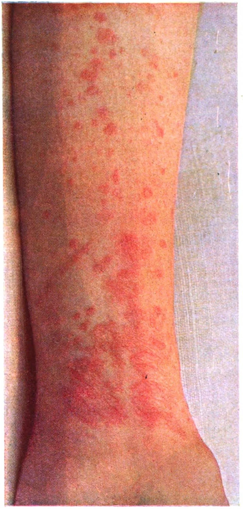
Long description
Plate 44, lichen planus: eruption of lilac-coloured, flat-topped papules on the front of the forearm, with silvery Wickham striae on close examination.

Figure 2Acanthosis nigricans, axillär, ©WIKIDERM
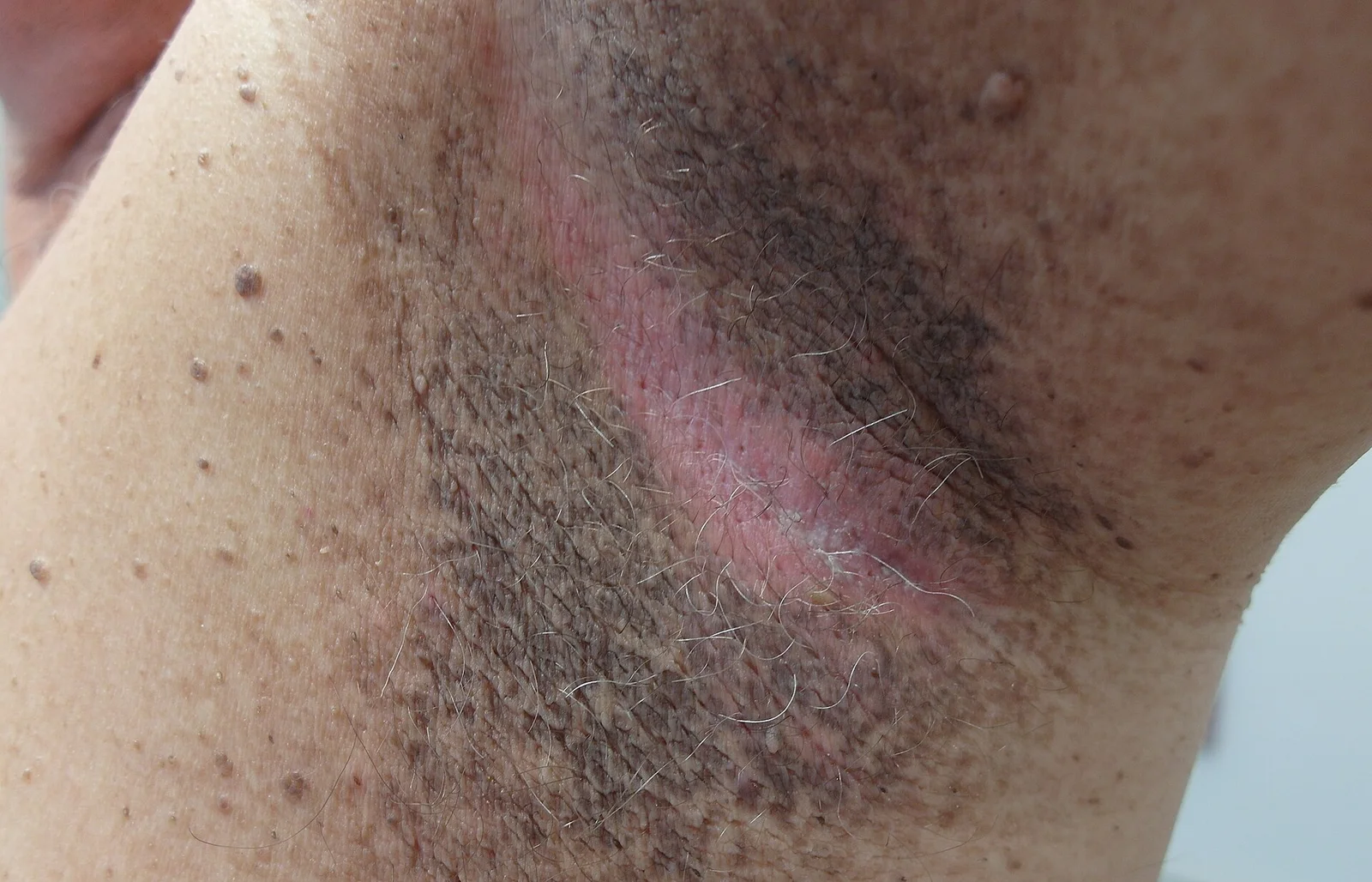
Long description
Clinical photograph labelled 'Acanthosis nigricans, axillar' (German for axillary), showing acanthosis nigricans at the axilla (photograph taken 16 May 2013).

Look at the flexures: posterior neck most often, then axillae, groin, antecubital and popliteal fossae, knuckles, elbows. The skin is dry, brown to grey-black, with accentuated skin lines. Generalised or acral variants and mucosal papillomatosis raise the question of malignancy.
Skin: pruritic, polygonal, purple, flat-topped papules and plaques. A white network (Wickham striae) is seen on close inspection. Sites: flexor forearms and wrists, shins, sacrum and trunk. New lesions often appear at sites of minor trauma (Koebner phenomenon).
Other sites: oral mucosa (buccal mucosa, tongue, gingivae), genitalia (vulvo-vaginal-gingival syndrome when vulva, vagina and gums are involved), nails (longitudinal ridging, splitting, pterygium in severe cases) and scalp (lichen planopilaris, which can scar and cause permanent hair loss).
Oral forms: reticular (white lacy streaks, often asymptomatic), erosive (red, painful erosions with white borders), atrophic, plaque-like, papular and bullous. The erosive form causes burning pain, worse with spicy or hot food.
For acanthosis nigricans the diagnosis is clinical, but the work-up is metabolic. The table lists useful tests and how to interpret them. There is no single cut-off that applies to every laboratory; fasting insulin and HOMA-IR are interpreted against local reference ranges.
| Test | When it is used | What the result means |
|---|---|---|
| Body mass index and waist circumference | Every patient | Measures excess adiposity, the commonest driver of insulin resistance. |
| Fasting plasma glucose | Baseline metabolic screen | High values point to impaired fasting glucose or diabetes. |
| HbA1c | Baseline metabolic screen | Reflects recent average glucose; raised in prediabetes and diabetes. |
| Oral glucose tolerance test | When glucose/HbA1c are borderline | Detects impaired glucose tolerance and early diabetes. |
| Fasting insulin and HOMA-IR | To quantify insulin resistance | Raised values support insulin resistance. Calculated as insulin x glucose / 22.5 in SI units; interpret against the local reference range. |
| Lipid profile and blood pressure | Metabolic syndrome screen | Dyslipidaemia and hypertension complete the metabolic risk picture. |
| Testosterone, LH, FSH, and pelvic ultrasound | In women with irregular periods or hirsutism | Supports polycystic ovary syndrome as the driver. |
| Endoscopy and age-appropriate cancer screening | Sudden adult-onset, extensive or mucosal AN | Looks for an occult gastric or other adenocarcinoma. |
For lichen planus the diagnosis is usually clinical. A biopsy (histopathology with direct immunofluorescence) is taken when the lesions are atypical, unilateral, or when malignancy or a lichenoid reaction is suspected. Oral lesions follow the modified WHO (2003) clinical and histopathological criteria, including bilateral, more or less symmetrical lesions with a reticular white pattern, and absence of epithelial dysplasia histologically. [6]
Investigations commonly include: full blood count, erythrocyte sedimentation rate, hepatitis B and C serology, liver function tests, thyroid function tests (when clinically indicated) and, for oral disease, candidal culture when steroids are used. [6]
| Condition | Feature that separates it |
|---|---|
| Intertrigo, tinea or candida | Erythema, satellite pustules, scaling; hypopigmented or not symmetrical; KOH positive. |
| Seborrhoeic dermatitis | Greasy yellow scale on scalp, brows and nasolabial folds; no velvety plate. |
| Lichenoid drug reaction | History of a drug started weeks to months earlier; unilateral or asymmetric distribution; mixed eosinophilic/plasma cell infiltrate on biopsy. |
| Lichen planus (female variant: pityriasis lichenoides) | Scaly papules that resolve leaving hypopigmented macules; no Koebner and no Wickham striae. |
| Psoriasis | Well-demarcated erythematous plaques with silvery scale on extensor surfaces; nail pitting. |
| Discoid lupus erythematosus | Scaly plaques with follicular plugging, atrophic scarring and pigment change in sun-exposed skin. |
| Oral candidiasis | White plaques that wipe off; KOH or culture positive; often after antibiotics or inhaled steroids. |
| Oral leukoplakia | White plaque that cannot be scraped off; may arise on a plaque-like oral lichen planus background; biopsy needed. |
| Secondary syphilis | Mucous patches and a maculopapular rash including palms and soles; serology positive. |
- Treat the cause. Weight reduction, activity and dietary change improve insulin sensitivity and often fade the plaques within months. Drug-induced cases improve when the offending agent is stopped.
- Metabolic management. Optimise glycaemic control in diabetes; metformin is commonly used when type 2 diabetes or prediabetes is present; treat dyslipidaemia and hypertension as part of metabolic syndrome care.
- Skin-directed measures are adjuncts only: emollients, topical keratolytics (urea or salicylic acid preparations), topical retinoids and, for resistant localised disease, chemical peels or laser. These change the surface but not the driver.
- Malignant AN is managed by finding and treating the underlying cancer; the rash often regresses when the tumour is treated.
Topical corticosteroids are the usual first-line anti-inflammatory treatment for limited skin and oral disease, with higher-potency preparations used for plaques and milder ones for the face and flexures. [6] Oral disease with pain or erosion may be treated with a high-potency topical steroid such as clobetasol propionate 0.025% oral gel applied to the lesions, as tested in recent trials. [7]
Systemic options are kept for widespread, severe or erosive disease: systemic prednisone regimens differ by source and guideline (about 10 to 20 mg in the morning, tapered when treatment extends beyond 2 weeks, in the cited review; 30 to 80 mg/day for 4 to 6 weeks in the European S1 guidance; 30 to 60 mg daily tapered over 2 to 6 weeks in other standard texts), with dosing under specialist care, together with agents such as topical tacrolimus for oral lesions and, under specialist care, acitretin, methotrexate, ciclosporin, hydroxychloroquine or phototherapy for skin disease. [6]
Phototherapy and systemic therapy should be supervised by dermatology; monitor for steroid side-effects including oral candidiasis during treatment.
Approach to lichen planus
- Suspected lichen planusPruritic purple polygonal papules/plaques, Wickham striae, or oral white lacy lesions
- Confirm and classifyClinical diagnosis; biopsy with DIF if atypical, unilateral, or malignancy suspected; modified WHO criteria for oral disease
- Check for triggers and associationsDrug history, dental metals, hepatitis C serology where indicated, thyroid disease
- Question: Disease extent and severity
Limited skin or oral disease
- Topical corticosteroid matched to site and potencyHigher potency for thick plaques; milder for face and flexures
- Oral disease with pain or erosionHigh-potency topical steroid; clobetasol propionate 0.025% oral gel is one tested option
Widespread or erosive disease
- Specialist dermatologySystemic steroids, acitretin, methotrexate, ciclosporin, hydroxychloroquine or phototherapy
- Monitor for candidiasis, skin atrophy and drug toxicity
- Follow upOral lichen planus needs long-term review; review for dysplasia and treatment response
Current management of these two dermatoses rests on clinical dermatology guidance rather than one single protocol: the StatPearls chapters, the BAD and AAD guidance routes, and the NICE CKS topics on acanthosis nigricans and lichen planus. [1][2][3][8][9][10][11] Where routes differ, the exact potency, site rules and follow-up interval should be taken from the local route.
- The underlying insulin resistance progresses to type 2 diabetes if untreated.
- In malignant AN, the underlying cancer (most commonly gastric adenocarcinoma, then pancreas, ovary, lung and others) is often advanced at diagnosis. [5]
- Tripe palm (acanthosis palmaris) and oral papillomatosis often accompany malignant AN and usually regress when the cancer is treated. [5]
- Erosive oral disease can impair eating and quality of life.
- Squamous cell carcinoma is a rare but real long-term risk in longstanding erosive oral lichen planus; regular review and biopsy of changing areas are needed.
- Lichen planopilaris can produce permanent scarring alopecia.
- Nail disease can destroy the nail matrix.
- Lichenoid drug reactions can be mistaken for lichen planus and recur if the drug is continued.
- AN often fades when weight and insulin sensitivity improve; malignant AN tracks the underlying cancer.
- Lichen planus of the skin is often self-limiting over months to years, but it may relapse. Oral lichen planus tends to be chronic and relapsing, and erosive forms rarely fully resolve.
- AN: case-find for metabolic disease. A person with new acanthosis nigricans deserves weight assessment and diabetes screening. There is no population screening test; the skin sign is the prompt.
- LP: identify and remove triggers (drugs, dental materials, infection); there is no screening test. Oral lichen planus needs long-term surveillance of the mucosa.
- In malignant AN, early recognition of the skin changes can bring forward cancer diagnosis. [5]
AN in children is most often the obesity-related type and is a marker for future type 2 diabetes. Management is family-based: activity, diet and, where appropriate, metformin, with attention to self-esteem. Screening for prediabetes and type 2 diabetes should follow local criteria.
AN may first appear or worsen in pregnancy. Skin treatment stays simple (emollients, topical keratolytics) and metabolic assessment continues. Lichen planus in pregnancy is uncommon; topical corticosteroids at appropriate potency are usually used and systemic therapy is avoided unless needed.
Older adults with malignant AN need malignancy work-up even if they are otherwise asymptomatic. In lichen planus, atrophic or erosive oral disease, polypharmacy and drug-related lichenoid reactions are more common; review medications carefully.
| Feature | Acanthosis nigricans | Lichen planus | Common mimic |
|---|---|---|---|
| Look and feel | Velvety, hyperpigmented plaques in folds | Flat-topped purple papules/plaques; Wickham striae | Psoriasis: sharp plaques with silvery scale and less itch. |
| Site | Neck, axillae, groin, knuckles | Flexor wrists, shins, sacrum, mouth, nails | Intertrigo/candida in the folds: red, moist, satellite pustules. |
| Itch | Usually absent | Intense itch in skin disease | Prurigo: scratched nodules without lacy surface change. |
| Onset | Gradual with weight gain; sudden in malignant forms | Weeks to months; may be relapsing | Lichenoid drug reaction: time-linked to a drug. |
| Cause test | Insulin resistance/metabolic work-up | Hepatitis C serology, drug review, biopsy | Discoid lupus: biopsy and serology. |
The histopathology of oral lichen planus is the gold standard for diagnosis when biopsy is needed. Typical findings include hyperkeratosis or parakeratosis, saw-tooth rete ridges, thickening of the granular layer, acanthosis of the spinous layer, intercellular oedema, Max-Joseph spaces from liquefaction necrosis of the basal layer, a dense band of T lymphocytes at the epithelial-connective tissue junction and Civatte (colloid) bodies, which are degenerating keratinocytes. [6]
Clinically the diagnosis is more confident when Wickham striae are present. The modified WHO (2003) criteria ask for bilateral, more or less symmetrical lesions, with a lace-like reticular white pattern, and for histopathology that shows a well-defined band-like lymphocytic infiltrate confined to the superficial connective tissue, basal cell liquefaction degeneration and absence of epithelial dysplasia. Later criteria from the American Academy of Oral and Maxillofacial Pathology (2016) refine rather than replace these. [6]
- Acanthosis nigricans is a clinical sign: no test is needed to name it. A waist measurement and a fasting glucose or HbA1c, where available, are the most useful next steps. Where laboratory testing is unavailable, the shape of the epidemic is still visible: central obesity plus velvety plaques in the folds. [4]
- Management follows the same principle: treat the metabolic risk with diet, activity and weight change. Skin creams help the surface but not the cause.
- Lichen planus can be recognised by its purple polygonal papules and Wickham striae, and a biopsy is needed when the picture is asymmetric, single or atypical. [6]
- High-potency topical corticosteroids are inexpensive and effective for flares; systemic agents and phototherapy need specialist supervision. Watch for oral candidiasis whenever topical steroids are used in the mouth. [6]
- Acanthosis nigricans is a sign of insulin resistance, not a skin disease on its own; the commonest form is linked to obesity and metabolic risk. [4]
- Insulin resistance showed the highest mean prevalence of acanthosis nigricans among metabolic syndrome criteria (72.6 percent) in a 2026 systematic review. [4]
- Malignant acanthosis nigricans is paraneoplastic, most often gastric adenocarcinoma; sudden adult-onset disease, especially with mucosal or nail change, needs cancer work-up. [5]
- Lichen planus is a chronic T-cell-mediated inflammation; its classic papules are planar, purple, polygonal, pruritic papules and plaques with Wickham striae. [6]
- Oral lichen planus affects about 1 to 2 percent of people; lichen planus overall is uncommon and reported prevalence varies widely; it is more common in middle-aged women. [6]
- Wickham striae are a fine lace-like network of white lines and help make the diagnosis with confidence. [6]
- Oral lichen planus needs long-term follow-up; erosive forms carry a small risk of squamous cell carcinoma and need prompt biopsy of changes. [6]
- Topical corticosteroids are the usual first-line anti-inflammatory treatment for lichen planus, with site-appropriate potency. [6]
- Oral lichen planus with pain or erosion can be treated with a high-potency topical steroid; clobetasol propionate 0.025% oral gel is one tested option. [7]
- Systemic prednisone for widespread symptomatic oral disease is dosed according to the chosen guideline (commonly about 10 to 20 mg in the morning in one review, 30 to 80 mg/day in the European S1 guidance) and tapered as the course extends. [6]
- Acanthosis nigricans improves when insulin resistance improves; lichen planus is self-limiting in many but can relapse, and oral disease often persists.
Answer each one in your head first, then open the answer.
A 16-year-old with obesity has dark, velvety thickening on the back of the neck. What is the skin finding, what is it telling you about metabolism, and what should you do?
Show answerHide answer
Acanthosis nigricans. It is a marker of insulin resistance and raises the risk of future type 2 diabetes. Assess weight, fasting glucose/HbA1c and other metabolic risk factors, and start family-based lifestyle change. [4]
A 50-year-old woman has intensely itchy, flat-topped purple papules on the flexor wrists with a fine white lacy pattern. Name the condition and the surface finding, and say why they occur.
Show answerHide answer
Lichen planus. The surface pattern is Wickham striae, produced by alternating hypergranulosis and acanthosis. The disease is T-cell-mediated damage to basal keratinocytes. [6]
A 55-year-old has a reticular white network on both buccal mucosae with no symptoms. In a second patient the gums are bright red and painful with white borders. Which oral forms are these and how does management differ?
Show answerHide answer
The first is reticular oral lichen planus, usually needing no treatment but long-term review. The second is erosive oral lichen planus, which needs anti-inflammatory treatment (topical corticosteroids) and closer follow-up for pain and change. [6]
A 47-year-old man with normal body mass develops rapid, widespread velvety darkening of the folds plus ridged thickening of the palms over 6 months, with unintentional weight loss. What is the skin picture and what must you do?
Show answerHide answer
Malignant acanthosis nigricans with tripe palm: a paraneoplastic presentation. Investigate for an underlying malignancy, most often gastric adenocarcinoma, even if he has no gastrointestinal symptoms. [5]
A patient with oral lichen planus has painful erosions. Outline a steroid-based plan and name one drug you would avoid giving repeatedly without follow-up.
Show answerHide answer
Use a high-potency topical corticosteroid (for example clobetasol propionate 0.025% oral gel) and review response; systemic prednisone at about 10 to 20 mg in the morning is one option in the cited review, with other guidelines using 30 to 80 mg/day, and treatment is tapered rather than stopped abruptly. Avoid prolonged unsupervised steroid use because of oral candidiasis and atrophy, and keep long-term follow-up. [6][7]
Dermatology and metabolism topics relevant to this page include atopic dermatitis, psoriasis, insulin resistance and type 2 diabetes mellitus, metabolic syndrome, polycystic ovary syndrome, oral candidiasis, hepatitis C infection, squamous cell carcinoma of the oral cavity, and drug-induced skin reactions.
- Hughes EK, Brady MF, Rawla P. Acanthosis Nigricans. StatPearls Publishing, Treasure Island (FL). StatPearls 2023. Bookshelf NBK431057.. Accessed .
- Arnold DL, Syed HA, Krishnamurthy K. Lichen Planus. StatPearls Publishing, Treasure Island (FL). StatPearls 2024. Bookshelf NBK526126.. Accessed .
- Akhtar N, Brizuela M. Oral Lichen Planus. StatPearls Publishing, Treasure Island (FL). StatPearls 2025. Bookshelf NBK578201.. Accessed .
- Gillespie J, Roland-McGowan J, Diehl K, Latour E, Nelson J, Tobey T, et al. Is Acanthosis Nigricans a Marker for Metabolic Syndrome? A Systematic Review. Am J Clin Dermatol 2026;27(5):945-957. doi:10.1007/s40257-026-01058-7. PMC13577971.. Accessed .
- Gartaula M, Khadka A, Chapagain L, Sharma A, Gupta P. Cutaneous Clues to a Silent Malignancy: Tripe Palm and Malignant Acanthosis Nigricans Unveiling Gastric Adenocarcinoma. Clin Case Rep 2026;14(7):e73195. doi:10.1002/ccr3.73195. PMC13381729.. Accessed .
- Gururaj N, Hasinidevi P, Janani V, Divynadaniel T. Diagnosis and management of oral lichen planus - Review. J Oral Maxillofac Pathol 2021;25(3):383-393. doi:10.4103/jomfp.jomfp_386_21. PMC8859620.. Accessed .
- Gränse F, Kroona L, Truedsson A, Larsson Wexell C, Götrick B. Topical clobetasol propionate 0.025% oral gel in the treatment of oral lichen planus: a double-blind, randomized placebo-controlled trial. BMC Oral Health 2026;26:910. doi:10.1186/s12903-026-08599-7. PMC13217672.. Accessed .
- National Institute for Health and Care Excellence (NICE). Acanthosis nigricans. Clinical knowledge summary (CKS), accessed in summary form.. Accessed .
- National Institute for Health and Care Excellence (NICE). Lichen planus. Clinical knowledge summary (CKS), accessed in summary form.. Accessed .
- British Association of Dermatologists. British Association of Dermatologists (BAD) clinical guidance. Dermatology clinical guidance portal.. Accessed .
- American Academy of Dermatology. American Academy of Dermatology (AAD) clinical guidelines. Clinical guidance portal.. Accessed .
Figure credits
Educational content for medical students and doctors. It is not a substitute for clinical judgement or local guidelines.