Pemphigus vulgaris and bullous pemphigoid
Pemphigus vulgaris and bullous pemphigoid: how intraepidermal and subepidermal blistering differ, how to diagnose them, and how to treat following current.
- Pemphigus vulgaris and bullous pemphigoid are both autoimmune blistering diseases, but they attack different adhesive structures, so their blisters form at different levels of the skin.
- Pemphigus vulgaris is an intraepidermal disease. Antibodies against desmogleins slacken the cement between keratinocytes, so the skin separates inside the epidermis and forms flaccid, easily ruptured blisters with a positive Nikolsky sign. [1][3][4]
- Bullous pemphigoid is a subepidermal disease. Antibodies against hemidesmosomal proteins (BP180 and BP230) detach the epidermis from the dermis at the basement membrane, producing tense blisters with a negative Nikolsky sign. [5]
- The single most useful bedside separator is Nikolsky sign, backed by histology (where the split sits) and direct immunofluorescence (a fishnet pattern versus a linear band). [4][5]
- Pemphigus vulgaris is usually treated first with prednisolone 0.5 to 1.5 mg/kg/day plus a steroid-sparing agent; rituximab at 1 g twice, 2 weeks apart, allows much faster steroid tapering. [3][4]
- Bullous pemphigoid is usually treated first with superpotent topical corticosteroids, with oral prednisolone 0.5 mg/kg/day or doxycycline 200 mg/day as first-line systemic options in extensive or frail disease. [5][6]
By the end of this page you should be able to:
- Explain why pemphigus vulgaris produces intraepidermal blisters and bullous pemphigoid subepidermal blisters, from the target antigens.
- Elicit and interpret Nikolsky sign, and use the history and examination to tell the two diseases apart.
- Choose the right biopsy and interpret histology and direct immunofluorescence in blistering disease.
- Read a diagnostic criteria table and order the differential diagnosis of autoimmune blistering disease.
- Plan treatment for pemphigus vulgaris by severity, including prednisolone dosing, rituximab dosing, pregnancy and paediatric adjustments.
- Plan first-line treatment for bullous pemphigoid, including topical, corticosteroid and doxycycline strategies and frail-patient modifications.
- Recognise the key differences from dermatitis herpetiformis and other blistering diseases.
Classification turns on where the blister sits. [3][4][5]
- Intraepidermal (within the epidermis): pemphigus (vulgaris, foliaceus, vegetans, paraneoplastic, IgA pemphigus). Desmosomes are broken, the epidermis itself splits.
- Subepidermal (below the epidermis, at the dermal-epidermal junction): bullous pemphigoid, dermatitis herpetiformis, linear IgA bullous dermatosis, epidermolysis bullosa acquisita, cicatricial pemphigoid.
The distinction is not academic. An intraepidermal split gives a thin, fragile roof, a flaccid blister, a positive Nikolsky sign and poor resistance to lateral pressure. A subepidermal split leaves the whole epidermis as the roof, so the blister is tense and Nikolsky is negative. [4][5]
Pemphigus vulgaris is a rare disease, and its epidemiology is region-dependent. Indian guidelines note that in their population about one-third of deaths in dermatology inpatients are attributed to pemphigus, that disease onset is under 40 years of age in about half of patients, that women of childbearing age make up about one-quarter of patients, and that there is a higher frequency of certain desmoglein-3 haplotypes (DSG3). [4] Outside high-prevalence regions, pemphigus is uncommon and usually presents in middle-aged adults. [1]
Pemphigus is not contagious, though triggers such as drugs (for example D-penicillamine, angiotensin-converting enzyme inhibitors, angiotensin receptor blockers, and cephalosporins) and other autoimmune disorders can precipitate it. [1][3]
Bullous pemphigoid predominantly affects older adults and is the most common autoimmune blistering disease of the dermal-epidermal junction in that group; it is rarely reported in minors. [8] It carries substantial comorbidity: reviews describe associations with cardiovascular disease, diabetes, stroke and neurological disease, particularly multiple sclerosis, Parkinson disease and Alzheimer disease, and an increased first-year mortality. [5]
- Pemphigus vulgaris: autoantibodies against desmoglein 3, and additionally desmoglein 1 in mucocutaneous disease. [3][4]
- Drug-induced pemphigus: penicillamine and certain antihypertensive and antibiotic drugs have been implicated. [1][3]
- Bullous pemphigoid: autoantibodies against BP180 (BPAG2/type XVII collagen) and BP230 (BPAG1/dystonin). [5]
- Dermatitis herpetiformis: triggered by gluten in genetically predisposed people, linked to coeliac disease. [7] It is included here because it is the other great autoimmune blistering disease a student meets early.
Desmogleins are the adhesion glycoproteins holding desmosomes together. [4] Antibodies bind them, acantholysis (loss of keratinocyte-keratinocyte adhesion) follows, and the epidermis separates at the suprabasal level, leaving the basal layer attached to the basement membrane like a row of tombstones. [4] That is why the blister is intraepidermal and the blister roof is thin. Histology shows the split and acantholytic cells; direct immunofluorescence shows intercellular IgG and C3 in the intercellular spaces in a fishnet or chicken-wire pattern. [3][4] Desmoglein antibody levels usually track with disease activity. [3][4]
BP180 and BP230 are components of hemidesmosomes, the structures that anchor basal keratinocytes to the basement membrane. [5] Antibody binding fixes complement, recruits eosinophils and neutrophils, and produces a split at the dermo-epidermal border with an eosinophil-predominant infiltrate. [5] Because the whole epidermis is the blister roof, the blister is tense and resistant; histology shows a subepidermal split; direct immunofluorescence shows a linear band of IgG and/or C3 along the basement membrane. [5]
Nikolsky sign is positive when gentle lateral pressure on the skin adjacent to a blister extends it, exposing raw dermis, because the adhesion within the epidermis has failed. [4] It is positive in pemphigus vulgaris. It is negative in bullous pemphigoid because the epidermis itself is intact. [5]
History. Painful mouth ulcers are usually the first complaint; desquamative gingivitis, odynophagia, dysphonia, genital erosions and nasal or conjunctival involvement may follow. [4] Skin lesions appear later: flaccid blisters that rupture into painful erosions, often in a seborrhoeic distribution. [4]
Examination. Flaccid blisters with a thin roof, easily ruptured; Nikolsky positive; erosions with peripheral spread. [4] Acantholysis on Tzanck smear. [4] Severity is scored with indices such as the Pemphigus Disease Activity Index, the Autoimmune Bullous Skin Disorder Intensity Score and, for oral disease, the Pemphigus Oral Lesions Intensity Score. [4]
History. A prodromal, intensely pruritic phase of urticarial or eczematous plaques is common, followed by tense bullae. [5] Extensive disease, poor mobility and frailty dominate in older patients. [5]
Examination. Large, tense bullae on an erythematous or urticated base, often in flexures; Nikolsky negative. [5] Mucosal involvement occurs but is usually mild. [5]
Infection of erosions and blisters, pain, dehydration and malnutrition from mucosal disease, and steroid-related harm all dominate the acute course. [3][4][5]
| Condition | Level of blister | Typical lesions and patients | Separator |
|---|---|---|---|
| Pemphigus vulgaris | Intraepidermal | Flaccid blisters, oral erosions; adults of any age | Nikolsky positive; fishnet IgG and C3 on DIF; anti-desmoglein 1/3 antibodies |
| Bullous pemphigoid | Subepidermal | Tense bullae in older adults; pruritus | Nikolsky negative; linear IgG/C3 at basement membrane; BP180/BP230 antibodies |
| Pemphigus foliaceus | Intraepidermal (subcorneal) | Superficial crusts in seborrhoeic areas; no mucosal disease | Anti-desmoglein 1 antibodies |
| Dermatitis herpetiformis | Subepidermal | Grouped itchy papules on elbows, knees, buttocks; young adults | Gluten sensitivity; granular IgA deposits; responds to gluten-free diet and dapsone [7] |
| Linear IgA bullous dermatosis | Subepidermal | Annular blisters, often in children | Linear IgA, not IgG, on DIF |
| Epidermolysis bullosa acquisita | Subepidermal | Blisters at trauma sites, often mucosal scarring | Antibodies to type VII collagen |
| Erythema multiforme / Stevens-Johnson syndrome | Subepidermal (apoptotic) | Target lesions, mucosal erosions after infection or drugs | Acute onset, target lesions, Nikolsky negative at the edges |
| Erythema nodosum (present on the same book page) | Not a blistering disease | Tender red nodules on the shins; young adults | Panniculitis, not a split in the skin; associations include infection, sarcoidosis, inflammatory bowel disease and drugs |
Note: erythema nodosum is a panniculitis on the same page, not an autoimmune blistering disease; it is dealt with fully in its own topic. [4]
Figure 1Arms from a case of dermatitis herpetiformis
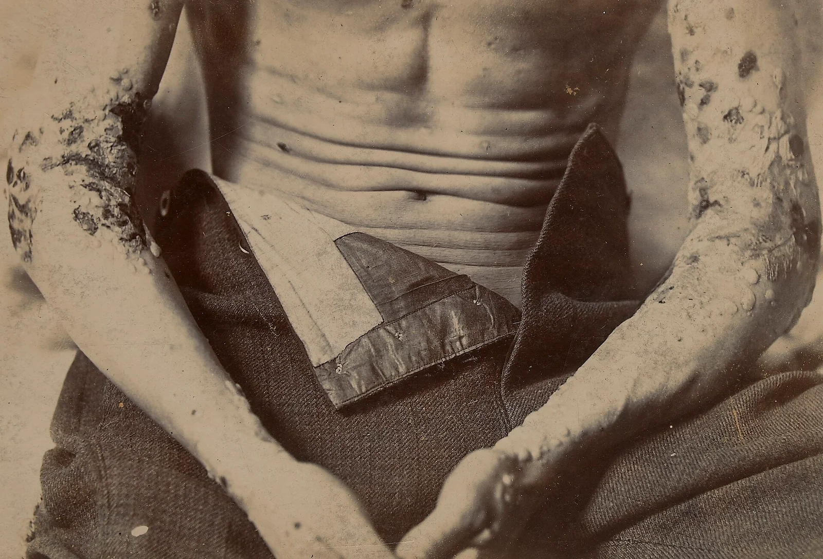
Long description
Black-and-white photograph described on its file page as showing a male patient's arms, from a case of dermatitis herpetiformis (St Bartholomew's Hospital Photographic Society, 1893).

Figure 2Pemphigus vulgaris
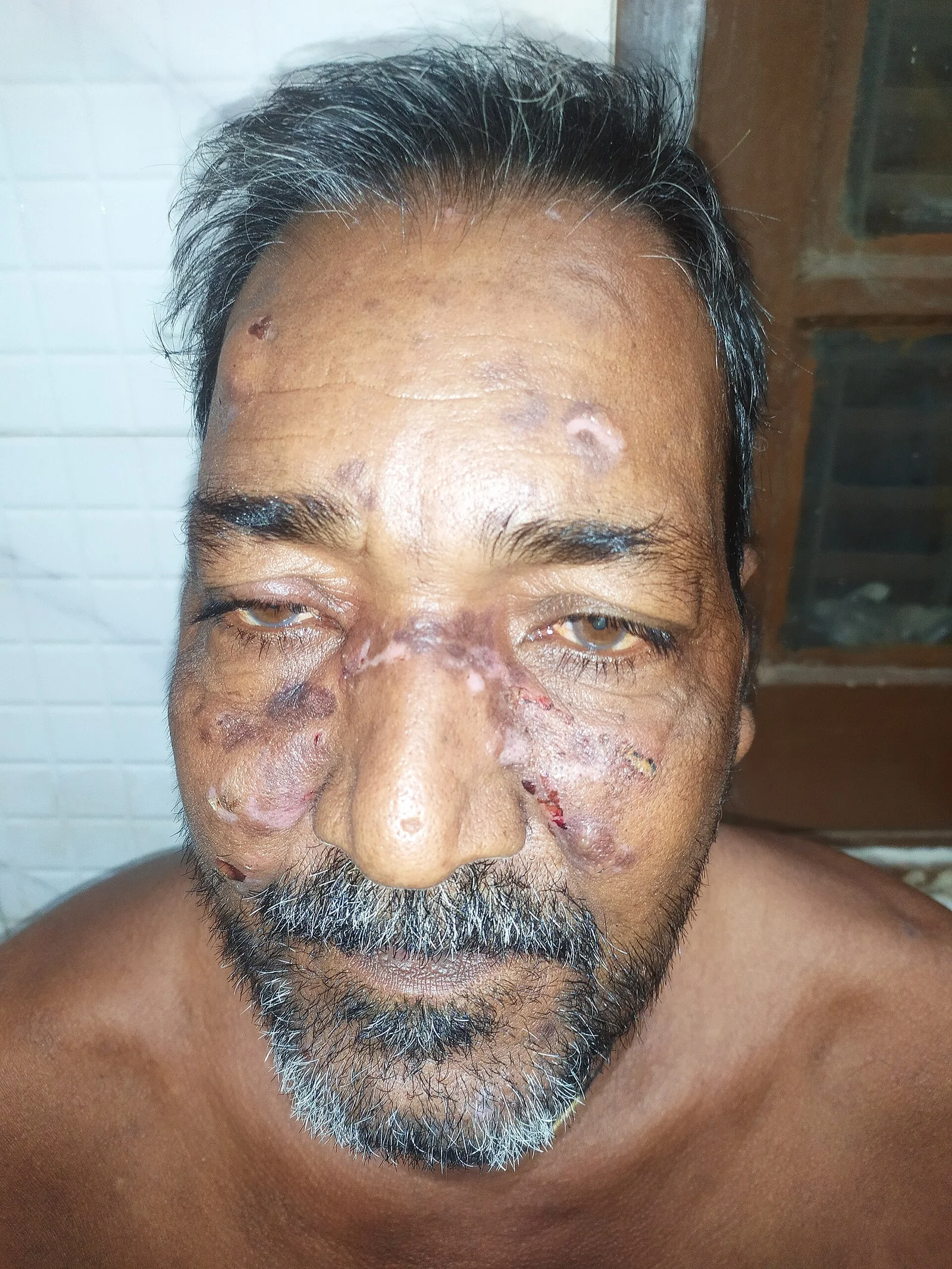
Long description
Wikimedia Commons file titled "Pemphigus vulgaris", self-published as own work by User:সাজিদ শরীফ on 2024-11-24 and placed in the pemphigus vulgaris category, with no further clinical detail on the file page.

Figure 3Bullous pemphigoid new image
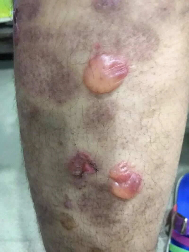
Long description
An uploaded image titled 'Bullous pemphigoid new image', whose attached French description of bullous pemphigoid identifies tense blisters as the characteristic finding and is used on multiple Wikipedias to illustrate pemphigoid.

Figure 4Anchoring junctions
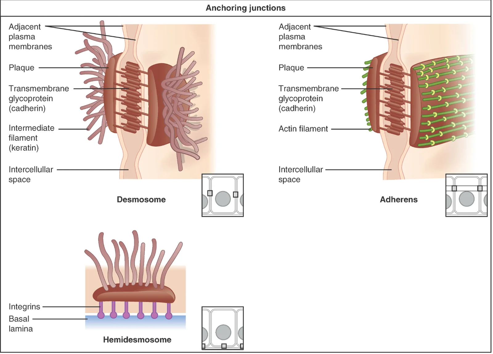
Long description
OpenStax 'Figure 4.5: Types of Cell Junctions' diagram of epithelial cell-cell and cell-matrix junctions, including a desmosome with keratin intermediate filaments, an adherens junction, and a hemidesmosome attaching to the basal lamina.

A biopsy is mandatory for a first episode. The key distinction is made by where the split sits and by immunofluorescence. [3][4]
| Test | What it shows | Result or cut-off in pemphigus vulgaris or bullous pemphigoid |
|---|---|---|
| Nikolsky sign | Level of the split (bedside) | Positive in pemphigus vulgaris; negative in bullous pemphigoid |
| Tzanck smear | Acantholytic cells | Positive in 71.4-100% of pemphigus; sensitivity 73-100%; specificity 43.4-83.3% [4] |
| Histopathology (3-5 mm punch from edge of a fresh blister under 24 hours old) | Level of split | Pemphigus vulgaris: suprabasal split with row of tombstones. Pemphigus foliaceus: subcorneal split. Bullous pemphigoid: subepidermal split with eosinophils |
| Direct immunofluorescence (DIF) of perilesional skin | Deposited antibodies and complement | Pemphigus vulgaris/foliaceus: intercellular fishnet or chicken-wire IgG and C3. Sensitivity 82-95%; specificity 98% [4]. Bullous pemphigoid: linear IgG and/or C3 along the basement membrane [5] |
| Serology: anti-desmoglein 1 and 3 ELISA | Circulating autoantibodies | Above the reference threshold for the kit supports pemphigus vulgaris (anti-desmoglein 3) and/or pemphigus foliaceus (anti-desmoglein 1); ELISA is positive in more than 90% of cases [3][4] |
| Anti-BP180 / anti-BP230 antibodies | Circulating autoantibodies | Support bullous pemphigoid |
| Indirect immunofluorescence | Circulating antibodies | Pemphigus: intercellular pattern. Bullous pemphigoid: linear basement-membrane pattern |
| Bullous pemphigoid diagnosis | Confirmation of subepidermal disease | Clinical picture confirmed by positive direct or indirect immunofluorescence for IgG or C3 at the dermal-epidermal junction [6] |
Diagnosis requires a clinical picture and histology consistent with an autoimmune blistering disease, plus either a positive direct immunofluorescence microscopy or serologic detection of the relevant autoantibodies. [3] Direct immunofluorescence is the gold-standard antibody test. [4]
Before starting systemic treatment, baseline tests include full blood count, liver and renal function, fasting glucose, electrolytes, urinalysis, chest radiograph, infection screen (hepatitis B and C, HIV), and pregnancy testing where relevant. [3][4] Bullous pemphigoid review further links the disease to multiple internal comorbidities. [5]
Pemphigus can be classified as possible, probable or definite depending on clinical features, Tzanck smear, histopathology and DIF or serology. [4] Severity of pemphigus vulgaris is stratified as below. [4]
| Severity | Cutaneous PDAI | Cutaneous ABSIS | POLIS (oral) | Clinical correlate |
|---|---|---|---|---|
| Mild | under 15 | under 17 | under 12 | up to 5 discrete skin lesions; up to 3 oral erosions [4] |
| Moderate | 15 to 45 | 17 to 53 | 12 to 24 | 5 to 20 skin lesions; 3 to 10 oral erosions, or generalised desquamative gingivitis [4] |
| Severe | above 45 | above 53 | above 24 | more than 20 lesions, confluent erosions, above 5% body surface area; more than 10 oral erosions or extensive confluent erosions [4] |
A diagnosis-first algorithm:
Approach to a patient with blistering skin or mucosa
- Blistering rash or mucosal erosionsHistory, full skin and mucosal examination, Nikolsky sign
- Level of the blister?Nikolsky positive suggests intraepidermal; negative suggests subepidermal
- Question: Mouth or other mucosa involved?
Yes; flaccid blisters
- Pemphigus vulgarisSuprabasal split, fishnet DIF, anti-desmoglein 1/3 antibodies
- Start prednisolone 0.5-1.5 mg/kg/day with steroid-sparing therapy
No mucosal disease; tense bullae
- Bullous pemphigoidSubepidermal split, linear IgG/C3 at basement membrane, BP180/BP230 antibodies
- Superpotent topical steroid; systemic therapy by severity and frailty
Pruritic grouped papules on extensors
- Dermatitis herpetiformisGranular IgA on DIF; gluten-free diet; dapsone
- Confirm with histology and DIF before immunosuppressionAdd baseline infection screen and metabolic tests
Suppress the autoantibody-driven inflammation, heal erosions, control pain, prevent infection and infection-related harm, and withdraw therapy as soon as remission allows. [3][4][5]
First-line by severity (Indian consensus, 2024; consistent with the international 2020 consensus). [3][4]
- Mild pemphigus vulgaris: oral prednisolone 0.5 mg/kg/day plus a steroid-sparing agent. [4]
- Moderate to severe pemphigus vulgaris: oral prednisolone 1 mg/kg/day plus rituximab 1 g intravenously twice, 2 weeks apart. [3][4]
- If not controlled by 3 to 4 weeks, increase prednisolone to 1.5 mg/kg/day or give an intravenous corticosteroid pulse, and repeat rituximab (1 g twice, 2 weeks apart) at month 6. [4]
- Third-line options are intravenous immunoglobulin and plasmapheresis. [4]
- Cyclosporine and methotrexate are not recommended for pemphigus. [4]
| Drug | Dose and route | Notes |
|---|---|---|
| Prednisolone | 0.5 to 1.5 mg/kg/day orally | 0.5 for mild, 1 for moderate-severe, up to 1.5 if not controlled [3][4] |
| Rituximab | 1000 mg IV twice, 2 weeks apart, or 375 mg/m^2 weekly for 4 weeks | First-line in moderate-severe; may allow faster steroid taper [3][4] |
| Low-dose rituximab | 500 mg IV twice, 2 weeks apart | For cost constraints; may give shorter remission [4] |
| Azathioprine | 1 to 3 mg/kg/day orally (2 mg/kg/day typical; up to 2.5 in moderate-severe) | Start at 50 mg daily for the first week to detect idiosyncratic reactions; check TPMT activity where polymorphism prevalence is high [3][4] |
| Mycophenolate mofetil | 1 to 2 g/day orally (30-45 mg/kg/day allowed) | Wait 4 days before dose changes; gastrointestinal intolerance common [3][4] |
| Mycophenolate sodium (enteric-coated) | up to 1440 mg/day orally | Equivalent enteric form [4] [3] |
| Cyclophosphamide | 2 mg/kg/day orally | Considered for severe or economic constraints; counsel on bone marrow suppression, gonadal failure and bladder cancer [4] |
| Intravenous immunoglobulin | 2 g/kg total, given over 2 to 5 days each month | Rescue in severe or refractory disease; pregnancy-safe option [3][4] |
| Dapsone (pemphigus foliaceus, with topical corticosteroids) | combined with topical corticosteroids; about 50% need systemic corticosteroids later | Useful first-line for limited pemphigus foliaceus [4] |
Consolidation and tapering. Maintain induction doses until no new lesions for at least 2 weeks and about 80% of lesions have healed. [4] Then taper prednisolone over 3 to 4 months in mild disease and about 6 months in moderate or severe disease. [4] A maintenance dose of prednisolone 10 mg/day or less is acceptable in international practice. [4]
Maintenance rituximab. In severe baseline disease or persistently elevated desmoglein antibody levels, infusions at 12 and 18 months can be given even without clinical relapse; otherwise treat relapse reactively. [4] Stopping conventional adjuvants is usually considered 6 to 12 months after complete remission on minimal therapy. [4]
Rituximab in practice. Rituximab is given with short-term systemic corticosteroids for less than 4 months and long-term immunosuppressive adjuvant therapy for more than 12 months; screening for hepatitis B and tuberculosis precedes it. [3][4] Hepatitis B prophylaxis with entecavir or tenofovir is mandatory in patients with occult hepatitis B who receive rituximab. [4]
Monitoring. Pemphigus requires structured monitoring of drug toxicity and of disease activity; the international panel supplies a detailed work-up. [3][4]
The British Association of Dermatologists 2012 guideline recommends superpotent topical corticosteroids as first-line in localised or moderate bullous pemphigoid, and oral prednisolone as the only validated systemic treatment in generalised disease. [5] Other reviewers recommend early steroid-sparing treatment because prednisolone (especially prolonged or higher-dose use) increases morbidity in older patients. [5]
- Localised or moderate disease: superpotent topical corticosteroids once or twice daily to lesions for some weeks. [5]
- Extensive or impractical disease, or living alone with poor application support: oral prednisolone 0.5 mg/kg/day. [5][6]
- Frail, diabetic or very old patients: reviewers advise omitting oral prednisolone where possible and using potent topical corticosteroids plus oral doxycycline 200 mg/day or dapsone. [5]
- Doxycycline 200 mg/day was non-inferior to prednisolone 0.5 mg/kg/day for short-term blister control in the BLISTER trial and was significantly safer long-term, so it is a strong option in older, frail or comorbid patients. [6]
Steroid-sparing and second-line agents. Bullous pemphigoid relapses are managed with azathioprine, dapsone, methotrexate, mycophenolate mofetil or mycophenolic acid, cyclophosphamide, ciclosporin, IVIG, plasmapheresis, or rituximab, sometimes combined. [5] Higher-dose prednisone monotherapy (above about 0.5 mg/kg) is an independent risk factor for fatal outcome in the first year, so it should not be used to patch poor control. [5] [4]
Dermatitis herpetiformis is gluten-driven and does not respond to immunosuppression alone. [7]
- Diagnosis: granular IgA deposits in the dermal papillae on direct immunofluorescence of perilesional skin; histology shows neutrophilic microabscesses in dermal papillae with or without a subepidermal blister. [7]
- False-negative DIF occurs in about 5%, especially if biopsy is taken from a blister or inflamed skin. [7]
- Treatment: a strict lifelong gluten-free diet is basic treatment; dapsone 25 to 50 mg/day (up to 100 mg/day) controls itch and rash within days, with a starting dose of 0.5 mg/kg/day in children. [7]
- Dapsone caution: methaemoglobinaemia and haemolysis (worse with G6PD deficiency), peripheral neuropathy with continuous doses at or above 100 mg/day; monitor blood counts and liver and renal function. [7]
- Pemphigus vulgaris: pain, superinfection of erosions, sepsis, failure to eat and drink, and the adverse effects of high-dose corticosteroids and adjuvants (diabetes, osteoporosis, infection, malignancy risk with azathioprine and cyclophosphamide). [3][4]
- Bullous pemphigoid: frailty, skin infection, and a 7.6-fold increase in mortality compared with an age- and sex-matched population in one Finnish study, strongly associated with comorbidity and with systemic corticosteroid use. [5]
- Rituximab and infection: the incidence of unforeseen fatal infections such as progressive multifocal leukoencephalopathy cannot be estimated because pemphigus is rare. [3]
Pemphigus vulgaris is potentially fatal if untreated but is controllable. [4] In the rituximab era, complete remission off therapy is now a realistic aim: real-world studies, including Indian cohorts, report 73 to 87% of patients attaining complete remission off therapy after a single cycle of rituximab. [4] Desmoglein antibody levels, when available, are used to guide tapering and maintenance. [3][4]
Bullous pemphigoid often remits, but relapses and treatment-related harm are common in older patients, so any withdrawal of therapy is gradual. [5]
There is no primary prevention. Useful measures: [3][4][5]
- Screen for tuberculosis and viral hepatitis before systemic corticosteroids or rituximab; antiviral prophylaxis for hepatitis B reactivation risk. [4]
- Steroid safety: ocular screening for cataract and glaucoma, osteoporosis risk assessment (FRAX) and bone protection, glucose monitoring, infection vigilance. [4]
- Medication review: avoid known drug triggers where possible. [3]
- Nursing and wound care: gentle cleansing, non-adherent dressings, analgesia, nutrition support; avoiding deroofing of blisters or aggressive cleansing, which can induce new erosions. [4]
- Vaccination status should be reviewed. [4]
Pemphigus in pregnancy is a high-risk condition. Systemic corticosteroids, especially prednisolone, are first-line and relatively safe. Azathioprine and intravenous immunoglobulin are second-line. Mycophenolate mofetil and methotrexate are contraindicated because they are teratogenic; cyclophosphamide is also not safe. Rituximab lacks pregnancy safety data and should be avoided 12 months before conception. [4]
Topical corticosteroids may be used for limited disease, with caution over potent formulations and large cumulative doses. [4]
Women with pemphigus should plan conception after clinical remission, and discontinuation of methotrexate, mycophenolate mofetil and cyclophosphamide for 12 weeks, 6 weeks and 4 weeks respectively is advised. [4]
Pemphigus is rare in children. [4][8] Systemic corticosteroids cause growth retardation and other effects more readily in children, so the lowest effective dose with close monitoring is needed. [4]
- Adjuvant therapy: azathioprine 2 to 2.5 mg/kg/day or mycophenolate mofetil 25 to 30 mg/kg/day. [4]
- Rituximab: body-surface-area dosing using the lymphoma protocol, 375 mg/m^2 weekly for 4 weeks, or two doses of 750 mg/m^2 2 weeks apart. [4]
- Intravenous immunoglobulin: a second-line option with low adverse-event rates in children. [4]
Bullous pemphigoid is rare in minors. [8]
Bullous pemphigoid is mainly a disease of the elderly, and treatment should be chosen to minimise cumulative corticosteroid exposure, preferring topical therapy and doxycycline where control allows. [5][6]
- Bedside diagnosis: Nikolsky sign and a Tzanck smear are cheap and immediate; histology should follow. [4]
- Transport of immunofluorescence samples: up to 24 hours in Michel's medium or normal saline at room temperature, processed or frozen on receipt. [4]
- Where ELISA/DIF is unavailable: a clinical diagnosis plus histology plus Tzanck supports treatment; desmoglein levels improve precision but are not mandatory before starting therapy. [4]
- Cost constraints: lower-dose rituximab (500 mg twice, 2 weeks apart), cyclophosphamide in selected severe cases, and whole-patient discussion of adverse effects are documented adaptations. [4]
- Tuberculosis screening is essential before immunosuppression in endemic regions. [4]
Pemphigus vulgaris, bullous pemphigoid and dermatitis herpetiformis are all autoimmune blistering diseases, but their levels, antibody targets, direct immunofluorescence and first-line treatments are completely different, so they should never be managed interchangeably. [3][4][5][7]
- Pemphigus vulgaris is an intraepidermal blistering disease caused by antibodies against desmogleins; Nikolsky sign is positive. [3][4]
- Bullous pemphigoid is a subepidermal disease caused by antibodies against BP180 and BP230; Nikolsky sign is negative. [5]
- Histology shows where the skin splits; direct immunofluorescence shows fishnet IgG/C3 in pemphigus and a linear band in bullous pemphigoid. [4][5]
- Diagnosis requires consistent clinical and histological findings plus positive DIF or relevant autoantibodies. [3]
- Pemphigus severity (PDAI, ABSIS, POLIS) drives treatment. [4]
- Prednisolone 0.5 to 1.5 mg/kg/day is used in pemphigus; taper over 3 to 4 months in mild and about 6 months in moderate to severe disease. [3][4]
- Rituximab 1 g intravenously twice, 2 weeks apart, is first-line in moderate to severe pemphigus and permits faster steroid tapering. [3][4]
- Bullous pemphigoid first-line systemic options include prednisolone 0.5 mg/kg/day and doxycycline 200 mg/day, with topical superpotent corticosteroids for localised or moderate disease. [6]
- Pregnancy: prednisolone first, azathioprine or IVIG second, mycophenolate and methotrexate contraindicated; rituximab avoided 12 months before conception. [4]
- Dermatitis herpetiformis is treated with a strict gluten-free diet plus dapsone, not with immunosuppression. [7]
Answer each one in your head first, then open the answer.
A 52-year-old man has painful oral erosions and flaccid skin blisters that rupture on light touch. Nikolsky sign is positive. What level of split and what antibody are expected, and how would pemphigus vulgaris differ from bullous pemphigoid?
Show answerHide answer
Intraepidermal split with acantholysis and anti-desmoglein antibodies. Direct immunofluorescence would show a fishnet or chicken-wire pattern of IgG and C3. Bullous pemphigoid would instead have a subepidermal split, a negative Nikolsky sign, a linear IgG and C3 band at the basement membrane and anti-BP180/BP230 antibodies.
A 78-year-old woman has tense, pruritic bullae on her thighs after a prodrome of urticarial plaques. Which first-line systemic options are supported, and which would be favoured in a frail patient with diabetes?
Show answerHide answer
Oral prednisolone 0.5 mg/kg/day or doxycycline 200 mg/day are the systemic options, with superpotent topical corticosteroids as first-line for localised or moderate disease. In a frail or diabetic patient, reviewers favour omitting oral prednisolone and using potent topical corticosteroids plus oral doxycycline 200 mg/day or dapsone.
What dose of prednisolone is used initially in mild and in moderate to severe pemphigus vulgaris, and how is the taper timed?
Show answerHide answer
Mild pemphigus vulgaris: 0.5 mg/kg/day. Moderate to severe: 1 mg/kg/day, increasing to 1.5 mg/kg/day if not controlled by 3 to 4 weeks. Taper begins after there are no new lesions for 2 weeks and about 80% of lesions have healed, over 3 to 4 months for mild and about 6 months for moderate to severe disease.
How should rituximab be dosed in moderate to severe pemphigus vulgaris, and how does pregnancy change planning?
Show answerHide answer
Rituximab 1 g intravenously twice, 2 weeks apart, or 375 mg/m^2 weekly for 4 weeks. A low-dose regimen of 500 mg twice, 2 weeks apart, is an option with cost constraints. Rituximab lacks adequate pregnancy safety data, so it should be avoided 12 months before conception; prednisolone is first-line in pregnancy, with azathioprine or IVIG as second-line options.
A patient with itchy grouped papules on the elbows has granular IgA deposits in the dermal papillae on DIF. What is the treatment?
Show answerHide answer
Dermatitis herpetiformis: a strict lifelong gluten-free diet as basic treatment, plus dapsone 25 to 50 mg/day (up to 100 mg/day) to control itch and rash within days, starting at 0.5 mg/kg/day in children.
Plain text only: systemic lupus erythematosus; urticaria and hypersensitivity reactions; paediatric dermatology; infections in immunocompromised hosts; pregnancy complications.
- Ingold CJ, Sathe NC, Khan MAB. Pemphigus Vulgaris. StatPearls [Internet]. Treasure Island (FL): StatPearls Publishing; 2024.. Accessed .
- Baigrie D, Nookala V. Bullous Pemphigoid. StatPearls [Internet]. Treasure Island (FL): StatPearls Publishing; 2023.. Accessed .
- Murrell DF, Peña S, Joly P, et al. Diagnosis and management of pemphigus: Recommendations of an international panel of experts. J Am Acad Dermatol 2020;82:575-585.e1. doi:10.1016/j.jaad.2018.02.021. Accessed .
- De D, Mehta H, Shah S, et al. Consensus Based Indian Guidelines for the Management of Pemphigus Vulgaris and Pemphigus Foliaceous. Indian Dermatol Online J 2025;16:3-24 (epub 2024). doi:10.4103/idoj.idoj_1059_24. Accessed .
- Bech R, Kibsgaard L, Vestergaard C. Comorbidities and Treatment Strategies in Bullous Pemphigoid: An Appraisal of the Existing Literature. Front Med 2018;5:238. doi:10.3389/fmed.2018.00238. Accessed .
- Williams HC, Wojnarowska F, Kirtschig G, et al. Doxycycline versus prednisolone as an initial treatment strategy for bullous pemphigoid: a pragmatic, non-inferiority, randomised controlled trial. Lancet 2017;389:1630-1638. doi:10.1016/S0140-6736(17)30560-3. Accessed .
- Reunala T, Hervonen K, Salmi T. Dermatitis Herpetiformis: An Update on Diagnosis and Management. Am J Clin Dermatol 2021;22:329-338. doi:10.1007/s40257-020-00584-2. Accessed .
- Nanda A, Tedbirt B, Bodemer C, et al. European S2k guidelines on management of autoimmune blistering diseases in children and adolescents. J Eur Acad Dermatol Venereol 2026;40:1137-1161. doi:10.1111/jdv.70308. Accessed .
Figure credits
- Figure 1: https://www.calmview.co.uk/BartsHealth/CalmView/Record.aspx?src=CalmView.Catalog&id=SBHB%2fMU%2f14%2f35%2f18%2f1&pos=1, via Wikimedia Commons, CC BY 4.0, CC BY 4.0. resized.
- Figure 2: সাজিদ শরীফ, via Wikimedia Commons, CC0, CC0. resized.
- Figure 3: Mohammad2018, via Wikimedia Commons, CC BY-SA 4.0, CC BY-SA 4.0.
- Figure 4: OpenStax Anatomy and Physiology, via Wikimedia Commons, CC BY-SA 4.0, CC BY-SA 4.0.
Educational content for medical students and doctors. It is not a substitute for clinical judgement or local guidelines.