Erythema nodosum and other reactive skin conditions
Tender inflammatory skin reactions that flag an underlying infection, sarcoidosis, bowel disease or drug trigger; covers erythema nodosum, pyoderma.
- Erythema nodosum is a painful, symmetrical, deep red rash of the shins: the commonest form of panniculitis (inflammation of the fat under the skin). It is a reaction, not a disease in its own right. [2]
- It is most often idiopathic, but it can be the first visible clue to streptococcal infection, sarcoidosis, tuberculosis, inflammatory bowel disease, pregnancy, or a drug reaction.
- Pyoderma gangrenosum and Sweet syndrome are two related "neutrophilic dermatoses" in the same family; they look completely different (ulceration and tender plaques respectively) but share the same mechanism of runaway neutrophil inflammation. [3]
- No test diagnoses erythema nodosum itself; it is recognised clinically, and the real work is finding the trigger. Pyoderma gangrenosum and Sweet syndrome usually need a biopsy to exclude infection and malignancy.
- Erythema nodosum is self-limited and managed supportively, except when the trigger needs specific treatment (for example, antituberculous therapy). Pyoderma gangrenosum and Sweet syndrome need systemic immunosuppression.
By the end of this page you should be able to:
- Recognise erythema nodosum, pyoderma gangrenosum and Sweet syndrome on the skin and say what mechanism links them.
- List the common causes of erythema nodosum and the systemic conditions found with pyoderma gangrenosum and Sweet syndrome.
- Plan a stepwise work-up for erythema nodosum and interpret the tests.
- Manage each condition with the correct first-line drugs and doses, and when to use topical rather than systemic treatment.
- Tell these conditions apart from infection, vasculitis, venous ulceration and other panniculitides.
- Modify your approach in pregnancy, children and older adults, and in places with limited tests or drugs.
DefinitionErythema nodosum
Erythema nodosum is a specific form of panniculitis (inflammation of the subcutaneous fat) seen as tender, red to violet, deep, palpable nodules or plaques, mainly on the pretibial shins. It is a non-ulcerating, reactive process that usually leaves bruise-like discolouration and heals without scarring.
DefinitionPyoderma gangrenosum
Pyoderma gangrenosum is a rare, non-infectious neutrophilic dermatosis that begins as an inflamed papule, pustule or nodule and rapidly ulcerates, forming a painful wound with a swollen necrotic base and a raised, undermined, violaceous border.
DefinitionSweet syndrome
Sweet syndrome (acute febrile neutrophilic dermatosis) is an acute inflammatory skin disease marked by the sudden appearance of tender, well-defined erythematous plaques or nodules, usually with fever and arthralgia, and a dense neutrophilic infiltrate in the dermis.
These three conditions are "reactive" or "neutrophilic dermatoses": the skin changes are driven by inflammation rather than by infection of the skin itself. The unifying mechanism is an inappropriate accumulation of neutrophils in skin tissue. Biopsy shows neutrophil-rich infiltrates without true vasculitis, and the conditions respond to immunosuppression rather than to antibiotics. Pyoderma gangrenosum and Sweet syndrome are widely regarded as lying on the same disease spectrum, and lesions can overlap or evolve from one to the other, particularly in the setting of myeloid malignancy.[4]
Broadly, neutrophilic dermatoses divide into:
- idiopathic or primary disease, versus disease secondary to an underlying disorder or drug;
- non-ulcerative forms (Sweet syndrome, pustular psoriasis-type presentations) versus ulcerative pyoderma gangrenosum;
- localised versus generalised disease, with or without extracutaneous involvement (eyes, joints, lungs, spleen).
Pyoderma gangrenosum has subtypes that change both the picture and the likely trigger:
- ulcerative (classic), about 85% of cases: rapidly progressive painful ulcers with an undermined violaceous edge, usually on the lower leg or trunk.[4]
- bullous (atypical): superficial vesicles and bullae that erode, mainly on the arms and face; haematological malignancy is found in up to 70% of these cases.[4]
- pustular: tender pustules on erythematous skin, classically with a flare of inflammatory bowel disease.[4]
- vegetative (superficial granulomatous pyoderma): a single indolent superficial plaque or ulcer, usually on the head or neck, less often linked to systemic disease.[4]
- peristomal: ulcers around a stoma, most often in inflammatory bowel disease.[4]
Sweet syndrome is usually classified as classical (idiopathic), malignancy-associated, or drug-induced; the somatic UBA1 mutation responsible for VEXAS (vacuoles, E1 enzyme, X-linked, autoinflammatory, somatic) syndrome is now an important fourth category in older men with relapsing nodules, cytopenias or macrocytosis.[3]
Two autoimmune blistering diseases named on the same book page are not part of this group: bullous pemphigoid and pemphigus vulgaris are autoantibody-mediated disorders of adhesion molecules, not neutrophilic dermatoses, and their diagnosis and treatment are different.
Erythema nodosum affects roughly 1 to 5 people per 100,000 per year.[5] A UK patient leaflet gives a higher local figure of about 2.4 cases per 10,000 per year; the two sources do not agree, and both are estimates.[8] In adults it is far more common in women, with a male-to-female ratio of about 1 to 6; in children the sexes are equally affected.[5] The peak age is 20 to 30 years, although it can occur at any age, and a BAD leaflet puts the more common adult age range at 25 to 40 years.[5][8] About a third of patients relapse, and in some the condition becomes chronic and persists for months or years.[8] [1]
Pyoderma gangrenosum is rarer, with an incidence of approximately 0.63 per 100,000, a median age at presentation of 59 years, and a female predominance of up to 76% in some series.[4] Most patients are at least 55 years old.[7]
Sweet syndrome is rare; no reliable population incidence is available in the sources used here.
Erythema nodosum is a delayed (type IV) hypersensitivity reaction to a wide range of antigenic stimuli; it is frequently accompanied by granulomatous disease such as sarcoidosis and tuberculosis.[5][6] About half of cases are idiopathic even after investigation.[6] The commonly reported causes, with approximate frequencies from one large review, are:
- idiopathic: up to 55%; [5]
- streptococcal pharyngitis: 28 to 48% (the most common identifiable cause overall, especially in children); [5]
- sarcoidosis, usually with bilateral hilar lymphadenopathy: 11 to 25%; [5]
- drugs: 3 to 10% (sulfonamides, amoxicillin, oral contraceptives, proton-pump inhibitors, leukotriene modifiers); [5]
- pregnancy: 2 to 5%; [5]
- inflammatory bowel disease, both Crohn's disease and ulcerative colitis: 1 to 4% (more often during an acute colitis flare); [5]
- tuberculosis and atypical mycobacteria;
- fungal infections such as coccidioidomycosis and histoplasmosis;
- other bacteria and viruses (Yersinia in Europe, Salmonella, Campylobacter, Mycoplasma, Chlamydia, herpes viruses, HIV, hepatitis B and C);
- malignancy (lymphoma, leukaemia, and rarely carcinoid or colorectal tumours).[5][6][8]
NICE Clinical Knowledge Summaries recognise erythema nodosum as an extra-intestinal manifestation of Crohn's disease, and a BAD leaflet lists tuberculosis and inflammatory bowel disease alongside streptococcal throat infection and sarcoidosis as the common triggers.[10][8] The American Academy of Dermatology notes that many people with sarcoidosis develop erythema nodosum on the shins as the first sign, and that when this happens the sarcoidosis usually clears on its own within weeks to months (Löfgren syndrome).[9]
Streptococcal infection accounts for up to 44% of cases in adults and 48% in children; erythema nodosum usually appears 2 to 3 weeks after the sore throat.[5] In tuberculosis it may occur with primary infection and can even precede a positive tuberculin skin test.[5] Women are more affected; the adult male-to-female ratio of 1 to 6 has been partly attributed to oestrogen, since oral contraceptives, pregnancy and the cycle can all trigger episodes, although a firm link with oestrogen alone has not been proven.[5] [6]
Pyoderma gangrenosum is idiopathic in many patients. An underlying systemic condition is present in up to 50% of cases, most often: [4][7]
- inflammatory bowel disease in up to 30%;
- rheumatoid arthritis and other seronegative arthritides in up to 10%;
- haematological malignancy or monoclonal gammopathy (especially IgA) in about 5%; [4]
- other malignancy in about 5%.[4]
Drugs have been implicated (propylthiouracil, tyrosine kinase inhibitors, TNF-alpha inhibitors, granulocyte-colony stimulating factor), but the underlying disease for which they are given may be the real trigger.[4] There is a genetic component, with familial clustering and syndromes in which pyoderma gangrenosum is a feature (PAPA, PASH and PAPASH syndromes).[4][7] Trauma or skin injury triggers lesions in roughly 30% of patients (pathergy).[7]
Sweet syndrome is idiopathic in many patients and is divided into classical, malignancy-associated and drug-induced forms. Malignancy-associated disease (especially haematological, including acute myeloid leukaemia and myelodysplastic syndrome) is present in a substantial minority, and bullous lesions are more often linked with haematological malignancy.[3] Infection (including atypical organisms), inflammatory bowel disease, rheumatoid arthritis, pregnancy, vaccination and drugs (granulocyte-colony stimulating factor, all-trans retinoic acid, some antibiotics) are recognised triggers.[3] The biopsy-proven coexistence of Sweet syndrome and erythema nodosum has been documented.[5] [4]
In erythema nodosum the proposed mechanism is a delayed (type IV) hypersensitivity reaction to many different antigens, often in association with granulomatous disease; the process targets the septa of the subcutaneous fat rather than the adipocytes themselves. Histology shows a septal panniculitis with neutrophils, lymphocytes and the characteristic actinic (Miescher's) radial granulomas, and no primary vasculitis.[5]
Pyoderma gangrenosum and Sweet syndrome are driven by innate immune over-activation. Neutrophils are the dominant cell, with over-expression of interleukin-8, interleukin-1 beta, interleukin-17, tumour necrosis factor alpha and other cytokines; increased matrix metalloproteinase expression may contribute to poor wound healing. In pyoderma gangrenosum the dermal neutrophilic infiltrate and pustules form, then ulceration follows. The syndromic forms (PAPA, PASH) are linked to mutations in PSTP1P1/CD2BP1, which restrain the normal anti-inflammatory action of pyrin.[4][7]
Sweet syndrome shows the same non-vasculitic neutrophilic dermal (or subcutaneous) infiltrate with papillary dermal oedema; it is an auto-inflammatory process rather than a reactive vasculitis. Less common variants include a bullous form that can ulcerate and resemble pyoderma gangrenosum, and a subcutaneous form with tender 2 to 3 cm nodules that can resemble erythema nodosum.[3]
- A prodrome of 1 to 3 weeks of malaise, low-grade fever, cough or arthralgia is common, whatever the cause.[5]
- Deep, firm, tender, erythematous nodules or plaques 1 to 10 cm across, usually symmetrical on the pretibial surfaces (ankles, thighs, forearms and trunk less often).[5][8]
- Nodules evolve from bright red to purple to bruise-like over weeks; individual nodules last about 2 weeks and new ones can appear for up to 6 weeks.[5]
- They do not ulcerate, rarely scar, and clear in roughly 3 to 6 weeks (up to 8 weeks in some patients).[5][6][8]
- Systemic features: fever, malaise, fatigue, joint pain and swelling (mainly knees, ankles, wrists). Arthralgias are seronegative and may last up to 2 years after the skin has healed.[5]
- Blood tests may show leukocytosis with a count above 10,000 per mm3 and a raised erythrocyte sedimentation rate and C-reactive protein.[5]
- Begins as an inflamed erythematous papule, pustule or nodule that ulcerates and expands rapidly.[7]
- The classic ulcer has a swollen necrotic base and a raised, undermined, dusky-to-violaceous border, and is very painful.[4][7]
- Lower extremities are most affected; the trunk, especially buttocks and perineum, is also common; lesions can coalesce and leave cribriform scars.[4][7]
- Pathergy: ulcers may be precipitated or worsened by minor trauma or surgery. Systemic symptoms (fever, malaise) are common.[4][7]
- Subtypes (bullous, pustular, vegetative, peristomal, malignant pyoderma) change the site, depth and systemic associations, as described above.[4][7]
- Abrupt onset of tender, well-defined erythematous plaques or nodules, often with fever, arthralgia, headache and eye inflammation (conjunctivitis, episcleritis, iridocyclitis).[3]
- Lesions favour the upper extremities, face, neck, chest and back; oral or genital lesions are uncommon but reported, especially with malignancy.[3]
- Fever is almost always present in drug-induced disease; extracutaneous involvement elsewhere is rare.[3]
- Variants: bullous (can ulcerate and mimic pyoderma gangrenosum) and subcutaneous (tender nodules mimicking erythema nodosum on the legs).[3]
Figure 1Erythema nodosum - Kolkata 2012-01-03 7753
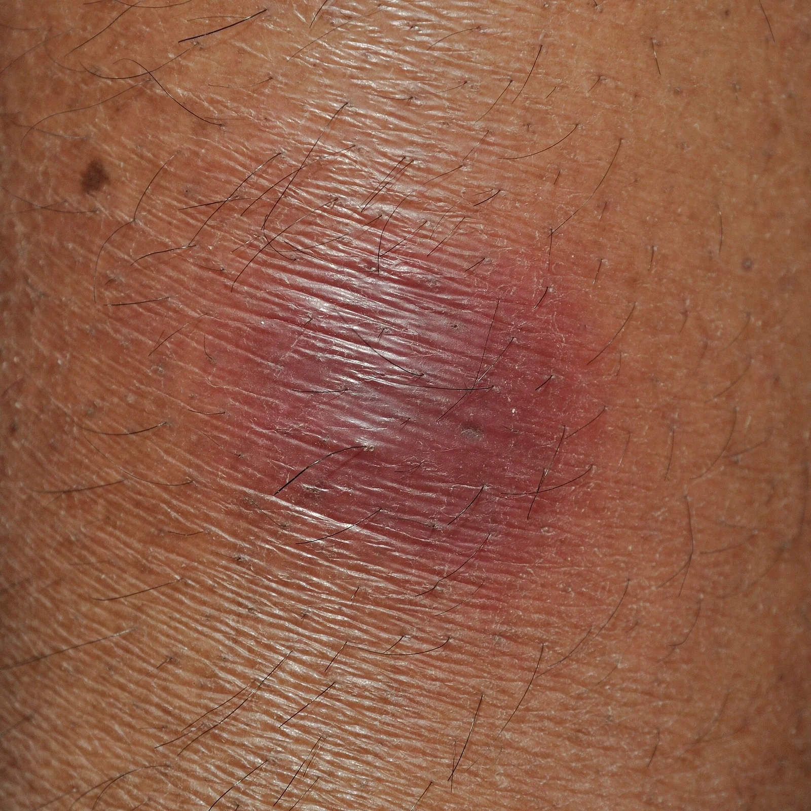
Long description
Clinical photograph of erythema nodosum presenting as tender red nodules on the right shin of a patient.

| Condition | First-line and confirmatory tests (what each adds) |
|---|---|
| Erythema nodosum | Diagnosis is clinical; deep incisional biopsy (wedge, not punch) when needed to show septal panniculitis without vasculitis. Basic screen: complete blood count, erythrocyte sedimentation rate and C-reactive protein; throat culture for group A streptococci with antistreptolysin-O titre, repeated within 4 weeks, and group A streptococcal PCR assay (sensitivity around 93%, specificity 98%). Streptococcal pharyngitis precedes the rash by 2 to 3 weeks. Test for tuberculosis exposure (purified protein derivative/Mantoux or interferon-gamma release assay; chest radiograph; acid-fast bacilli sputum if needed), stratified by risk. Stool cultures and ova/parasites if diarrhoea. Consider tests for sarcoidosis, inflammatory bowel disease and malignancy based on symptoms.[5][6] |
| Pyoderma gangrenosum | Clinical diagnosis, often by exclusion; biopsy of the active edge deep to subcutaneous fat to exclude infection, vasculitis and cancer. Recommended laboratory screen: full blood count, erythrocyte sedimentation rate, C-reactive protein, liver and renal function, protein electrophoresis, urinary Bence Jones protein, hepatitis screen, vasculitic screen, cryoglobulins if suggested, coagulation screen, faecal calprotectin if inflammatory bowel disease is suspected, chest radiograph, and computed tomography if malignancy is suspected. PARACELSUS score and the 2018 Delphi/MAVERAKIS criteria (one major: neutrophilic infiltrate at the ulcer edge; plus four minor criteria) can support the diagnosis.[4][7] |
| Sweet syndrome | Skin biopsy is required for diagnosis: dense dermal neutrophilic infiltrate with nuclear debris and papillary dermal oedema, without true vasculitis. Supporting tests: full blood count (neutrophilia), erythrocyte sedimentation rate/C-reactive protein, blood cultures, and screening for an underlying infection, inflammatory disorder, drug exposure or haematological malignancy based on the clinical picture. If the patient is older and male with cytopenias or macrocytosis, consider VEXAS.[3] |
Cut-offs and practical points that can be applied directly:
- erythema nodosum nodules are usually 1 to 10 cm across and rarely ulcerate.[5][8]
- leukocytosis above 10,000 per mm3 is a common finding in erythema nodosum.[5]
- In the 2018 Delphi criteria for ulcerative pyoderma gangrenosum, the single major criterion is a biopsy of the ulcer edge showing a neutrophilic infiltrate; at least four of eight minor criteria are then required (exclusion of infection, pathergy, history of inflammatory bowel disease or inflammatory arthritis, papule/pustule/vesicle ulcerating within 4 days, peripheral erythema with undermining border and tenderness, multiple ulcers with at least one on the anterior lower leg, cribriform scarring, and decreased ulcer size within 1 month of immunosuppression).[4]
| Condition | Feature that usually separates it |
|---|---|
| Erythema nodosum | Tender, deep, red-to-purple nodules symmetrically on the pretibial shins; no ulceration, no vasculitis on biopsy; heal with bruise-like colour. Usually an underlying trigger. |
| Pyoderma gangrenosum | Painful ulcer with swollen necrotic base and undermined violaceous border; pathergy (worsens with debridement); adult with inflammatory bowel disease, rheumatoid arthritis or haematological malignancy. |
| Sweet syndrome | Abrupt tender erythematous plaques/nodules with fever and arthralgia; upper-body and facial predominance; dense dermal neutrophilic infiltrate without vasculitis. |
| Erythema induratum (Bazin) | Nodules on the calves, classically in tuberculosis; may ulcerate and scar; a lobular rather than septal panniculitis. |
| Nodular panniculitis / lupus profundus | Lupus panniculitis and cytophagic histiocytic panniculitis appear on the differential list of erythema nodosum mimics and are separated by biopsy. |
| Cellulitis or deep infection | Usually unilateral, spreading, with warmth and fever but no symmetrical nodules; wounds worsen; cultures help. |
| Venous or arterial leg ulcer | Chronic, at the gaiter area or ankle; associated with venous disease or critical ischaemia; no undermined violaceous border and no pathergy. |
| Leukaemia or lymphoma of skin | Atypical, persistent, treatment-resistant lesions; always consider in bullous pyoderma gangrenosum. |
| Bullous pemphigoid / pemphigus vulgaris | Autoimmune blistering disease with tense or flaccid bullae, not deep nodules or ragged ulcers; diagnosis rests on direct immunofluorescence. |
Erythema nodosum itself has no formal severity score or staging system: the diagnosis is clinical, supported by biopsy when in doubt, and the central management question is how extensively to search for a trigger and how unwell the patient is (fever, arthritis, weight loss).[5][6]
Pyoderma gangrenosum is "no longer a diagnosis of exclusion" because of the Delphi international consensus criteria and the validated PARACELSUS score; both are aids, and the clinical picture remains decisive.[4][7] Severity is judged by the size, depth, number and rate of growth of ulcers, by pain, and by the presence of systemic disease.
Sweet syndrome is diagnosed on the combination of major clinical and histological criteria with at least two minor criteria; several minor schemes exist in the literature, so the safest practical rule is to require the characteristic histology (dense dermal neutrophilic infiltrate, no vasculitis) plus compatible clinical findings and exclusion of infection.[3]
The first principle for all three conditions is to treat the trigger wherever one is found. For erythema nodosum this is usually the only specific treatment needed. For pyoderma gangrenosum and Sweet syndrome, immunosuppression is added to gain control of the skin while the underlying disease is managed.
- Underlying cause: treat the identified disease (for example, antituberculous therapy is started presumptively in erythema nodosum patients with a positive purified protein derivative test with or without an identified focus; streptococcal pharyngitis is treated with antibiotics; active colitis is brought under firm control).[5]
- Supportive care: bed rest, leg elevation, cool compresses, avoidance of contact irritation, analgesia.[5][6]
- Pain: non-steroidal anti-inflammatory drugs as first-line analgesia; avoid them (or use with caution) when the trigger is Crohn's disease, because they can worsen a flare.[5]
- Potassium iodide as a supersaturated solution, 400 to 900 mg per day for 1 month, is a recognised option; another widely cited source gives 300 to 900 mg daily divided into 3 doses. The two sources give slightly different doses.[5][6] It is more likely to help if started early, and prolonged use carries a risk of hyperthyroidism.[5]
- Systemic glucocorticoids (for example, prednisone 60 mg each morning, about 1 mg per kg body weight per day) are effective but reserved as a last resort, and only once infection, risk of bacterial dissemination or sepsis, and malignancy have been excluded, because steroids can worsen an occult infection.[5][6]
- Disease-specific options: colchicine for coexistent Behcet's syndrome; combinations of steroids with hydroxychloroquine, cyclosporin or thalidomide for inflammatory bowel disease-associated disease.[5]
- Do not stop drugs suspected of causing the rash without discussing with the prescriber when the drug is essential; the balance is clinical.
There are no national or international guidelines; treatment is by review-level consensus and is chosen according to severity and rate of progression.[4]
- Wound and pain care is part of treatment, not an afterthought: moisture-retaining occlusive dressings for dry plaques, absorptive dressings for exudative ones; avoid wet-to-dry dressings. Adequate analgesia matters, and systemic antibacterial therapy is mandatory when infection is present. Do not debride the wound, which feeds pathergy.[4][7]
- Early, superficial or small lesions: high-potency topical glucocorticoids or tacrolimus on the ulcer surface, with or without intralesional corticosteroid injected into the active border.[4][7]
- Systemic first-line for more severe or rapidly progressive disease: prednisone (or prednisolone) 0.5 to 1 mg per kg per day, which is commonly written as 60 to 80 mg orally once daily in one source. The two sources frame the dose differently. Ciclosporin 3 to 4 mg per kg per day is an alternative or steroid-sparing agent, particularly in rapidly progressive disease; the trial dose was 4 mg per kg per day with a maximum of 400 mg.[4][7]
- The STOP GAP randomised controlled trial (121 patients) compared prednisolone 0.75 mg per kg per day (maximum 75 mg) with ciclosporin 4 mg per kg per day (maximum 400 mg): about half of all patients had healed ulcers at 6 months, with no difference between the two drugs and similar recurrence and adverse event rates.[4]
- Steroid-sparing and adjunctive agents: azathioprine, methotrexate, mycophenolate mofetil, dapsone, colchicine, apremilast, thalidomide, minocycline, sulphasalazine and high-dose intravenous immunoglobulin. Pulse intravenous methylprednisolone is useful to gain rapid control.[4][7]
- Biologics: infliximab has the strongest evidence (5 mg per kg intravenously; 69% of patients showed clinical improvement at 6 weeks after one infusion), and infliximab or adalimumab may heal faster than corticosteroids alone in observational studies. Adalimumab, etanercept, ustekinumab, anakinra and canakinumab are reported in case series; TNF-alpha inhibitors are preferred when inflammatory bowel disease is present. JAK inhibitors (tofacitinib, upadacitinib) and inhibitors of interleukin-1, interleukin-17 and interleukin-23 are used in refractory cases, with limited evidence.[4][7]
- Hydroxychloroquine, hyperbaric oxygen and split-skin or autologous keratinocyte grafts are adjunctive options in selected cases, alongside immunosuppression.
- Treat the underlying disease (for example, optimise inflammatory bowel disease therapy); idiopathic disease still needs control of the skin inflammation.
- Avoid unnecessary surgery in patients with a history of aggressive pyoderma gangrenosum because of pathergy.
- First-line: systemic corticosteroids (or topical if lesions are limited); colchicine or dapsone are options when steroids are contraindicated.[3] Oral corticosteroids are usually effective; resistant or frequently relapsing cases should be discussed with a specialist and may need steroid-sparing agents.[3]
- Treat any underlying condition (infection, inflammatory bowel disease, rheumatoid arthritis, malignancy) and withdraw an offending drug if present.
- Pregnancy, lactation and drug choice should be individualised.
- Erythema nodosum: no direct serious complication, but arthralgia may persist months to 2 years; chronic or recurrent disease occurs in about one third (with relapses lasting months to years in some). The main risk is missing a serious trigger such as tuberculosis, sarcoidosis with visceral involvement, or malignancy.[5][8]
- Pyoderma gangrenosum: pain (often severe), scarring, secondary infection, risk of limb loss when misdiagnosed as necrotising fasciitis, pathergic worsening with surgery, and extracutaneous disease (eyes, lungs, spleen). The condition is unpredictable and about half of patients are not healed at 6 months on conventional therapy.[4][7]
- Sweet syndrome: recurrence, chronic pain, skin breakdown and scarring of bullous lesions; extracutaneous involvement of eyes and joints. Bullous lesions can ulcerate and mimic pyoderma gangrenosum.[3]
Erythema nodosum is self-limited and almost always clears spontaneously in 3 to 6 weeks (sometimes up to 8 weeks), without ulceration or scarring; treating the trigger is the best way to shorten it.[5][6][8]
Pyoderma gangrenosum heals slowly, breaks down despite treatment and recurs; prognosis is said to be unpredictable, and deep or extensive disease carries real morbidity. Early diagnosis is the main modifiable factor.[4]
Sweet syndrome usually responds rapidly to systemic corticosteroids; most lesions resolve without scarring unless they have ulcerated. Recurrence is more likely when an underlying malignancy or drug trigger is present.[3]
- There is no specific prevention for these reactions. Avoid known triggering drugs where an alternative exists, treat streptococcal pharyngitis promptly, and keep inflammatory bowel disease, sarcoidosis and Behcet disease well controlled.[5][8]
- Screening is the essential clinical action when erythema nodosum is newly diagnosed: stratify for tuberculosis exposure; look for streptococcal infection, sarcoidosis and inflammatory bowel disease; and consider malignancy if the clinical picture suggests it. Chest imaging and, when relevant, stool cultures and tuberculosis testing form the minimum screen.[5]
- In pyoderma gangrenosum and Sweet syndrome, screen for systemic disease at diagnosis (inflammatory bowel disease, rheumatoid and seronegative arthritis, haematological malignancy) and for infection in any ulcer.[4][7]
- For bulky or atypical disease, and for older male patients with cytopenias, add appropriate haematological and (where available) genetic assessment for VEXAS.[3]
- Pregnancy: erythema nodosum is a recognised occurrence in pregnancy (2 to 5% of listed causes) and during use of oral contraceptives, possibly related to oestrogen/progesterone balance. In sarcoidosis, erythema nodosum with hilar lymphadenopathy and polyarthritis (Löfgren syndrome) usually resolves in 6 to 8 weeks. Treatment choices in pregnancy (non-steroidal anti-inflammatory drugs, steroids, potassium iodide) must be individualised and specialist advice sought; in coccidioidomycosis, erythema nodosum is a favourable prognostic sign.[5][9]
- Children: the male-to-female ratio of erythema nodosum is equal, streptococcal pharyngitis is the commonest identifiable cause, and up to 48% of cases are streptococcal. Pyoderma gangrenosum is rare in infants and children. Sweet syndrome in children is mainly described in small case series and usually warrants a search for an underlying infection or myeloid disorder. [3]
- Older adults: pyoderma gangrenosum predominantly affects those aged at least 55 years, and aggressive disease is more common. In older men with relapsing Sweet-syndrome-like nodules and cytopenias, consider VEXAS syndrome from a somatic UBA1 mutation.[7][3]
- Immunocompromised patients: always exclude infection before using systemic steroids or biologics.
- Erythema nodosum is a clinical diagnosis: no biopsy is needed when the picture is typical, and a low-cost screen (tuberculin skin test or interferon-gamma release assay where available, chest radiograph, throat swab, stool cultures, blood count, erythrocyte sedimentation rate/C-reactive protein) will find most triggers. Potassium iodide and non-steroidal anti-inflammatory drugs are inexpensive and effective for symptom control; keep systemic steroids reserved for refractory cases after infection is excluded.[5][6]
- Always suspect tuberculosis and endemic mycoses (coccidioidomycosis, histoplasmosis) from the local geography, and treat tuberculosis presumptively when the skin test is positive.[5]
- In pyoderma gangrenosum, avoid surgical debridement and use simple wound care plus prednisolone or ciclosporin (often available as generics) at the doses above; treat the underlying bowel disease. If cardiopulmonary bypass or specialised care is needed for complications, refer early.[4][7]
- In Sweet syndrome, a short course of systemic corticosteroids plus treatment of infection or malignancy is the backbone of care.[3]
Approach to a suspected reactive skin condition
- Patient with tender skin lesionsSymmetrical shin nodules, rapidly worsening ulcer with undermined border, or sudden tender plaques with fever
- Red flagsFever with neutrophilia, rapid ulcer expansion, pathergy, systemic illness, cytopenias, older male with relapsing nodules
- Question: What is the lesion morphology?
Tender deep shin nodules, no ulcer (erythema nodosum)
- Confirm clinically; biopsy only if atypical
- Treat the trigger (streptococcus, tuberculosis, sarcoidosis, inflammatory bowel disease, pregnancy, drug)
- Supportive care plus NSAIDs; potassium iodide early if prolonged; systemic steroids last resort at about 1 mg/kg/day
Ulcer with swollen necrotic base and undermined violaceous border (pyoderma gangrenosum)
- Biopsy the active edge; exclude infection, vasculitis and cancer
- Optimise wound care; do not debride
- Mild/superficial: topical high-potency steroid or tacrolimus; severe: prednisolone/prednisone 0.5-1 mg/kg/day or 60-80 mg daily, or ciclosporin 3-4 mg/kg/day
- Add TNF-alpha inhibitors or other immunosuppressants for refractory or bowel-associated disease
Tender erythematous plaques/nodules with fever and arthralgia (Sweet syndrome)
- Biopsy: dense neutrophilic infiltrate without vasculitis; exclude infection
- Systemic or topical corticosteroids first-line; colchicine or dapsone if steroids contraindicated
- Treat underlying infection, inflammatory bowel disease, rheumatoid arthritis or malignancy; withdraw offending drug
- Screen for systemic disease and arrange follow-upDocument response; watch for recurrence
Related skin and systemic conditions that help place this topic (MedGrasp topics on these are written or planned): sarcoidosis, inflammatory bowel disease, Behcet disease, tuberculosis, panniculitis, urticaria, vasculitis, pressure injury and wound healing. Bullous pemphigoid and pemphigus vulgaris are autoimmune blistering disorders rather than reactive neutrophilic dermatoses and are dealt with separately.
- Erythema nodosum is the commonest panniculitis: tender, non-ulcerating nodules on the shins that are a reaction to many antigens, most often streptococcal infection, sarcoidosis, drugs, inflammatory bowel disease or pregnancy.
- Around half of cases remain idiopathic; antibiotic, tuberculous, sarcoid and bowel triggers should be sought because they are treatable.
- Pyoderma gangrenosum and Sweet syndrome belong to the same neutrophilic dermatosis family; both require biopsy to exclude infection and cancer before immunosuppression.
- Pyoderma gangrenosum shows a rapidly progressive, very painful ulcer with an undermined violaceous border, often worsened by surgery (pathergy); systemic disease is present in up to half.
- Sweet syndrome presents abruptly with tender plaques or nodules, fever and arthralgia, and is diagnosed by dense dermal neutrophilic infiltrate without vasculitis.
- Erythema nodosum management is supportive (NSAIDs; potassium iodide 300 to 900 mg daily in divided doses or 400 to 900 mg daily for 1 month) with systemic steroids about 1 mg per kg per day only as a last resort after infection is excluded. [5]
- Pyoderma gangrenosum first-line systemic therapy is prednisolone 0.5 to 1 mg per kg per day (commonly 60 to 80 mg once daily) or ciclosporin 3 to 4 mg per kg per day, with only about half healed at 6 months; severe cases may need biologics such as infliximab 5 mg per kg intravenously. [4][7]
- Topical therapy and meticulous wound care treat early or superficial lesions; surgical debridement should be avoided.
- Sweet syndrome is treated first-line with systemic or topical corticosteroids; colchicine or dapsone are alternatives. [3]
- Older, treatment-resistant or atypical cases, and older men with cytopenias, need assessment for VEXAS syndrome and haematological malignancy.
- Prevention is trigger avoidance and good control of the underlying disease; there is no disease-specific vaccine or prophylaxis.
Answer each one in your head first, then open the answer.
A 25-year-old woman presents 3 weeks after streptococcal pharyngitis with painful red nodules on both shins. What is the most likely diagnosis, and what two simple tests support the streptococcal link?
Show answerHide answer
Erythema nodosum. Test the throat for group A streptococci (culture or rapid antigen/PCR) and take antistreptolysin-O titres at diagnosis and again within 4 weeks; the rash typically follows streptococcal infection by 2 to 3 weeks.
Why should a raised erythematous nodule on the shin not be debrided if pyoderma gangrenosum is possible?
Show answerHide answer
Pyoderma gangrenosum exhibits pathergy: trauma or surgery worsens the ulcer and enlarges it. Biopsy of the active edge is taken for diagnosis, but therapeutic debridement is avoided.
How does Sweet syndrome differ histologically and clinically from erythema nodosum?
Show answerHide answer
Sweet syndrome shows a dense dermal neutrophilic infiltrate without vasculitis and presents with abrupt tender plaques or nodules, fever and arthralgia, often on the upper body or face. Erythema nodosum is a septal panniculitis on the shins with no primary vasculitis and a much lower rate of fever at presentation; biopsy is only needed when in doubt.
Which first-line dose of ciclosporin is supported for severe pyoderma gangrenosum, and what did the STOP GAP trial find?
Show answerHide answer
Ciclosporin 3 to 4 mg per kg per day (trial dose 4 mg per kg per day, maximum 400 mg). In STOP GAP, prednisolone 0.75 mg per kg per day (maximum 75 mg) and ciclosporin 4 mg per kg per day produced similar results, with about half of patients healed at 6 months and no difference between the two drugs.
A 60-year-old man has relapsing Sweet-syndrome-like nodules and new cytopenias. What syndrome should be considered and what mutation underlies it?
Show answerHide answer
VEXAS syndrome, caused by a somatic mutation in UBA1, which produces an auto-inflammatory neutrophilic dermatosis with cytopenias or macrocytosis in older men.
What is the difference between erythema nodosum and erythema induratum, and which infection is classically involved?
Show answerHide answer
Erythema nodosum is a septal panniculitis of the pretibial shins that does not ulcerate. Erythema induratum (Bazin) favours the calves, more often ulcerates and scars, and is classically associated with tuberculosis.
Please note: always check your local formulary and follow current national guidelines. This is a teaching summary, and practice recommendations evolve. Treatment doses above are typical adult doses from the cited sources and need individual adjustment for weight, organ function, pregnancy, breastfeeding and comorbidity.
- Hafsi W, Badri T. Erythema nodosum. StatPearls [Internet]. Treasure Island (FL): StatPearls Publishing; 2022. Accessed .
- Schmieder SJ, Krishnamurthy K. Pyoderma gangrenosum. StatPearls [Internet]. Treasure Island (FL): StatPearls Publishing; 2023. Accessed .
- Vashisht P, Goyal A, Hearth Holmes MP. Sweet syndrome. StatPearls [Internet]. Treasure Island (FL): StatPearls Publishing; 2022. Accessed .
- George C, Deroide F, Rustin M. Pyoderma gangrenosum: a guide to diagnosis and management. Clinical Medicine (London) 2019;19(3):224-228. Accessed .
- Schwartz RA, Nervi SJ. Erythema nodosum: a sign of systemic disease. American Family Physician 2007;75(5):695-700. Accessed .
- Benedetti J. Erythema nodosum. Merck Manual Professional Version (updated September 2026). Accessed .
- Benedetti J. Pyoderma gangrenosum. Merck Manual Professional Version (updated September 2026). Accessed .
- British Association of Dermatologists. Erythema nodosum (patient information leaflet). BAD Patient Information Leaflet (April 2021). Accessed .
- Ludmann P. Sarcoidosis and your skin: signs and symptoms. American Academy of Dermatology (reviewed 2023). Accessed .
- NICE Clinical Knowledge Summaries. Crohn's disease: extra-intestinal manifestations. CKS (NICE). Accessed .
Figure credits
Educational content for medical students and doctors. It is not a substitute for clinical judgement or local guidelines.