Rosacea and pityriasis rosea
Rosacea and pityriasis rosea: definitions, diagnosis, treatment and differentials for two common dermatoses.
- Rosacea is a chronic inflammatory skin disease of the central face: recurrent flushing and fixed redness, papules and pustules, telangiectasia and, in some, thickening of the skin (phyma) and eye involvement. It is diagnosed clinically after excluding mimics. [1]
- There is no cure, but each feature responds to a different treatment: persistent redness to alpha-agonist creams and vascular laser, papules and pustules to ivermectin, metronidazole, azelaic acid, or doxycycline, phyma to isotretinoin or surgery, and flushing to trigger avoidance and, occasionally, vasoconstrictive oral drugs. [2][3]
- Pityriasis rosea is an acute, self-limited papulosquamous rash: a single herald patch is followed days later by smaller lesions on the trunk in a "Christmas tree" pattern along skin cleavage lines. It lasts 2 to 12 weeks. [4][5]
- A herald patch and Christmas-tree pattern suggest pityriasis rosea, but secondary syphilis can look identical; a papulosquamous rash in the right setting needs non-treponemal (RPR or VDRL) and treponemal serology before the diagnosis is accepted. [6]
By the end of this page you should be able to:
- Describe the clinical features of rosacea and of pityriasis rosea, and state why one is chronic and the other self-limited.
- Distinguish the major phenotypes of rosacea and match each to its evidence-based treatment.
- Explain why ivermectin and metronidazole are used for papules and pustules, and what role doxycycline has at antibacterial and sub-antibiotic doses.
- Recognise the herald patch and Christmas-tree pattern of pityriasis rosea and explain why secondary syphilis must be excluded.
- Interpret the findings of the Cochrane review on antiviral and antibiotic treatment for pityriasis rosea.
- Advise a patient on prognosis, relapse, self-care, and when to refer.
Rosacea is a chronic inflammatory dermatosis of the facial skin (and often the eyes) with a characteristic central (centrofacial) distribution. [2] It was formerly described as four subtypes (erythematotelangiectatic, papulopustular, phymatous, ocular, plus a granulomatous variant) defined by the National Rosacea Society Expert Committee in 2002. [2][3] Those subtypes grouped several different features into one label, which caused confusion and did not describe the individual patient well. [2] Current guidance (the global ROSacea COnsensus 2019 panel, the American Acne & Rosacea Society and the National Rosacea Society update) uses a phenotype approach: rosacea is described by the features actually present in that patient at that visit. [1][2][3]
Diagnostic criteria in the phenotype approach: [2]
- One of two diagnostic features alone is enough: (i) fixed central facial erythema that may periodically intensify, or (ii) phymatous changes (thickened skin, most often a bulbous nose).
- In the absence of those, two or more major features establish the diagnosis: papules/pustules, flushing/transient erythema, telangiectasia, or ocular features (lid margin telangiectasia, interpalpebral conjunctival injection, spade-shaped corneal infiltrates, scleritis or sclerokeratitis).
- Secondary (minor) features - burning, stinging, dryness, oedema, peripheral location - can occur but do not, on their own, diagnose rosacea.
The cutaneous rosacea features, as described by consensus: [1]
| Feature | Description |
|---|---|
| Persistent erythema (diagnostic) | Background ongoing central facial redness that may intensify with variable triggers |
| Phymatous changes (diagnostic) | Facial skin thickening from fibrosis and/or sebaceous gland hyperplasia; most often the nose |
| Flushing / transient erythema (major) | Temporary increase in central facial redness, with warmth, heat, burning or pain |
| Papules and pustules (major) | Red papules and pustules, usually central, sometimes larger and deeper |
| Telangiectasia (major) | Visible vessels in the central face, not limited to the alar region |
| Burning, stinging, dryness, oedema (minor) | Sensory and mild oedematous changes in the central face |
Ocular features define a phenotype of their own (blepharitis, conjunctivitis, keratitis, anterior uveitis) and warrant co-management with ophthalmology; evidence-based guidelines for ocular rosacea are still lacking. [1][2]
Pityriasis rosea (PR, "rose-coloured scale") is an acute, self-limited, papulosquamous skin disease of unknown cause, most common in young adults. [4][5] Older names include pityriasis circinata, roseola annulata and herpes tonsurans maculosus. [4] Its course is strikingly "programmed": one larger herald patch, then a secondary eruption that follows skin cleavage lines in a "Christmas tree" pattern, then spontaneous resolution over weeks. [5]
A diagnosis of pityriasis rosea is clinical, usually made on morphology and distribution. [4] There are no specific tests; no bacterial or viral cause is established, and serology is used to exclude mimics rather than to confirm PR. [5]
Rosacea is one of the most common facial dermatoses. It is reported in at least 10 percent of Caucasian adults and affects people of many racial backgrounds, including Latin-American, African, African-American and Asian populations; erythema can be camouflaged by melanin, leading to under-diagnosis in darker skin. [3][1] Peak presentation is in adults who may have had disease for years, and prevalence estimates vary by region.
Pityriasis rosea is relatively common. The estimated incidence is 0.5 to 2 percent (one community study reported about 170 cases per 100,000 people per year), and it is diagnosed in about 0.3 to 1.2 percent of patients seen by dermatologists worldwide, with higher reported rates (2.2 to 4.8 percent of dermatology patients) in several African countries. [5] It peaks between ages 15 and 30 years. [5] Most series report a female predominance (male to female ratio about 1 to 1.1 to 1.4), although some report more male cases in Singapore and India. [5] Seasonal incidence is debated but tends to be higher in colder months in many regions. [5] Most people have only one episode in life, although recurrence is possible. [5]
Rosacea. The cause is not fully understood, but it is believed to be multifactorial: dysregulation of the cutaneous innate and adaptive immune response, neurovascular hyperreactivity, and changes in the skin microbiome. [1][3] Triggers are individual: ultraviolet radiation and sun exposure, heat, cold, hot drinks, spicy food, alcohol, stress, strenuous exercise, and skin-care products or topical steroids. [1][3] Rosacea is associated with Demodex mite proliferation in some patients, and ivermectin both reduces mites and suppresses relevant inflammatory pathways. [3] Flu (flushing) is a vascular event; triggers produce an exaggerated vasodilator response in a facial skin that is already hyperreactive. [3]
Pityriasis rosea. The exact cause is unknown. Epidemiological features (a "programmed" course, prodromal viral-type symptoms, seasonal and clustering patterns, and near-lifelong immunity after one episode) suggest an infectious agent. [5] Human herpesvirus 6 and 7 (HHV-6, HHV-7) reactivation is the leading, though still controversial, hypothesis: positive studies support a causative link and negative studies exist. [5] Herpes simplex virus 1 and 2, Epstein-Barr virus and cytomegalovirus are not associated. [5] PR-like eruptions can be induced by drugs (different in nature from true PR) and have followed vaccinations (smallpox, tuberculosis, H1N1 influenza, HPV, others). [5]
Rosacea. The rosacea pathway from cause to sign runs as follows. [1][3] Repeated flushing produces fixed dilation and remodelling of facial vessels (telangiectasia) and background erythema. Innate immune overactivity generates antimicrobial peptides (including cathelicidin/LL-37), matrix metalloproteinases and cytokines that recruit neutrophils and produce papules and pustules. Sebaceous hyperplasia and fibrosis produce phymatous thickening late in disease. Ocular disease arises from Meibomian gland dysfunction and conjunctival inflammation. Melanin can mask erythema in darker skin. [1][3] Inflammation is chronic and relapsing, which explains why treatment must be maintained rather than given once.
Pityriasis rosea. In PR there is no generally accepted mechanism beyond a probable reactivation of HHV-6/HHV-7 leading to a delayed hypersensitivity-type papulosquamous reaction. [5] Histologically PR is a superficial perivascular dermatitis with scaling. Because the condition resolves spontaneously in weeks, biopsy is rarely needed and is reserved for atypical cases or for excluding mimics (when it shows a non-diagnostic pattern). [5]
History (symptoms in the patient's words):
- Episodic facial flushing triggered by sun, heat, alcohol, spicy food or stress, with warmth or burning.
- Persistent central facial redness between flares.
- Papules and pustules on the central face.
- Stinging, burning, dryness, or ocular dryness and grittiness.
- Aesthetically and psychologically burdensome disease even when apparently "mild" clinically.
Examination (signs, elicit them well):
- Fixed centrofacial erythema (cheeks, nose, chin, forehead), sometimes extending to the neck or chest.
- Telangiectasia over the same distribution (not only the alar area).
- Papules and pustules in the central face without comedones (this absence is an important separator from acne).
- Phymatous change, most often rhinophyma.
- Ocular: lid margin telangiectasia, blepharitis, conjunctival injection, keratitis, anterior uveitis - inspect eyelids, lid margins and conjunctiva in every patient.
- Dry, rough or oedematous skin. Melanin can mask erythema; palpate and use good lighting in darker skin. [1]
Severity is recorded per feature rather than as a subtype, since different features can respond differently. [1][2]
History:
- The herald patch is often the first sign and usually precedes the widespread rash by days to about 2 weeks. Mild prodromal symptoms (malaise, headache, fever, sore throat or fatigue) can precede the herald patch in some patients, but many feel well. [4][5]
- The herald patch appears first: a single, slightly raised, oval, scaly patch, commonly on the trunk. It is reported at 1 to 3 cm in Cochrane; other references report larger lesions (about 2.5 cm in AAD, with some reaching about 10 cm). It has a rose-coloured, scaly border and a paler centre. [5][8]
- One to two weeks later, the secondary eruption arrives abruptly over trunk and proximal limbs.
Examination:
- Herald patch as above; may be absent in many patients (its absence does not exclude PR). [5]
- Multiple smaller oval or round lesions with a fine collarette of scale, following skin cleavage lines in the "Christmas tree" pattern on the trunk, extending to upper arms and upper thighs; forearms and legs are rarely involved. [5]
- Palms and soles are only occasionally involved; the herald patch itself can occur there. [5]
- Face and scalp involvement is rare, more often reported in darker skin. [5]
- Itching in 80 to 90 percent of patients, moderate to severe in one-third to one-half. [5]
- Darkening or lightening of the skin can remain for months after the rash clears. [5]
Atypical PR variants (localized, vesicular, purpuric, urticarial, inverse) occur and should not be missed; when in doubt, exclude secondary syphilis. [5][6]
Contact dermatitis, seborrhoeic dermatitis, photodamage, acne vulgaris, cutaneous lupus erythematosus, carcinoid syndrome and perioral dermatitis can all mimic rosacea; each is separated by history, distribution and, where needed, biopsy. [3] Acne vulgaris has comedones and usually a younger patient. Cutaneous lupus often shows photosensitivity and scarring. Perioral dermatitis lacks telangiectasia and phyma. [3]
Pityriasis rosea is most often confused with secondary syphilis, tinea corporis, pityriasis lichenoides, guttate psoriasis, nummular dermatitis, pityriasis versicolor, drug eruption and viral exanthem. [5][6] Syphilis is the mimic that matters most to exclude.
| Condition | Distinguishing features |
|---|---|
| Secondary syphilis | Identical PR-like papulosquamous rash; look for palm/sole involvement, mucous patches, condylomata lata, lymphadenopathy; diagnose with nontreponemal (RPR/VDRL) and treponemal serology. [5][6] |
| Tinea corporis | Often annular with a raised, scaly, advancing border and central clearing; potassium hydroxide preparation shows hyphae. [5] |
| Guttate psoriasis | Drop-like, salmon-pink, silvery-scaled papules, often after streptococcal infection; elbows/knees/scalp often involved. [5] |
| Pityriasis lichenoides | Recurrent crops of papules that crust and scar; persists for months to years. [5] |
| Pityriasis versicolor | Hypo- or hyperpigmented macules with fine scale, not in a Christmas-tree pattern; Malassezia on KOH. |
| Drug eruption / exanthem | Temporal relationship to a drug or vaccine; often pruritic; may be atypical or generalised. [5] |
Syphilis exclusion rule (apply this): a patient with a PR-like rash and any risk factor for sexually transmitted infection, mucous membrane or palm/sole lesions, lymphadenopathy, or treatment failure should be tested with a nontreponemal test (RPR or VDRL, reported quantitatively) confirmed by a treponemal test. [6] A negative nontreponemal with a reactive treponemal screen needs adjudication with a second treponemal assay and, where indicated, presumptive treatment and repeat testing. [6]
Figure 1Sequeira Plate 42
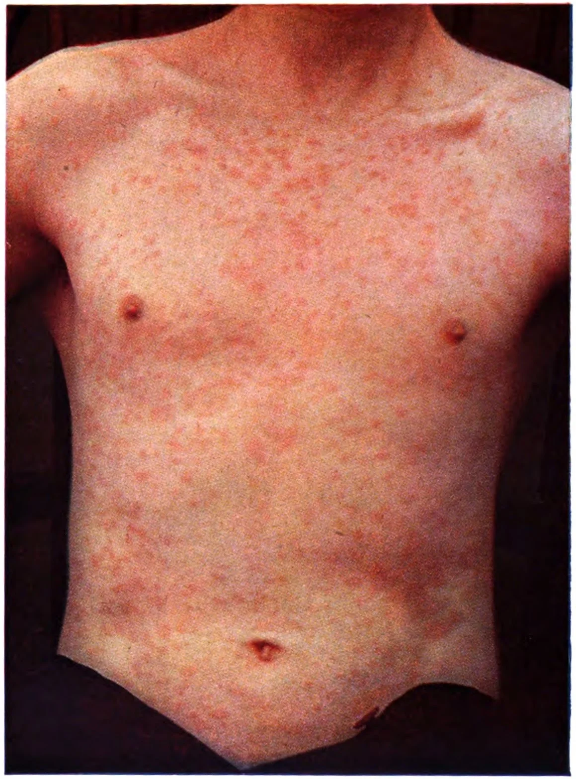
Long description
Colour plate (1919) of pityriasis rosea in which the eruption was on the trunk and upper segments of the limbs, consisting of a primary or herald patch on the left flank and medallions and smaller spots covered with a fine scale.

Figure 2Rhinophyma 01
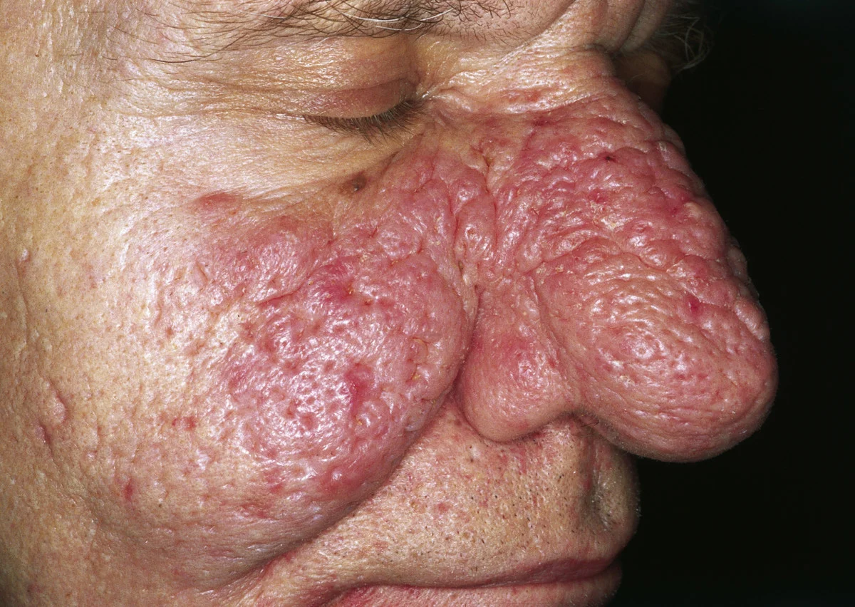
Long description
Clinical photograph described as 'Rhinophyma. Large exophytic, pink, lobulated mass over the nose with superficial vascular dilation. The lesion is spreading to the cheeks; however, it can also be limited to the nose.'

Both conditions are clinical diagnoses. Tables summarise when tests are used.
| Test | Rosacea | Pityriasis rosea |
|---|---|---|
| Clinical diagnosis | Yes; phenotype-based, after excluding mimics. [1][3] | Yes; on morphology and distribution. [4] |
| Skin biopsy | Not diagnostic; helps exclude lupus or sarcoid. | Rarely needed; non-diagnostic; excludes mimics. [5] |
| Serology (RPR/VDRL +/- treponemal) | Not indicated | Indicated when secondary syphilis is possible. [5][6] |
| KOH / mycology | Exclude tinea if suspected | Exclude tinea corporis if annular scaly. |
| Patch testing | Consider in suspected allergic mimics. | Not routine. |
| Ocular slit-lamp | For ocular phenotype or refractory eye symptoms. [1] | Not indicated. |
Diagnostic criteria and scores:
- Rosacea: no numeric severity score is mandatory. The phenotype approach records each feature present (persistent erythema, flushing, papules/pustules, telangiectasia, phyma, ocular) and its severity; the Rosacea Tracker tool formalises this. [1]
- Pityriasis rosea: no diagnostic criteria or scoring system with cut-offs; diagnosis is clinical, and "pathognomonic" herald patch may be absent. [4][5]
- Ocular disease: slit-lamp measures (Schirmer test, tear break-up time) quantify tear dysfunction; these guide treatment rather than diagnose. [2]
General measures (for every patient): gentle skin care, regular emollients, photoprotection (sunscreen, protective clothing, behavioural avoidance of triggers), and identification and avoidance of individual trigger factors (sun, heat, alcohol, spicy food, stress, exercise). [3][1] These measures are not optional extras; they remain the substrate on which drug therapy is built. [3]
Evidence-based treatments by feature (phenotype-led): [2][3]
| Clinical feature | Management options (evidence quality noted) |
|---|---|
| Persistent central facial erythema without papules/pustules | Topical alpha-agonist (brimonidine or oxymetazoline); intense pulsed light, KTP laser or pulsed-dye laser. [3] |
| Diffuse central facial erythema with papules/pustules | Topical metronidazole, azelaic acid or ivermectin; oral tetracyclines; topical alpha-agonists; oral isotretinoin for refractory disease. [3] |
| Flushing (episodic vasodilation) | Trigger avoidance and photoprotection; low-dose oral vasoconstrictive drugs (mirtazapine, propranolol, carvedilol); intradermal botulinum toxin (limited data). [3] |
| Telangiectasia | Vascular lasers / intense pulsed light (low-to-moderate certainty evidence). [2] |
| Phyma (early or fibrotic) | Oral isotretinoin for early soft changes; surgical/laser debulking (CO2 laser, erbium YAG, electrosurgery, dermabrasion) for established fibrotic phyma. [3] |
| Ocular rosacea | Lid hygiene, sunglasses, ocular lubricants; cyclosporin ophthalmic emulsion; topical metronidazole or ivermectin to external eyelid skin for blepharitis; oral doxycycline, erythromycin or azithromycin. [3] |
| Granulomatous rosacea | Oral tetracyclines, oral isotretinoin (0.7 mg/kg/day for 6 months), oral dapsone, topical pimecrolimus; combined device options (limited data). [3] |
Pharmacological doses and notes: [2][3]
- Topical ivermectin 1% cream, once daily: high-certainty evidence for improving papules/pustules; 615 of 910 participants rated good-to-excellent improvement vs 169 of 461 with vehicle; in head-to-head 1% ivermectin once daily was superior to metronidazole 0.75% cream twice daily, with a longer median time to first relapse (115 vs 85 days). [2][3]
- Topical metronidazole, available as 0.75% gel or cream and 1% cream or gel; 0.75% cream was used twice daily in trials. Moderate-certainty evidence of effectiveness vs vehicle. [2][3]
- Topical azelaic acid 15% gel or 15% foam, twice daily, or 20% cream. High-certainty evidence. Adverse events (burning, stinging) are common but usually mild and managed by gentle skin care. [2][3]
- Sub-antibiotic (modified-release) doxycycline 40 mg once daily: moderate-to-high-certainty evidence for papules/pustules and probably slight erythema reduction; 40 mg is at least as effective as 100 mg with fewer side effects. 40 mg MR achieved "clear" or "almost clear" in 91 of 353 vs 53 of 354 with placebo. Because it is sub-antibiotic it is the preferred initial oral therapy (avoids bacterial selection pressure). [2][3]
- Oral tetracycline 250 mg twice daily: low-certainty evidence. [2]
- Oral isotretinoin 0.25 to 0.3 mg/kg/day: high-certainty evidence for improvement in papules/pustules in difficult, antibiotic-refractory or relapsing disease; best for early soft phyma. [2][3]
- Topical alpha-agonists: brimonidine 0.33% gel, oxymetazoline 1% cream, once daily in the morning. Brimonidine reduces persistent erythema for up to 12 hours after application (high certainty); oxymetazoline onset 1 to 3 hours, duration 8 to 10 hours (moderate certainty). [2][3] Combined therapy (topical alpha-agonist for erythema plus ivermectin or metronidazole for papules) improves both features. [2][3]
- Minocycline 1.5% foam: moderate-certainty evidence; oral azithromycin is an alternative when tetracyclines fail or are poorly tolerated (caution in patients with cardiac risk). [2][3]
Combination therapy: combining rationally chosen treatments increases the chance of clearing the skin and lengthens remission, because different features respond to different mechanisms. [1][2] Desired outcomes: "clear" (IGA clear/almost clear) rather than "almost clear" as treatment goal. [1][2]
Red flags in rosacea: rapidly progressive papulopustular disease on the background of immunosuppression, nodulocystic acne-like disease, or any lesion not typical of rosacea (biopsy to exclude lupus or a malignant process). [1][3]
Treatment is symptomatic because the condition resolves spontaneously. Current standard of care controls itch and inflammation: emollients, antipruritic lotions, topical corticosteroids (such as hydrocortisone or betamethasone of appropriate potency, under professional guidance) and oral antihistamines. [5]
Evidence from the Cochrane systematic review (14 RCTs, 761 patients): [5]
- Oral acyclovir (800 mg 5 times daily for 7 days, or 400 mg 5 times daily for 7 days in one trial) probably increases good-to-excellent medical practitioner-rated rash improvement compared with placebo, no treatment or vitamins (RR 2.45, 95% CI 1.33 to 4.53; 3 studies, 141 participants), but evidence on itch was inconclusive and, in one trial, itch resolved more often with placebo than with acyclovir. [5]
- Oral acyclovir added to standard care (calamine plus cetirizine) may improve itch resolution (RR 4.50, 95% CI 1.22 to 16.62) and reduce itch score (MD 1.26, 95% CI 0.74 to 1.78; low-certainty evidence). [5]
- Erythromycin probably reduces itch more than placebo (MD 3.95 on a 0 to 10 scale; one study, 34 participants, moderate certainty) but its effect on rash improvement is uncertain (low certainty).
- Clarithromycin probably does not differ from placebo in itch resolution (RR 0.84, 95% CI 0.47 to 1.52) or rash improvement (RR 1.13, 95% CI 0.89 to 1.44). [5]
- Azithromycin probably does not differ from placebo or vitamins in itch resolution (RR 0.83, 95% CI 0.28 to 2.48) or rash improvement (RR 1.02, 95% CI 0.52 to 2.00). [5]
- No serious adverse events were reported; side effects were minor (gastrointestinal upset, mild abdominal pain, headache).
- Small study sizes, heterogeneity and bias in blinding and selective reporting limit these conclusions; further research should examine different acyclovir doses and the role of antivirals.
Practical interpretation: explain to the patient that pityriasis rosea is self-limited and that treatment is for comfort (emollients, topical steroid, antihistamine). A course of oral acyclovir may be offered within the first weeks to speed rash resolution, with the evidence being moderate quality for rash outcome and uncertain for itch. Antibiotics are not reliably helpful and should not be used routinely. [5] (Check local guidance before prescribing any systemic treatment.)
Approach to rosacea in the clinic
- Patient presents with facial redness, flushing or papulopustular lesionsTake history of triggers; examine distribution and exclude acne, lupus, seborrhoeic dermatitis and perioral dermatitis
- Apply phenotype-based diagnostic criteriaFixed central erythema or phyma alone is diagnostic; otherwise need two or more major features (papules/pustules, flushing, telangiectasia, ocular)
- Identify which features are presentPersistent erythema / flushing / papules and pustules / telangiectasia / phyma / ocular
- Question: Which feature dominates management?
Persistent erythema (no or few PP lesions)
- Topical alpha-agonist once each morningBrimonidine 0.33% gel or oxymetazoline 1% cream; add vascular laser or IPL for telangiectasia
Papules/pustules present
- Topical anti-inflammatoryIvermectin 1% cream once daily, or metronidazole 0.75% gel/cream twice daily, or azelaic acid 15% gel/foam twice daily
- If moderate-severe or refractoryAdd sub-antibiotic doxycycline 40 mg modified release once daily; reserve isotretinoin 0.25-0.3 mg/kg/day for difficult or phymatous disease
Phymatous changes
- Early/soft: oral isotretinoin; established fibrotic: surgical or laser debulkingRefer to dermatology/plastic surgery for phyma
Ocular features or eye symptoms
- Lid hygiene, lubricants; refer to ophthalmology if severeTopical cyclosporin or doxycycline/erythromycin/azithromycin as appropriate
- Combine treatments when more than one feature is presentAlpha-agonist for erythema plus ivermectin or metronidazole for PP lesions; re-assess at 8-12 weeks
- Maintenance and low resource pathwayMaintenance topical therapy after control; where drugs are unavailable, use emollients, sun protection, trigger avoidance and the most affordable of metronidazole/azelaic acid
Rosacea. [3] Ocular rosacea can threaten vision if keratitis or sclerokeratitis progress; blepharokeratoconjunctivitis, sclerokeratitis and anterior uveitis warrant ophthalmology referral. [1] Established phyma (especially rhinophyma) causes cosmetic and functional impairment. Chronic disease affects quality of life, self-esteem and psychosocial wellbeing; "clear skin" rather than "almost clear" is the therapeutic target because it demonstrably delays relapse. [1][2] Complications of isotretinoin (cheilitis, dryness, myalgia, possible teratogenicity) require counselling and monitoring. Device therapies and alpha-agonists can produce rebound erythema in a minority of patients. [2]
Pityriasis rosea. It is essentially benign and self-limited. Potential problems: moderate-to-severe pruritus, temporary pigmentary change (especially in dark skin), anxiety and under-diagnosis in primary care. In pregnancy, PR has been reported to be followed by premature delivery, neonatal hypotonia or fetal death, with an overall abortion rate of 13 percent and up to about 60 percent if the rash develops within the first 15 weeks of pregnancy; such cases warrant specialist, multidisciplinary care. [5]
Rosacea. Rosacea is chronic and relapsing: features vary over time and management is long-term, requiring ongoing dialogue between patient and clinician. [1] Periods of control are possible, especially with maintenance topical therapy (metronidazole 0.75%, ivermectin 1% or azelaic acid 15% gel), but phymatous and fixed vascular change are largely irreversible without procedural treatment. [2] Sub-antibiotic doxycycline maintains control in many patients. [2][3]
Pityriasis rosea. Lesions appear over the first 2 to 6 weeks and then clear spontaneously; the whole illness lasts 2 to 12 weeks (occasionally up to 7 months). [5] Most people never have a second episode. [5] Pigmentary change may linger for months. Itch is present in 80 to 90 percent during the acute phase; moderate-to-severe itch occurs in one-third to one-half. [5]
Neither condition has a curable or vaccine-preventable cause in day-to-day practice.
- Rosacea: rigorous trigger identification and avoidance (sun protection is the single most consistent measure), gentle skin care from the first, and avoidance of mid-potency or high-potency topical corticosteroids on the face (they can cause or worsen perioral dermatitis/rosacea). Ocular screening (ask about dryness and redness) should be routine in rosacea, with early referral. [1][3]
- Pityriasis rosea: no prophylaxis or screening exists. Education (reassurance, hand/soap hygiene, avoiding sharing of clothing while under investigation in atypical cases) and exclusion of secondary syphilis are the preventive actions. [5][6]
Pregnancy.
- Rosacea: general measures and topical metronidazole, azelaic acid and ivermectin are generally acceptable, but oral tetracyclines (including doxycycline) and isotretinoin are contraindicated; management should follow local formularies. [3]
- Pityriasis rosea: reassuring the majority, but monitor pregnancies closely when PR appears early (under 15 weeks), given the reported association with prematurity, neonatal hypotonia and fetal death. [5]
Children.
- Rosacea can occur, and in children or young adults with a myxoma-like (familial) pattern or recurrent tumour, think of Carney complex rather than rosacea mimics (not applicable); rosacea itself is treated with the same phenotypes but with paediatric doses. Tetracyclines stain developing teeth and are avoided while the teeth are developing (up to about 8 years); for a clear indication, short courses may be used on specialist advice. [7]
- PR most commonly affects 15-30 year-olds but occurs in children too; treatment remains symptomatic (emollients, topical steroid, antihistamine). [5]
Older adults.
- Phymatous and persistent vascular change are more prominent with long disease duration; surgical options need cardiovascular and healing assessment. [3]
- Rosacea can first present in older adults; exclude cutaneous lupus and malignancy in atypical lesions. [3]
Immunocompromised patients. Consider unusual, disseminated or nodulocystic variants and biopsy any atypical lesion before settling on a rosacea diagnosis.
Where dermatologists, lasers or newer drugs are not available: focus on clinical diagnosis and mimic exclusion; use emollients, sun protection and trigger avoidance for all; metronidazole 0.75% (inexpensive, widely available) is the first-choice topical agent for papules and pustules; oral doxycycline 100 mg daily is a reasonable alternative where 40 mg modified release is unavailable, with the caveats above; for itch in pityriasis rosea use emollients, calamine or a topical corticosteroid plus an oral antihistamine. Refer atypical or treatment-resistant cases and any ocular involvement. [2][3][5]
- Rosacea is a chronic, relapsing, central-facial inflammatory dermatosis diagnosed clinically by feature (phenotype), not by historical subtype.
- Diagnostic features: fixed central erythema or phyma; major features: papules/pustules, flushing, telangiectasia, ocular features; secondary features alone are not diagnostic.
- Evidence-based match: erythema to alpha-agonists and lasers; papules/pustules to ivermectin, metronidazole or azelaic acid topically and doxycycline 40 mg modified-release orally; early phyma to isotretinoin; established phyma to surgery.
- Topical ivermectin 1% once daily is more effective than metronidazole 0.75% twice daily and has a longer relapse-free interval; both are good choices for papules/pustules.
- Sub-antibiotic doxycycline 40 mg modified release is the preferred first oral agent because it works without driving antibacterial resistance; 40 mg is as effective as 100 mg with fewer side effects.
- Aim for "clear" rather than "almost clear" skin, because complete clearance lengthens remission and improves quality of life.
- Pityriasis rosea is an acute, self-limited papulosquamous disease in young adults, lasting 2 to 12 weeks, usually after one herald patch.
- Herald patch (reported 1-3 cm in Cochrane, sometimes larger, rose, scaly, paler centre, often trunk) then smaller lesions along skin cleavage lines in a "Christmas tree" pattern; herald patch may be absent. [5][8]
- Itch affects 80-90 percent of patients; moderate-to-severe itch in one-third to one-half; post-inflammatory pigment change may linger.
- Probable cause is HHV-6/HHV-7 reactivation; drugs and vaccines can produce PR-like eruptions.
- Secondary syphilis can be identical: exclude with nontreponemal (RPR/VDRL) and treponemal serology in all atypical, high-risk or treatment-resistant cases.
- Pityriasis rosea in early pregnancy (under 15 weeks) is linked to premature delivery, neonatal hypotonia and fetal loss; arrange specialist care.
- Acyclovir may speed rash clearance in PR (moderate-quality evidence) but has uncertain effects on itch; clarithromycin and azithromycin are probably not helpful; erythromycin may reduce itch.
- Neither condition is curable, but rosacea needs long-term phenotype-based management; pityriasis rosea needs reassurance and symptomatic care.
Answer each one in your head first, then open the answer.
A 28-year-old woman has recurrent facial flushing, a fixed redness over the cheeks and nose, and dome-shaped papules without comedones. What is the diagnosis and why is it not acne vulgaris?
Show answerHide answer
Rosacea in the papulopustular phenotype: the central facial distribution, fixed erythema and telangiectasia, history of flushing and absence of comedones. Acne vulgaris has comedones, a different distribution (including trunk), and no telangiectasia or phyma, and usually begins in adolescence.
Why is doxycycline 40 mg modified release the preferred first oral agent for moderate rosacea papules and pustules, rather than 100 mg?
Show answerHide answer
At 40 mg modified release it is sub-antibiotic: it reduces inflammation without bacterial selection pressure. It is at least as effective as 100 mg, with fewer side effects, and it improved "clear/almost clear" outcomes in randomised trials.
Which drug would you choose for a rosacea patient whose main problem is persistent background erythema, and which for papules and pustules?
Show answerHide answer
Persistent erythema: a topical alpha-agonist (brimonidine 0.33% gel or oxymetazoline 1% cream in the morning) and, for telangiectasia, vascular laser or IPL. Papules/pustules: ivermectin 1% cream once daily, or metronidazole 0.75% gel/cream twice daily, or azelaic acid 15% twice daily; add doxycycline 40 mg modified release if needed.
A 22-year-old has a solitary 2 cm rose-coloured scaly patch on the trunk, then ten days later a widespread truncal rash following skin lines in a 'Christmas tree' pattern. PR is likely, but what must you exclude first?
Show answerHide answer
Secondary syphilis, which can look identical. Test with a nontreponemal test (RPR or VDRL, with titre) and confirm with a treponemal test; if screening and clinical picture disagree, use a second treponemal assay and treat presumptively where appropriate.
The Cochrane review on pityriasis rosea found that oral acyclovir probably improves rash clearance but what about itch?
Show answerHide answer
Evidence on itch with acyclovir was inconclusive, and in one trial itch resolved more often with placebo. Erythromycin probably reduces itch; clarithromycin and azithromycin probably do not. Advise patients that relief comes from supportive care; acyclovir is optional evidence-based add-on therapy.
A pregnant woman presents with a typical PR rash that began early in pregnancy. What is the key management point?
Show answerHide answer
Arrange obstetric awareness and closer fetal monitoring, because PR starting before 15 weeks has been associated with premature delivery, neonatal hypotonia and fetal loss (up to about 60% in that window); reassure where appropriate and treat itch symptomatically.
A 45-year-old has rhinophyma. What is the treatment pathway?
Show answerHide answer
Established fibrotic phyma needs procedural reduction (CO2 laser, erbium YAG, electrosurgery or dermabrasion) with dermatology/plastic surgery referral; oral isotretinoin is useful for early, soft, inflammatory phymatous change before fibrosis.
Differential diagnoses mentioned above (acne vulgaris, cutaneous lupus, tinea corporis, secondary syphilis) and the broader topic of dermatological drug safety and pregnancy. Where separate MedGrasp topics exist for them, they would be linked here via their slugs once verified.
- Schaller M, Almeida LMC, Bewley A, Cribier B, Del Rosso J, Dlova NC, Gallo RL, Granstein RD, Kautz G, Mannis MJ, Micali G, Oon HH, Rajagopalan M, Steinhoff M, Tanghetti E, Thiboutot D, Troielli P, Webster G, Zierhut M, van Zuuren EJ, Tan J. Recommendations for rosacea diagnosis, classification and management: update from the global ROSacea COnsensus 2019 panel. Br J Dermatol 2020;182(5):1269-1276. doi:10.1111/bjd.18420. Accessed .
- van Zuuren EJ, Fedorowicz Z, Tan J, van der Linden MMD, Arents BWM, Carter B, Charland L. Interventions for rosacea based on the phenotype approach: an updated systematic review including GRADE assessments. Br J Dermatol 2019;181(1):65-79. doi:10.1111/bjd.17590. Accessed .
- Del Rosso JQ, Tanghetti E, Webster G, Stein Gold L, Thiboutot D, Gallo RL. Update on the Management of Rosacea from the American Acne & Rosacea Society (AARS). J Clin Aesthet Dermatol 2019;12(6):17-24. Accessed .
- Litchman G, Nair PA, Syed HA, Le JK. Pityriasis rosea (StatPearls chapter). StatPearls Publishing, Treasure Island (FL), 2024. Accessed .
- Contreras-Ruiz J, Peternel S, Jiménez Gutiérrez C, Culav-Kosçak I, Reveiz L, Silbermann-Reynoso M de L. Interventions for pityriasis rosea. Cochrane Database Syst Rev 2019; (10):CD005068. doi:10.1002/14651858.CD005068.pub3. Accessed .
- Centers for Disease Control and Prevention. Sexually Transmitted Infections Treatment Guidelines, 2021 (Syphilis section). CDC, last reviewed 2024. Accessed .
- Centers for Disease Control and Prevention. Research: Doxycycline and Tooth Staining (warning on use of tetracycline-class antibiotics in children under 8 years). CDC Rocky Mountain Spotted Fever (RMSF) resource. Accessed .
- American Academy of Dermatology. Pityriasis rosea: signs and symptoms. AAD patient information. Accessed .
Figure credits
Educational content for medical students and doctors. It is not a substitute for clinical judgement or local guidelines.