Contact, seborrhoeic and stasis dermatitis
Irritant and allergic contact dermatitis, seborrhoeic dermatitis, and stasis dermatitis: diagnosis, patch testing, and treatment.
- Contact dermatitis is one of the commonest skin problems. Irritant contact dermatitis (ICD) damages the skin barrier directly; allergic contact dermatitis (ACD) is a delayed type IV hypersensitivity to a small chemical hapten. Both can look identical, so the mechanism - and keeping or discarding the possibility of allergy - matters more than the rash.
- Seborrhoeic dermatitis (SD) is a chronic, greasy, scaly rash in sebaceous-rich sites (scalp, eyebrows, nasolabial folds, chest). It is driven by cutaneous lipids and Malassezia yeasts, together with host skin-barrier and immune factors. It affects around 5% of people worldwide. [4]
- Stasis dermatitis is an eczematous rash of the lower legs produced by chronic venous insufficiency. It affects around 6% of people older than 50 years, about twice as common as psoriasis, and can progress to dermatoliposclerosis and venous ulcers. [6]
- ICD is a default diagnosis: there is no confirmatory test, so allergic contact dermatitis must be excluded with patch testing before you call a rash "irritant".
- SD is a clinical diagnosis with no universally accepted guideline; expert consensus favours combined antifungal and anti-inflammatory treatment for flares and maintenance, and systemic escalation for refractory disease. [5]
- Stasis dermatitis is managed by treating the underlying venous disease: topical anti-inflammatory care, short-stretch compression bandaging, leg elevation, and, where available, adjunct intermittent pneumatic impulse compression.
By the end of this page you should be able to:
- Distinguish irritant from allergic contact dermatitis by mechanism, history, morphology, and distribution, and explain why the distinction matters for treatment and prognosis. [1]
- List the thresholds that define wet work and the conditions that lead to cumulative irritant contact dermatitis.
- Explain how haptens produce a delayed type IV sensitisation, and recognise the three phases (sensitisation, elicitation, resolution).
- Describe the indications, technique, timing of readings, and contraindications for patch testing, and interpret a result.
- Explain the role of Malassezia, sebum lipids, skin barrier, and host immune and oxidative factors in seborrhoeic dermatitis, and outline its treatment ladder.
- Recognise stasis dermatitis clinically and relate it to chronic venous insufficiency, stating the basic compression-based management and its safety checks.
- Separate these rashes from psoriasis, atopic dermatitis, tinea, cellulitis, and other leg rashes using a structured comparison.
- Apply the diagnostic and management algorithm in the flowchart and recognise low-resource adaptations.
A dermatitis is an inflammation of the skin. The three conditions grouped on this page share an eczematous morphology but have different causes, sites, behaviour, and treatment. Defining them precisely is the first step in reasoning about them.
DefinitionContact dermatitis
Contact dermatitis is an inflammatory skin reaction caused by direct contact with noxious agents in the environment. It may be an allergic, delayed (type IV) hypersensitivity (allergic contact dermatitis) or a non-specific innate immune injury to the skin barrier (irritant contact dermatitis), or a mixture of the two. [3]
- Irritant contact dermatitis (ICD) is a non-specific response to direct chemical damage. The irritant disrupts the skin barrier, releases primary cytokines (among them interleukin-1 alpha, interleukin-1 beta, and tumour necrosis factor alpha), activates innate immune cells, and recruits inflammatory cells. There is no antigen-specific sensitisation and no immunological memory in the ordinary sense; severity rises with concentration, dose, and duration of exposure. [1]
- Allergic contact dermatitis (ACD) is a delayed (type IV) hypersensitivity to an exogenous contact allergen. The allergens are reactive chemicals, usually of molecular weight under 500 daltons and exceptionally in the range 500 to 1000 daltons. They are haptens that bind to skin protein to become immunogenic. A sensitised person develops ACD on re-exposure, and the condition persists until the allergen is avoided. [3] [2]
The two are classified into clinical variants. ICD includes: acute ICD after a single exposure to a potent irritant; delayed acute ICD, where substances such as benzalkonium chloride, anthralin, calcipotriol or tretinoin take 8 to 24 hours to produce visible inflammation; cumulative (chronic) ICD after repeated subthreshold insults; an irritant reaction that begins under occlusive jewellery and may spread; subjective or sensorial irritation without visible signs; non-erythematous irritation with barrier change only; frictional, traumatic, pustular or acneiform, asteatotic, and airborne irritant dermatitis. [1] ACD includes contact eczema in acute, subacute, and chronic stages, photo-contact dermatitis (lesions confined to sun-exposed areas even though the allergen has also touched covered skin), allergic contact urticaria, and protein contact dermatitis. [3]
Seborrhoeic dermatitis (SD) is a common, chronic, recurrent inflammatory skin disease. It presents as pink or red, greasy, pruritic plaques with fine scaling on sebaceous-rich skin of the nasolabial folds, eyebrows, chin, scalp, retroauricular regions, and presternal area. [4] A patchy, petaloid variant with polycyclic rings can occur, and SD on darker skin may present with hypopigmented patches that usually improve with treatment. [4]
Stasis dermatitis is an inflammatory skin condition of the lower extremities caused primarily by chronic venous insufficiency. It is characterised by pruritus, pain, oedema (swelling), and skin discolouration, progressing in some patients to dermatoliposclerosis and venous ulcers. [6] It is diagnosed clinically when bilateral erythema, scaling, and swelling of both lower legs are present, with pruritus, hyperpigmentation, or ulceration also possible. [6]
ACD affects 15 to 20% of individuals in industrialised regions. [2] ICD is more common than ACD overall and is the most common presentation of occupational skin disease. [1] In one tertiary referral occupational dermatology clinic in Australia, ICD was diagnosed in 71% of patients with occupational skin disease. [1] Half of the cases of occupational contact dermatitis are observed to appear within the first 2 years of employment. [1]
High-risk occupations for ICD include healthcare workers, food service workers, metal workers, hairdressers, and construction workers. [1] Within contact dermatitis generally, men and women are both affected; women show higher rates of occupations involving wet work. [1]
Risk factors for ICD include atopy, especially a personal history of atopic dermatitis rather than mucosal atopy, because of impaired epidermal barrier function with higher transepidermal water loss and increased skin permeability. [1] Filaggrin loss-of-function mutations raise risk when combined with atopy. [1] Genetic variants in the major histocompatibility complex and a tumour necrosis factor alpha gene polymorphism (the A allele at position P308) are linked to lower irritant thresholds. [1] Asteatotic irritant dermatitis is common in older people with dry skin, particularly in low humidity. [1]
SD affects approximately 5% of the global population, with great regional variation. [4] Reported figures include 0.25% in rural India and 30.30% of patients in dermatology centres in Jamaica; 0.50% of imprisoned men in Italy and 16.30% in Italian geriatric dermatology clinics. [4] SD occurs more often in men than in women, appears in infants within the first 3 months of life and in adolescents and young adults, and becomes more prevalent over the age of 50 years. [4]
Immunocompromised patients are especially susceptible, including organ transplant recipients, patients with lymphoma, and people with human immunodeficiency virus (HIV) infection. The prevalence of SD in HIV-positive patients reaches approximately 83%, most commonly when CD4+ T-lymphocyte counts are between 200 and 500 per cubic millimetre. [4] Other associations given weight by expert consensus are insulin resistance and metabolic syndrome, cancer (with no firm consensus), neurological conditions (Parkinson disease), and psychiatric conditions and stress. [5] Alcohol consumption is associated with severity. [5] There are no universally accepted guidelines for the diagnosis, classification, or treatment of SD, which is part of why it is managed inconsistently. [5]
Stasis dermatitis affects approximately 6% of individuals older than 50 years in the United States, making it about twice as common as psoriasis. [6] It is a problem of older adults and of chronic venous insufficiency; a significant proportion of affected inpatients also have lymphoedema, cardiac insufficiency, lipedema, or arterial disease rather than uncomplicated venous disease. [6] The burden is heavy: in one inpatient cohort the mean Wound Quality of Life score was 44.5. [6]
The conditions that worsen or mimic it - obesity, prolonged standing, previous deep vein thrombosis, and poorly controlled cardiac, renal, or hepatic disease - should be sought in every patient. From the consensus literature on contact dermatitis, occupational wet-work thresholds help quantify risk elsewhere: exposure of skin to liquid for more than 2 hours per day, use of occlusive gloves for more than 2 hours per day or a change of gloves more than 20 times per day, and hand washing or disinfectant use more than 20 times per day. [1]
ICD was once thought to be non-immunological; it is now recognised that immune mechanisms drive it. The irritant disrupts epidermal cells and the epithelial barrier, allowing increased penetration. Keratinocytes convert the insult into cytokine and chemokine release; T-lymphocytes recruited to the skin carry the cutaneous lymphocyte antigen. [1] Pattern recognition receptors (toll-like and NOD-like receptors) activate innate responses via the inflammasome and NF-kappa B pathways, and oxidative stress with reactive oxygen species contributes. [1] Cumulative ICD results when the interval between exposures is too short for the barrier to recover; the threshold for inflammation falls as the disease progresses. [1] Delayed acute and subjective irritation explain how a patient can react 8 to 24 hours after an exposure and still have "irritant" disease. [1]
ACD is a delayed (type IV) hypersensitivity. Haptens (reactive chemicals, usually under 500 daltons) covalently modify endogenous skin proteins to form immunogenic neoantigens. Dendritic cells and Langerhans cells process the neoantigen, migrate to lymph nodes, and prime naive T cells during a clinically silent sensitisation (induction) phase. On re-exposure the effector T cells (Th1 and Th17, with cytotoxic T lymphocytes) produce cytokines and damage keratinocytes through perforin/granzyme and Fas/Fas ligand pathways. Resolution depends on regulatory T cells and on interleukin-10 and transforming growth factor beta. [2] [3]
The immunogenicity of haptens is dose dependent and follows high-zone tolerance patterns, so sensitising thresholds vary by allergen and by route of exposure. [2] Pattern recognition receptors, including toll-like receptor 4 (for nickel), the NLRP3 inflammasome, and C-type lectin receptors such as Mincle, link barrier damage and danger signals to the inflammatory cascade. [2]
A helpful clinical mnemonic is the direction of change over time: ACD tends to worsen after removal of the allergen (a "crescendo" pattern) because the immune response continues, whereas acute ICD tends to peak and then resolve (a "decrescendo" pattern) once the irritant is gone. [1]
SD has a multifactorial aetiology combining Malassezia colonisation, sebaceous activity, and host immune response. [5] The Malassezia genus comprises 18 yeast species that live on human skin; they are opportunistic organisms with virulence factors including indoles, reactive oxygen species, hyphae formation, and biofilm production. [4] Sebum lipids are hydrolysed by Malassezia to produce skin-irritating metabolites (oleic acid, arachidonic acid, malassezin, indole-3-carbaldehyde). These interfere with keratinocyte differentiation and induce pro-inflammatory cytokines, including interleukin (IL)-1 alpha, IL-1 beta, IL-6, IL-12, interferon gamma, and tumour necrosis factor alpha. Arachidonic acid is further converted to prostaglandins, recruiting neutrophils. [4]
At the same time, three broad factors - skin barrier dysfunction, altered immune responses, and Malassezia overgrowth - underlie SD. Whether Malassezia is a primary cause or an incidental passenger has not been definitively established. [4] Systemic and local molecular changes in SD include a Th1/Th17-skewed cutaneous response, elevated beta-endorphin correlating with severity and itch, thiol/disulfide redox imbalance, oxidative stress and lipid peroxidation, genetic susceptibility signals (heritability around 14%, loci such as MAST4), and epigenetic microRNA dysregulation. [4] SD is therefore not simply "yeast and sebum"; it is a disorder in which sebaceous lipids feed a yeast, the yeast produces irritants, and the host response determines whether disease develops.
Stasis dermatitis is produced by chronic venous insufficiency, with venous hypertension, inflammatory cell accumulation, and extravasation across the endothelium. [6] It is not purely a skin disease: the underlying problem is venous return and microcirculation. The RCT evidence in this dossier shows that additional intermittent pneumatic impulse compression on top of standard compression improved tissue oxygen saturation and reduced ankle oedema where wrapping alone did not. [6] Contributors listed in that cohort included chronic venous insufficiency in most patients, lymphoedema in several, and cardiac, lipedema or arterial disease in others. [6]
Figure 1Stasis dermatitis (Gravitational eczema)
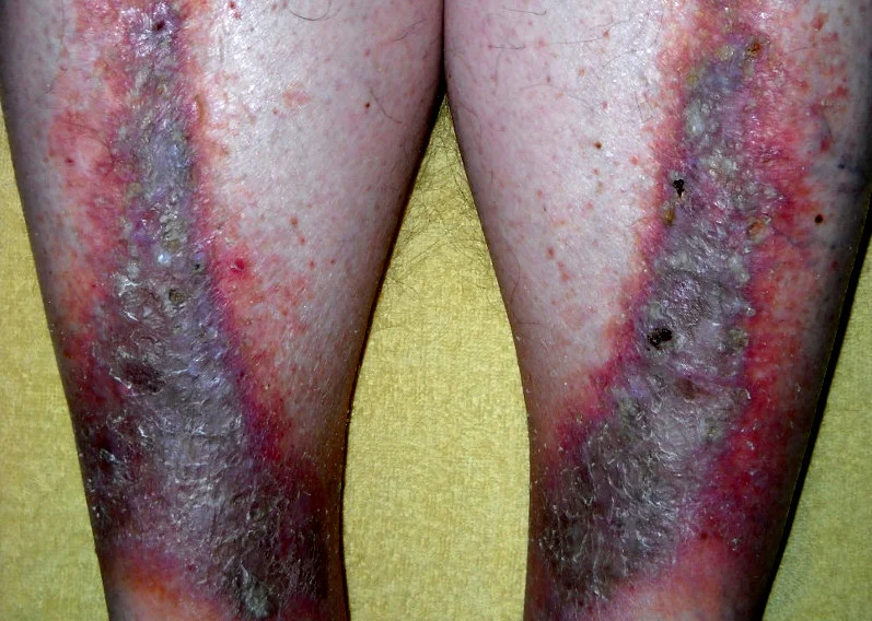
Long description
Clinical photograph titled 'Stasis dermatitis (Gravitational eczema)' showing stasis dermatitis (gravitational eczema).

Figure 2Seborrhoeic dermatitis highres
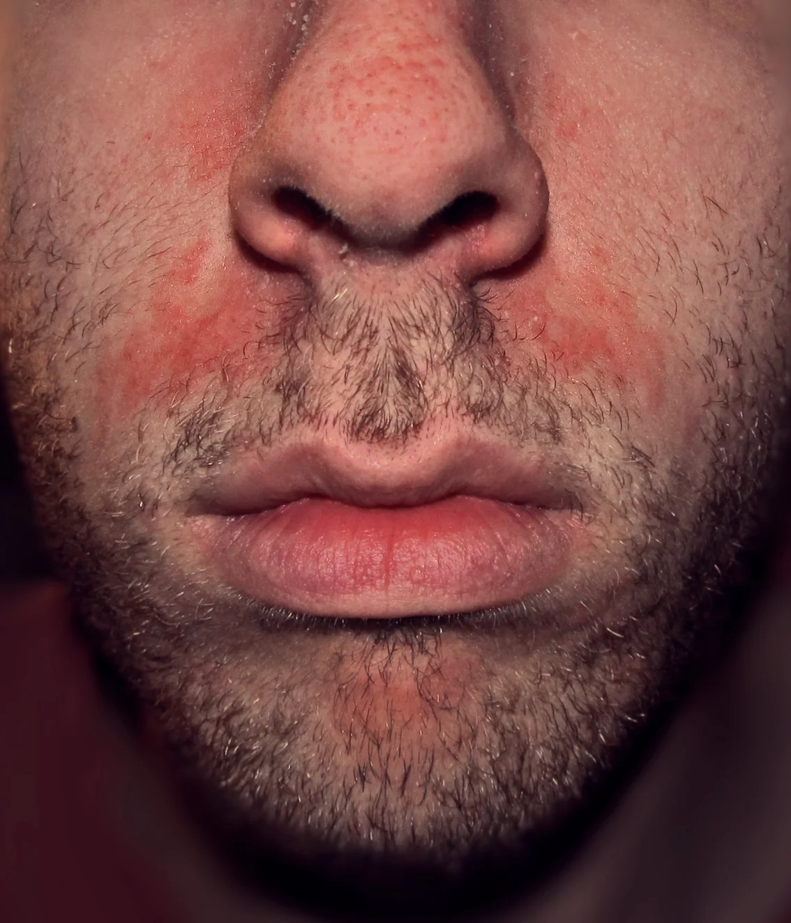
Long description
Self-published clinical photograph titled 'Seborrhoeic dermatitis' in Commons category 'Stubble', i.e. a facial photograph of seborrhoeic dermatitis; the file description itself does not name the nasolabial folds.

Figure 3Cradle cap new photo help in diagnosis
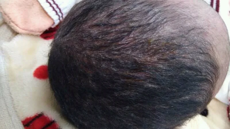
Long description
Clinical photograph used in the Wikipedia article on seborrhoeic dermatitis to illustrate cradle cap, with yellowish greasy scales on an infant scalp; the file caption describes cradle cap as a benign seborrhoeic dermatitis of the infant scalp.

History should probe occupation, hobbies, cosmetics, medications, topical products, metals, plant exposure, wet work, and the time course. ACD is suspected when the rash is restricted to the contact area of a discrete object or product, stops at a clear edge (for example where a watch strap or clothing ends), and recurs after re-exposure. ICD follows repeated contact; it often starts where an irritant pools (between the fingers, on the dorsum of the hand from a ring, under a wet glove). Symptoms differ: ICD tends to burn, sting, or feel sore rather than itch; ACD often itches and is accompanied by acute vesicles, oedema, and weeping in the acute stage. [1] [3]
Examination records distribution, symmetry, morphology, and any linear or geometric pattern. Acute ACD presents with erythema, oedema, papules, vesicles, oozing and crusting; chronic disease shows scaling, fissures, lichenification, and hyperkeratosis. [3] Acute ICD produces erythema, oedema, bullae, and necrosis and heals in a decrescendo pattern; delayed acute ICD mimics allergic disease; cumulative ICD is dry, lichenified, and distributed over the hands and forearms. [1] Secondary infection, nail dystrophy, or post-inflammatory pigment change can complicate both.
SD preferentially occupies scalp (dandruff at the mild end, diffuse erythema and thick scale at the severe end), eyebrow, glabella and nasolabial folds, beard, retroauricular folds, ears, presternal chest, and intertriginous areas. Greasy yellowish scale rather than dry white scale is characteristic. Pruritus varies. In HIV or neurological disease the rash is more extensive, more severe, and more treatment resistant. [4] [5] On darker skin SD may leave hypopigmented macules or annular petaloid plaques. [4]
Stasis dermatitis is bilateral on the lower legs, usually from the ankle to the mid-calf. It presents as erythema, scaling, and oedema, with pruritus or pain, and commonly brown haemosiderin hyperpigmentation. Lipodermatosclerosis or varicose veins are supporting signs. Ulcers may be present. [6] The rash spares the foot, which helps separate it from cellulitis and allergic contact reactions from topical products used on the legs. [6]
Examine the patient standing and seated, record oedema, and measure leg circumference. In the study gown's inpatient setting trained staff measured ankle, 10 cm below the knee, and 15 cm above the knee and assessed transcutaneous oxygen pressure on the dorsum of the foot. [6]
There is no single diagnostic test for ICD or for SD. The diagnosis is clinical, with tests used to exclude look-alikes and, in contact dermatitis, to identify an allergen.
| Test | What it measures | Cut-off or key point | Source |
|---|---|---|---|
| Patch testing | Reproduces the elicitation phase of type IV allergy under occlusion on skin, with readings up to 7 days later | Applied as a baseline series in every patient suspected of contact allergy; a hapten allergy prevalence above 0.5 to 1.0% justifies inclusion in a baseline series | [3] |
| Measurement of wet work | Occupational/domestic exposure | Skin wet more than 2 hours per day; occlusive gloves more than 2 hours or more than 20 changes per day; hand washing or disinfectant more than 20 times per day | [1] |
| Postponement of patch testing | Conditions that distort results | Sufficient washout before patching: 7 days after low or medium potency topical steroid, 14 days after high potency topical steroid; active generalised dermatitis, recent sun or ultraviolet exposure, and pregnancy or lactation (postponed as a precaution) are reasons to defer | [3] |
| Clinical diagnosis of ICD | Exclusion diagnosis | No routine confirmatory test; dermatitis starts at the site of contact, generally does not spread; negative patch testing supports it | [1] |
| Bioengineering tests (transepidermal water loss) | Skin barrier function in ICD | Research tools, not routine in clinic | [1] |
| Clinical diagnosis of SD | Pattern recognition | Greasy scaly plaques in seborrhoeic sites; HIV test when appropriate; no universally accepted guideline or criterion | [4] [5] |
| Clinical diagnosis of stasis dermatitis | Recognised pattern | Bilateral lower leg erythema, scaling, swelling, pruritus or pain, hyperpigmentation; exclude infection and arterial disease | [6] |
| Assessment before compression | Arterial supply and contraindications to impulse compression | Check leg pulses before treating; intermittent pneumatic impulse compression is contraindicated in acute untreated infection (phlebitis, erysipelas), current thrombosis or pulmonary embolism, acute decompensated heart failure, severe peripheral arterial disease, and active bleeding or high haemorrhage risk | [6] |
No blood test diagnoses a contact or seborrhoeic dermatitis. Histology does not distinguish irritant from allergic disease reliably; reflectance confocal microscopy can show more superficial epidermal disruption in irritant disease in experimental settings but is not a routine clinical tool. [1] Allergic contact dermatitis is diagnosed once a relevant positive patch test reaction is found in a patient with compatible dermatitis. [3]
The cornerstone of management is removing the trigger, because neither ICD nor ACD heals while exposure continues.
For ICD: avoidance of the irritant, protection of the skin (appropriate gloves chosen for the specific task and chemical, changed frequently and with correct donning technique), and use of topical therapy, particularly moisturisers applied before and after shifts and after hand washing. [1] Soap-free cleansers are preferable to harsh detergents. Barrier creams have at best a slight protective effect of low quality evidence. [1] Moisturisers with high lipid content are more effective at preventing ICD than low lipid formulations. [1] For acute flare-ups, cool compresses reduce inflammation and surface temperature. [1] Identify and treat cumulative, frictional, or asteatotic variants by matching the management to the cause. [1]
For ACD: once patch testing identifies the allergen, the patient must avoid it permanently. Hapten sensitisation does not fade; re-exposure reproduces the dermatitis, usually faster and more fiercely than before. Teaching points include reading product labels, avoiding cross-reacting substances, and stopping the product rather than continuing it under the misconception that allergy can be "treated through". [2] [3] Where positive patch tests are relevant, patient education and written information at the time of testing are part of the guideline. [3] Escalation to potent or systemic therapy is sometimes needed, but the practical choice of drug, potency and duration should follow local dermatology and formulary practice, because the open sources used here do not set doses for contact dermatitis.
Photobiology is a related concern: photo-contact allergy produces dermatitis confined to sun-exposed skin even though the allergen also touches covered skin, so a light-exposed rash with an unexposed clear zone still warrants patch testing. [3]
Named practice resources that also inform this page include the British Association of Dermatologists (BAD) guidance on contact dermatitis, the American Academy of Dermatology (AAD) guidelines of care, NICE Clinical Knowledge Summaries (CKS), and the StatPearls chapters on contact, seborrhoeic, and stasis dermatitis; specific values quoted here come only from the open-access sources listed in the references. [3]
United Kingdom (NICE)
In the United Kingdom, NICE Clinical Knowledge Summaries offer a living summary of the primary care management of contact dermatitis and related eczemas. Matching descriptions and local formularies fill the gap where the present text does not set drug doses.
No universally accepted guideline exists for SD, so the recommendations below are those of a 2026 European expert Delphi consensus (33 dermatology experts, consensus on 44 statements). [5] The central message is to prioritise combined antifungal and anti-inflammatory therapy for both acute flares and long-term maintenance, and to escalate to systemic treatment in extensive or refractory cases. [5]
Short-term control:
- Scalp: antifungal shampoo (azole derivatives) alone is an option as first-line short-term treatment; ciclopirox olamine and/or piroctone olamine alone; selenium sulphide; pyrithione zinc; tar shampoo or keratolytics; cosmeceutical shampoo ingredients. [5]
- Scalp, more effectively: antifungal shampoo (for example ketoconazole, terbinafine) combined with a topical corticosteroid (hydrocortisone, betamethasone valerate, clobetasone butyrate, mometasone furoate, or clobetasol propionate). [5]
- Potent or very potent topical corticosteroids alone (mometasone furoate, betamethasone valerate, clobetasol butyrate, clobetasol propionate) for the short-term control of SD; mild or moderate topical corticosteroid (hydrocortisone) alone for short-term control. [5]
- Calcineurin inhibitors (tacrolimus, pimecrolimus) for short-term control and maintenance. [5]
- Face and body (other than scalp): antifungal cream or gel (for example ketoconazole); ciclopirox olamine or piroctone olamine; a topical corticosteroid; tacrolimus or pimecrolimus. [5]
- Lithium succinate or gluconate for maintenance. [5]
- Beta-glycyrrhetinic acid in combination with ciclopirox olamine and zinc pyrithione for scalp. [5]
Maintenance:
- Scalp: antifungal shampoo, ciclopirox olamine or piroctone olamine, tacrolimus or pimecrolimus as maintenance, selenium sulphide, pyrithione zinc, tar and keratolytics. [5]
- Face and body: antifungal cream or gel, ciclopirox olamine or piroctone olamine, tacrolimus or pimecrolimus, lithium succinate or gluconate. [5]
- Sequential and simultaneous combinations of topical agents can be used to improve response when a single agent fails. [5]
Escalation:
- Systemic treatment is considered for SD not responding to topical agents and for extensive lesions. [5]
- Low-dose isotretinoin is a potentially effective systemic treatment for refractory SD. [5]
- Itraconazole and fluconazole are potentially or actually effective systemic treatments for refractory SD. [5]
- Phototherapy is beneficial in SD. [5]
Risks and prognosis modifiers: HIV, neurological conditions (Parkinson disease), and stress or psychiatric conditions are significant risk factors for more severe disease. [5] SD is unlikely to be curable; it remits and relapses, so maintenance and patient education about chronicity are as important as clearance, and the choice of maintenance agent should reflect tolerability and cost on the local formulary.
Stasis dermatitis is managed by targeting the underlying disease. The practical elements used in the open source are standard topical and compressive care plus adjunctive impulse compression. [6]
Standard inpatient care described in the trial:
- Topical anti-inflammatory treatment: mometasone furoate or betamethasone cream applied to the affected erythematous area; the cream is absorbed in about 10 minutes. [6]
- Tubular bandage directly on the skin, followed by a layer of medical cotton wool. [6]
- Short-stretch compression bandages applied by trained wound specialists to decongest the leg; these are replaced daily throughout hospitalisation (typically 5 days). [6]
- Leg elevation. [6]
- Systemic assessment and treatment of the cause (chronic venous insufficiency, lymphoedema, cardiac insufficiency, lipedema, arterial disease). [6]
Adjunct intermittent pneumatic impulse compression (IIC), studied in 17 inpatients with bilateral stasis dermatitis:
- Applied to one randomised leg for 4 hours per day in standard mode, with parameters 130 millimetres of mercury per second, one impulse every 20 seconds, for 5 days, on top of standard care. [6]
- Outcome: a significant reduction in ankle circumference (mean difference minus 2.125 cm) and a significant improvement in tissue oxygen saturation (mean difference plus 19.87 mmHg) over time. [6]
- No significant improvement in calf or thigh circumference, and no worsening of pain (there was no significant difference in pain versus baseline in either group). [6]
- The authors conclude that compression bandaging alone produces less improvement than additional IIC. [6]
Contraindications to IIC must be respected: acute untreated infection (phlebitis, erysipelas), current thrombosis or pulmonary embolism, acute decompensated heart failure, severe peripheral arterial disease, and active bleeding or high haemorrhage risk. [6] Lower-limb arterial pulses should always be checked before prescribing any compression. The trial's standard care uses topical mometasone or betamethasone, which reminds us that patients with stasis dermatitis also need anti-inflammatory skin treatment in addition to the compression; skin disease and venous disease are treated together. [6]
Outpatient management in many settings will look different from this inpatient protocol. In low-resource or outpatient practice the principles remain the same: long-leg graduated compression, daily or alternate-day renewal, elevation, weight and footwear advice, and treatment of the skin with anti-inflammatory care. Branded devices and 5-day inpatient stays are not essential to the method.
- Contact dermatitis: chronicity, lichenification, fissuring, secondary infection, post-inflammatory pigment change, disability-related absence from work, and, in allergic disease, lifelong sensitisation with flares on re-exposure. Removing the allergen early is what prevents the cascade. [1] [2] [3]
- Seborrhoeic dermatitis: relapsing course, more severe disease and greater extent in HIV infection and in neurological and psychiatric conditions, pigmentary change on darker skin, and psychosocial burden. [4] [5]
- Stasis dermatitis: progression to dermatoliposclerosis and venous ulcers, with the heavy quality-of-life and economic burden described in the trial cohort (mean Wound-QoL 44.5 out of 68). [6]
- Disease that is called contact dermatitis but is really atopic dermatitis, psoriasis, or tinea will recur until the true diagnosis is made.
- ICD generally resolves well once the trigger is removed and repeated exposures do not occur; recovery can take weeks after a single severe acute insult. [1]
- ACD is chronic in the immunity: sensitisation persists, and re-exposure causes recurrence, usually faster and more severely than at first exposure. [2] [3]
- SD is chronic and relapsing; maintenance treatment is needed after clearance to keep it controlled, and beta-endorphin and oxidative stress correlate with severity and itch, suggesting biological readouts for future use. [4] [5]
- Stasis dermatitis depends on the underlying venous disease; worse venous haemodynamics predict worse skin outcomes. Treating the skin without the veins buys time only. [6]
-
ICD prevention rests on standard occupational hygiene: elimination or substitution of irritants, isolation and engineering controls, administrative controls, education in glove choice and use, and attention to sentinel signs such as interdigital dermatitis, soap-free cleansers, and moisturisation before and after work. [1]
-
Training in glove choice and use reduces user error and allergen exposure; barrier creams have at best a slight protective effect of low quality evidence. [1]
-
ACD prevention is avoidance of identified and cross-reacting allergens, reading product labels, and changing products. Sensitisation registers (national occupational skin disease schemes where they exist) support surveillance. [2] [3]
-
Screening for atopy, filaggrin carrier status, and MHC variants remains a research topic, not clinical practice; most clinicians screen by history. [1]
-
SD prevention is limited to trigger modification (stress, obesity, seborrhea severity factors) and consistent maintenance. [4] [5]
-
Stasis dermatitis prevention comprises weight control, leg elevation, compression adherence, prompt care of minor trauma, and early treatment of varicose veins. Compression adherence is often poor because of discomfort, application difficulty, and cost, which is itself a prevention problem. [6]
- Pregnancy and lactation: no patch testing harm is known, but most dermatologists postpone patch testing as a precaution because of limited data. [3] Treat the skin condition symptomatically and postpone definitive allergy work-up until after delivery. [3]
- Children: patch testing with a modified paediatric series is justified in suspected allergic contact dermatitis (for example medicaments, metals near pacemakers or tooth braces, and parental or school exposures). Small benzalkonium chloride and other delayed irritants produce irritant rather than allergic reactions in children too. [3]
- Older adults: stasis dermatitis and asteatotic irritant dermatitis are diseases of ageing; SD becomes more common after age 50. [1] [4] [6]
- Immunocompromised people: SD may be severe in HIV-positive patients and often first appears at CD4 counts of 200 to 500 per cubic millimetre. [4]
- Occupational groups exposed to wet work are at specific risk, and the thresholds above (>2 hours wet, >20 glove changes) give a measurable basis for workplace modification. [1]
The following are the commonest competing diagnoses. Features overlap, so several clinical and test characteristics are usually needed.
| Condition | Typical site and trigger | Key separating features |
|---|---|---|
| Contact dermatitis (ACD) | Follows a discrete exposure; edge of the rash matches the shape of the contact | Positive patch test to a relevant allergen; itch; acute vesicles and oedema; recurrent on re-exposure. [1] [2] [3] |
| Contact dermatitis (ICD) | Sites of contact and wet work; hands, face, perineum | No confirmatory test; negative patch tests; burning more than itching; cumulative chronic changes; delayed acute onset 8 to 24 hours for some chemicals. [1] |
| Seborrhoeic dermatitis | Sebaceous-rich central face, scalp, presternal skin | Greasy yellow scale, central facial and scalp distribution; worse in HIV, Parkinson disease, and stress; responds to antifungals plus anti-inflammatories. [4] [5] |
| Stasis dermatitis | Bilateral lower legs in adults over 50 with chronic venous insufficiency | Ankle-to-mid-calf erythema, scaling, oedema, brown hyperpigmentation, varicose veins; arterial pulses must be checked before compression. [6] |
| Psoriasis | Extensor surfaces, scalp, sacrum, nails | Well-defined erythematous plaques with silvery scale; nail pitting; Auspitz (pinpoint bleeding when scale is removed); often chronic; can coexist with stasis disease but is not caused by it |
| Atopic dermatitis | Flexures; childhood onset | Personal and family atopy, infant onset, xerosis, itch-scratch cycle; seborrhoeic dermatitis in infancy can be confused with it |
Points on the differential: psoriasis is not a form of dermatitis but is often confused with seborrhoeic dermatitis of the scalp and with stasis disease of the legs. Its plaques are sharply demarcated and extensor; treatment follows psoriasis-specific regimens. Contact allergy can also complicate chronic stasis dermatitis when topical products are applied long term, so a deteriorating "stasis" rash warrants patch testing. [1] [3]
Approach to a patient with eczematous skin disease
- History and examinationSites, triggers, occupation, products, onset, morphology, patient age, HIV or neurological risk, leg signs and pulses
- Is the rash where the skin is in contact with something?Product, metal, glove, handwashing, or wet work
- Question: Yes: consider contact dermatitis
Run patch tests when allergic contact dermatitis is possible
- Baseline series plus exposure-relevant allergensRead at day 2 and again by day 7; search for a relevant positive reaction
- Question: Patch test result
Relevant positive allergen
- Allergic contact dermatitis: avoid the allergen for lifeEducate on labels and cross-reactors; re-check any topical care sent by other clinicians
No relevant positive
- Irritant contact dermatitis or another diagnosisReduce and substitute irritants; protect skin; moisturise; reconsider atopy, psoriasis, tinea
Is the rash seborrhoeic?
- Sebaceous sites: scalp, eyebrows, nasolabial folds, presternal skinGreasy yellow scale; worse in HIV and neurological disease
- Start combined antifungal and anti-inflammatory treatmentScalp shampoos; face/body creams; maintenance and fungal-combinations before escalation
- Escalate when refractory or extensiveSystemic treatment (low-dose isotretinoin, itraconazole, fluconazole) or phototherapy; consider HIV and metabolic factors
Is the rash on the lower legs?
- Stasis dermatitis: erythema, scaling, oedema, hyperpigmentationCheck pulses; look for chronic venous insufficiency; exclude infection
- Treat skin plus veinsTopical anti-inflammatory care, short-stretch compression, elevation, daily bandage change; add impulse compression where available (4 h per day for 5 days, 130 mmHg/s every 20s)
- Monitor and arrange vascular follow-upProgress to dermatoliposclerosis or ulceration if venous disease is not controlled
- A careful history (products, occupation, wet work above the thresholds) and a clear examination are the real tests for contact dermatitis where patch testing is unavailable; document the trigger pattern.
- Seborrhoeic dermatitis responds to inexpensive antifungal shampoo (zinc pyrithione, selenium sulphide) and short courses of a mild topical corticosteroid; combined preparations are preferable if available.
- Stasis dermatitis can be treated with cotton tubular bandage and correctly sized crepe or elastic (short-stretch where possible) changed regularly, plus skin moisturiser and topical anti-inflammatory care. Simple leg elevation and weight-loss advice cost nothing.
- Check leg pulses before any compression; do not compress an ischaemic limb.
- In every setting, avoid long-term potent topical steroids on the face or thin skin, and avoid compression in patients with acute infection, thrombosis, decompensated heart failure, severe peripheral arterial disease, or active bleeding.
- Irritant contact dermatitis is direct toxic injury to the skin barrier; allergic contact dermatitis is a delayed type IV hypersensitivity to a hapten, usually under 500 daltons.
- Irritant contact dermatitis is more common than allergic contact dermatitis and more common as occupational skin disease.
- Allergic contact dermatitis affects 15 to 20% of people in industrialised regions and persists for life; re-exposure causes recurrent disease. [2]
- Wet work is defined by wet skin >2 hours/day, occlusive gloves >2 hours or more than 20 changes/day, or more than 20 hand washes/disinfections/day.
- Cumulative (chronic) irritant contact dermatitis results from repeated subthreshold insults, and its threshold falls as the disease progresses.
- Patch testing is the standard test for allergic contact dermatitis; read by 7 days; baseline series inclusion is guided by a contact allergy proportion above 0.5 to 1.0%; defer in pregnancy and after topical steroid use (7 days for low/medium potency, 14 days for high). [3]
- Seborrhoeic dermatitis is a chronic relapsing disease of sebaceous-rich sites with a three-factor model: barrier dysfunction, altered immunity, and Malassezia overgrowth.
- Malassezia species hydrolyse sebum lipids to irritants (oleic acid, arachidonic acid, malassezin, indole-3-carbaldehyde), which release pro-inflammatory cytokines.
- SD affects about 5% of the world; men more than women; prevalence rises after 50 years; it reaches ~83% in HIV infection with CD4 counts 200 to 500 per cubic millimetre.
- No universally accepted guideline exists for SD; 2026 expert consensus favours combined antifungal and anti-inflammatory treatment (topical azoles, ciclopirox, ketoconazole, tacrolimus, calcineurin inhibitors, topical corticosteroids by potency) and escalation to low-dose isotretinoin, itraconazole, fluconazole or phototherapy.
- Stasis dermatitis affects ~6% of people aged >50 years, about twice as common as psoriasis, and arises from chronic venous insufficiency.
- Compression-based management of stasis dermatitis (topical mometasone or betamethasone, short-stretch compression bandages with cotton wool, daily renewal, elevation, plus impulse compression 4 h/day over 5 days) improved oxygenation and oedema in a randomised trial. [6]
- Intermittent pneumatic impulse compression is contraindicated in infection, thrombosis or embolism, acute decompensated heart failure, severe peripheral arterial disease, and active bleeding; pulses must be checked first.
- Typical masqueraders are psoriasis, atopic dermatitis, tinea, cellulitis, and stable post-inflammatory pigment change; the clinical and test features in the differential table separate them.
Answer each one in your head first, then open the answer.
What is the difference between irritant and allergic contact dermatitis in a single sentence for each?
Show answerHide answer
Irritant contact dermatitis is direct chemical injury to the skin barrier activating innate immunity, with severity rising with concentration and exposure time. Allergic contact dermatitis is a delayed type IV hypersensitivity to a hapten, produced by sensitised T cells on re-exposure, and persists until the allergen is avoided.
A 24-year-old hairdresser has dry, fissured, slightly scaly hand dermatitis. Patch testing is negative. What is the most likely diagnosis, and what are the immediate practical steps?
Show answerHide answer
Irritant contact dermatitis. Steps: remove and substitute irritants (wet work thresholds are wet skin above 2 hours/day, gloves above 2 hours or more than 20 changes/day, more than 20 hand-washes or disinfections/day), protect skin with appropriate gloves and careful technique, use lipid-rich moisturisers before and after work, consider soap-free cleansers, and treat with topical anti-inflammatory care as per local formulary. Re-check for atopy and reassess if any relevant new allergen is introduced.
What concentration threshold justifies including an allergen in a routine patch test baseline series, and for how long are patch tests left on the skin before readings begin?
Show answerHide answer
An allergen is suggested for the baseline series when routine patch testing gives a proportion of contact allergy above 0.5 to 1.0%. Readings are taken after removal, with final readings up to 7 days later so that late reactions are not missed.
How does Malassezia contribute to seborrhoeic dermatitis, and why is the relationship described as uncertain?
Show answerHide answer
Malassezia (18 skin-colonising yeast species) hydrolyse sebum lipids to produce oleic acid, arachidonic acid, malassezin and indole-3-carbaldehyde. These interfere with keratinocyte differentiation and induce IL-1, IL-6, IL-12, interferon gamma, TNF alpha and prostaglandins. Whether Malassezia is a primary cause or an incidental passenger has not been definitively established, because barrier and immune factors also matter; the relationship is neither proven nor necessary-and-sufficient.
A 72-year-old man has bilateral lower-leg erythema, scaling, oedema, brown pigmentation, and varicose veins. Before prescribing any compression what must you check, and what is a reasonable management plan?
Show answerHide answer
Check arterial pulses and exclude contraindications to impulse compression (acute untreated infection, thrombosis or pulmonary embolism, decompensated heart failure, severe peripheral arterial disease, active bleeding). Management: treat the skin (topical mometasone or betamethasone, 10 minutes before bandages), apply short-stretch compression with a tubular bandage and cotton wool changed daily, elevate the legs, treat the underlying venous disease, add intermittent pneumatic impulse compression (4 hours per day for 5 days at 130 mmHg/s, one impulse every 20 seconds) where available, and arrange vascular follow-up. [6]
A couple planning a family asks when patch testing can be done. What do you say, and what steroid wash-out rules apply to sites that have been treated?
Show answerHide answer
Patch testing during pregnancy and lactation is not known to be harmful, but most dermatologists postpone testing as a precaution because the data are limited; treat the dermatitis symptomatically and test later. Patch test sites recently treated with topical steroids need 7 days free for low or medium potency preparations and 14 days for high potency preparations.
A patient with severe seborrhoeic dermatitis on 40% body surface, failing topicals, has chronic diarrhoea and no HIV test. What steps follow?
Show answerHide answer
Test for HIV (SD in HIV-positive patients can reach 83% at CD4 counts of 200 to 500 per cubic millimetre), assess for neurological or metabolic conditions and other risk factors, then step up to systemic treatment (for example low-dose isotretinoin, itraconazole, or fluconazole), with phototherapy as an alternative; decisions on dose and monitoring follow local formulary and specialist advice.
Differential and follow-on topics (plain names, no links): psoriasis, atopic dermatitis, chronic venous insufficiency and venous leg ulcer, lymphoedema, HIV skin disease, and occupational skin disease.
- Patel K, Nixon R. Irritant Contact Dermatitis - a Review. Current Dermatology Reports 2022;11(2):41-51. doi:10.1007/s13671-021-00351-4. Accessed .
- Tong Y, Chen H, Kou Q, He B, Tang Y, Ran K, Zhang L, Gan X. Immunopathogenesis and immunoregulatory mechanisms in allergic contact dermatitis. Frontiers in Immunology 2026;17:1835083. doi:10.3389/fimmu.2026.1835083. Accessed .
- Uter W, Aerts O, Agner T, et al. European Society of Contact Dermatitis Guideline for Diagnostic Patch Testing - Recommendations on Best Practice (Update 2026). Contact Dermatitis 2026;95(3):237-275. doi:10.1111/cod.70195. Accessed .
- Khimuk S, Andrusiewicz A, Mijas D, Nowicka D. Molecular Mechanisms in Seborrheic Dermatitis - Systematic Review. International Journal of Molecular Sciences 2026;27(6):2799. doi:10.3390/ijms27062799. Accessed .
- Naldi L, Kyrgidis A, Tiplica GS, et al. Practical recommendations for seborrheic dermatitis management: An expert consensus. Journal of the European Academy of Dermatology and Venereology 2026;40(9):1460-1468. doi:10.1111/jdv.70444. Accessed .
- Janssen S, Schmoelders J, Jansen TM, et al. Intermittent Pneumatic Impulse Compression in the Treatment of Stasis Dermatitis - A Monocenter Randomized Controlled Trial. Journal of Clinical Medicine 2025;14(10):3321. doi:10.3390/jcm14103321. Accessed .
Figure credits
Educational content for medical students and doctors. It is not a substitute for clinical judgement or local guidelines.