Acyanotic congenital heart disease
Left-to-right shunts and left-sided obstructive lesions in children: physiology, recognition, echocardiography, and treatment.
- Acyanotic congenital heart disease means structural heart lesions that do not cause central cyanosis at rest: the common left-to-right shunts (ventricular septal defect, atrial septal defect, atrioventricular septal defect, patent ductus arteriosus) and the left-sided obstructive lesions (coarctation of the aorta, aortic stenosis).
- Most babies look well at birth. As pulmonary vascular resistance falls over the first weeks and months, a large shunt pours extra blood into the lungs, straining the left heart and producing feeding difficulty, breathlessness and heart failure.
- Severity depends on shunt size and the resistance of the lung vessels; small defects often close or never need treatment, whereas large untreated shunts can permanently damage the pulmonary vasculature and reverse into Eisenmenger syndrome.
- Congenital coarctation and aortic stenosis often present later, with hypertension, a gradient between arm and leg, or exertional symptoms, unless they are critical in the newborn.
- Echocardiography is the test that names the lesion, measures the shunt, and guides whether to watch, treat medically, or close or relieve surgically.
By the end of this page you should be able to:
- Explain the left-to-right shunt and why an acyanotic baby becomes breathless weeks after birth.
- Recognise and distinguish ventricular septal defect, atrial septal defect, atrioventricular septal defect and patent ductus arteriosus clinically and on examination.
- Recognise coarctation of the aorta and aortic stenosis, and the gradients and symptoms that prompt intervention.
- Describe the natural history that leads from a large shunt to pulmonary hypertension and Eisenmenger syndrome.
- Manage heart failure in an infant and know where prostaglandin E1 and indomethacin fit.
- Use echocardiography and a murmur algorithm to decide who needs referral.
- Recognise the syndromic associations (Down, Turner, congenital rubella) and their consequences for timing.
DefinitionAcyanotic congenital heart disease
Structural heart defects in which systemic oxygen saturation is normal at rest because shunted blood still passes through the lungs before reaching the body. The group splits into left-to-right shunts, which add volume to the pulmonary circulation, and left-sided obstructive lesions, which obstruct blood leaving the heart.
The two groups behave differently. A left-to-right shunt loads the pulmonary circulation and the left heart, so the infant shows pulmonary overcirculation and heart failure when the shunt is large. A left-sided obstruction (coarctation or valve stenosis) loads the left ventricle, so the child shows pressure overload, hypertension or low cardiac output.
Acyanotic disease accounts for most congenital heart disease. Less than 1% of childhood heart murmurs are pathologic, but congenital heart disease overall occurs in roughly 0.8 to 1% of live births, and 15 to 25% of affected newborns have critical disease requiring intervention in the first year of life. [3][10]
Shunt lesions dominate. Approximate shares of all congenital heart disease are: [1]
- Ventricular septal defect 20 to 25%.
- Atrial septal defect 8 to 13%.
- Patent ductus arteriosus 6 to 11%.
- Atrioventricular septal defect 4 to 5%.
- Coarctation of the aorta 5 to 8%. [1]
- Aortic (valvar) stenosis 5 to 6%. [1]
There is marked geographic variation in both prevalence and access to care. The disease causes about 3% of neonatal deaths and 46% of deaths from all congenital malformations. [11] International guidelines also disagree on parts of management, and differences between the US cardiology societies (AHA/ACC) and European Society of Cardiology (ESC) frameworks are a recurring theme in modern overviews. [11]
Most lesions result from multifactorial disruption of cardiac development in the first 8 weeks of gestation. Recognised influences include maternal smoking, alcohol, infections and teratogens, alongside chromosomal and single-gene conditions. [11]
- Down syndrome (trisomy 21). Atrioventricular septal defect is the most common cardiac defect in infants with Down syndrome; ventricular septal defect is also common. Infants with Down syndrome develop pulmonary vascular damage earlier than others. [1]
- Turner syndrome (45,X or mosaic 45,X). Coarctation of the aorta (about 7.4% in 45,X versus 2.1% in mosaic or structural karyotypes), atrial septal defect, ventricular septal defect and bicuspid aortic valve occur more often than in the general population. [7]
- Congenital rubella. Patent ductus arteriosus and peripheral pulmonary artery stenosis are the cardiac features, usually with other findings. Before immunisation, as many as 4 in 1000 live births developed congenital rubella syndrome; rubella vaccine (as MMR/MMRV) is now given at 12 to 15 months and again at 4 to 6 years. [8]
- Prematurity. Patent ductus arteriosus is especially common and behaves differently from term infants.
| Condition | Cardiac defects that occur more often | Notes |
|---|---|---|
| Down syndrome (trisomy 21) | Atrioventricular septal defect (most common), ventricular septal defect | Pulmonary vascular damage develops earlier than in other infants [1] |
| Turner syndrome | Coarctation of the aorta (about 7.4% versus 2.1%), atrial septal defect (about 9.4% versus 3.6%), ventricular septal defect, bicuspid aortic valve | Echocardiography and ongoing aortic imaging [7] |
| Congenital rubella | Patent ductus arteriosus, peripheral pulmonary artery stenosis | Usually with other features of the syndrome [8] |
| Prematurity | Patent ductus arteriosus | Behaves differently from the term infant |
The key is the change in the circulation after birth. In the fetus the lungs are bypassed by the foramen ovale and ductus arteriosus, and pulmonary vascular resistance is high. At birth the placenta is removed, systemic resistance rises, the lungs expand, and pulmonary resistance falls to near-adult levels over 6 weeks to 3 months. [3]
In a baby with a large septal defect or patent ductus, this fall in resistance opens the shunt. Blood then runs left to right, producing three consequences.
- Pulmonary overcirculation. Extra blood returns to the lungs. In large defects the pulmonary-to-systemic flow ratio (Qp:Qs) rises well above 1. A moderate ventricular septal defect is defined by a Qp:Qs greater than 2:1. [1]
- Left heart volume overload. The left atrium and left ventricle receive the shunted volume, dilate and eventually fail.
- Right heart pressure overload later. Chronic high flow injures the pulmonary arterioles; resistance rises and, if nothing is done, the shunt reverses.
The direction and size of a shunt are governed by the balance between systemic and pulmonary resistance and the size of the defect. The pulmonary-to-systemic flow ratio can be estimated with oxygen saturations using the Fick relation: [3]
Qp:Qs = (arterial saturation − mixed venous saturation) / (pulmonary venous saturation − pulmonary arterial saturation)
In a balanced parallel circulation a post-ductal saturation of 75 to 85% often corresponds to a Qp:Qs near 1. [3]
The natural endpoint of an uncorrected large shunt is Eisenmenger syndrome: pulmonary arterial hypertension with reversal of the shunt, central cyanosis, and multi-organ complications such as haemoptysis, arrhythmia, cerebrovascular events and brain abscess. [9] Once Eisenmenger physiology is established, closing the defect is dangerous; management is then pulmonary vasodilator therapy and cardiac follow-up rather than closure.
Figure 1Ventricular septal defect
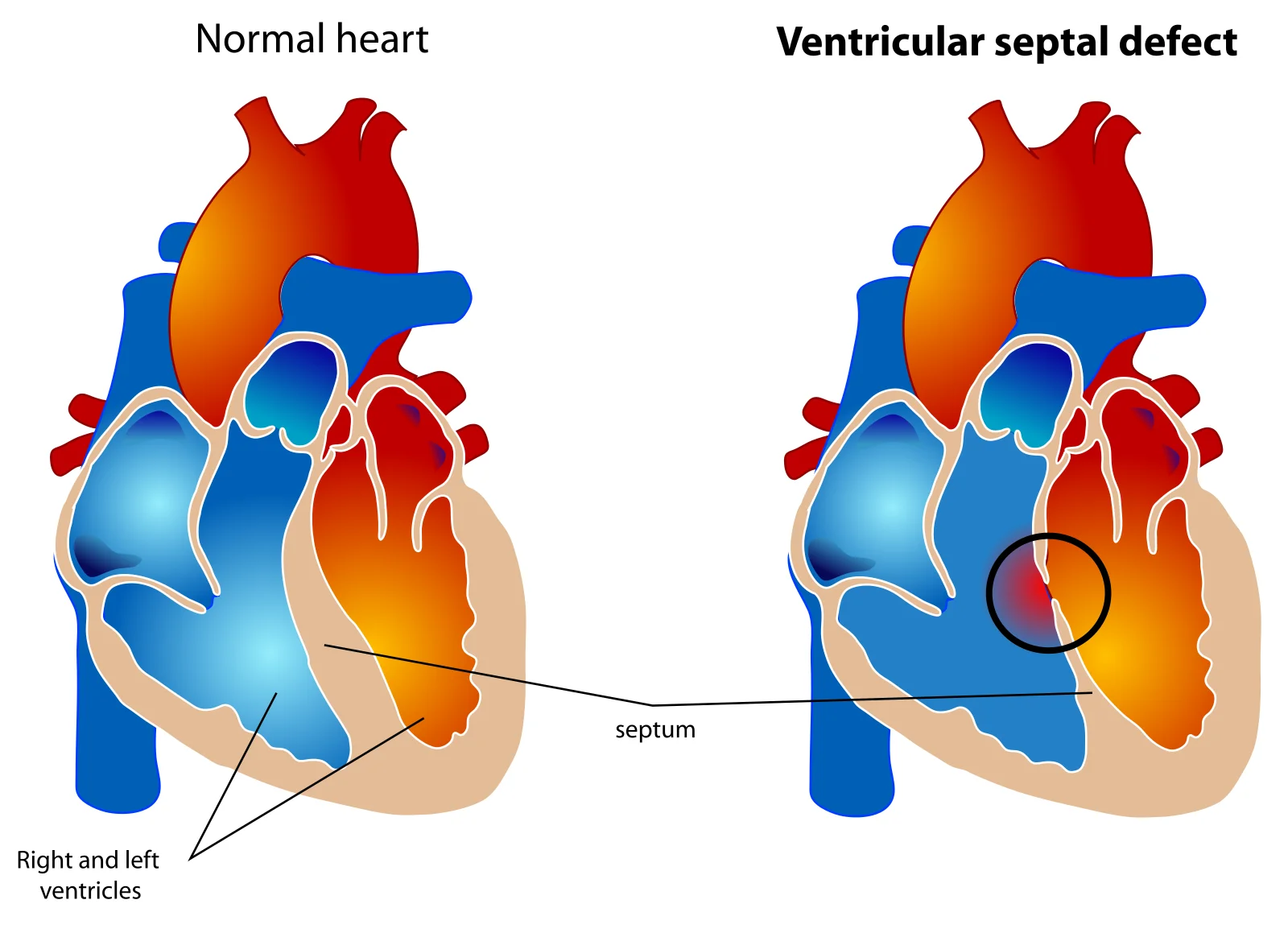
Long description
Two heart diagrams side by side. The normal heart has an intact septum between the right and left ventricles; the other has a circled opening in the septum where blood crosses from the higher-pressure left ventricle to the right.

A ventricular septal defect is an opening in the ventricular septum. It is the commonest congenital heart lesion. Perimembranous defects make up about 80% of them. [1]
- Classification by size and haemodynamics. Small (restrictive) defects generate a high-velocity jet and a loud murmur but little shunt; moderate defects have a Qp:Qs greater than 2:1; large non-restrictive defects equalise ventricular pressures and pour blood into the lungs.
- Course. Many small and muscular defects close spontaneously in the first two years. Large defects declare themselves with heart failure as pulmonary resistance falls.
- Examination. A harsh holosystolic murmur at the left lower sternal border, often with a thrill; a displaced apex if the left ventricle is volume overloaded; the second heart sound may be masked by the murmur.
- Complications. Pulmonary hypertension and Eisenmenger syndrome (earliest 18 months to 2 years if a large defect is not repaired; sooner in Down syndrome), aortic valve prolapse with regurgitation, infective endocarditis, and acquired right ventricular outflow obstruction. [1]
- Management. Small defects: observe. Moderate and large defects: close before 6 to 12 months of age, and certainly no later than 18 months, regardless of how well heart failure and weight are controlled; in Down syndrome close before 6 months. Muscular defects in very small infants who fail medical therapy may need pulmonary artery banding first. Anti-congestive medical therapy (diuretics, afterload reduction, digoxin) is used while awaiting closure. [1]
Figure 2Atrial septal defect
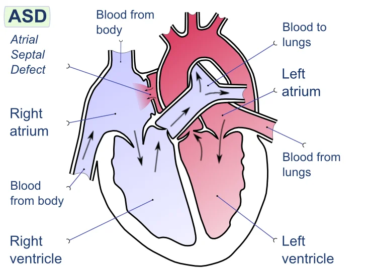
Long description
Labelled diagram of the four heart chambers. A gap in the wall between the two atria lets oxygenated blood from the left atrium flow into the right atrium, adding to the volume the right heart handles.

An atrial septal defect is an opening in the atrial septum, allowing left-to-right flow at atrial level. Defects are of three main types: ostium secundum (the majority), ostium primum (part of the atrioventricular septal defect spectrum) and sinus venosus, often with anomalous pulmonary venous drainage.
- Course. Most secundum defects are asymptomatic in childhood; many small defects under 5 mm close spontaneously. Presentation in adult life with right heart enlargement is common. [1]
- Examination. Wide, fixed splitting of the second heart sound with a systolic ejection murmur at the left upper sternal border from increased pulmonary flow; a low mid-diastolic rumble at the lower sternal border if the shunt is large.
- Indications for closure. Right ventricular volume overload and/or a Qp:Qs greater than 1.5:1. Elective closure is usually performed at 4 to 5 years of age, and not in infancy unless the infant is symptomatic. Closure of secundum defects with adequate rims can be achieved by catheter device; primum and sinus venosus defects need surgery. [1]
- Complications. Pulmonary arterial hypertension and Eisenmenger syndrome late in life, atrial arrhythmia, and paradoxical embolism or stroke when right-sided pressures rise.
- Turner association. Atrial septal defect occurs in about 9.4% (45,X) versus 3.6% (mosaic or structural) of patients with Turner syndrome. [7]
| Feature | Atrial septal defect | Ventricular septal defect |
|---|---|---|
| Associations | Turner syndrome [7] | Down syndrome, Turner syndrome [1] |
| Presentation | Usually asymptomatic in childhood; right heart enlargement in adult life [1] | Small: asymptomatic; large: heart failure as pulmonary resistance falls |
| Murmur | Wide fixed split second sound, systolic ejection murmur at the left upper sternal border, diastolic rumble if the shunt is large | Harsh holosystolic murmur at the left lower sternal border, often with a thrill |
| Chest radiograph | Cardiomegaly and plethoric lung fields if the shunt is large | Cardiomegaly and plethoric lung fields if the shunt is large |
| ECG | Right ventricular hypertrophy in a significant shunt | Right ventricular hypertrophy in a significant shunt |
| Echocardiogram | Site, size and direction of the shunt, with Doppler Qp:Qs [1] | Site, size and direction of the shunt, with Doppler Qp:Qs [1] |
| Closure | Qp:Qs above 1.5:1 or right ventricular volume overload, usually at 4 to 5 years [1] | Moderate or large: before 6 to 12 months [1] |
An atrioventricular septal defect is a combination of an ostium primum atrial septal defect, an inlet ventricular septal defect and abnormal atrioventricular valves with a common atrioventricular junction. It is the defect most strongly associated with Down syndrome. [1]
- Types. Complete (single common valve, large shunt, early heart failure), transitional (large primum atrial defect with small ventricular communication) and partial (primum atrial defect only, often presenting later).
- Course. Complete forms present in early infancy with heart failure and fail to thrive; without intervention, pulmonary vascular obstructive disease can be seen as early as 6 to 12 months, and sooner in Down syndrome. [1]
- Examination. A holosystolic murmur of mitral or common atrioventricular valve regurgitation at the apex, a systolic murmur of the ventricular component, and often a low diastolic rumble; marked cardiomegaly.
- Management. All defects require repair: complete forms usually in the first 6 to 12 months of life, with pulmonary artery banding in early infancy when the baby is very small or weighs under 5 kg. [1]
- Complications. Premature pulmonary vascular disease, heart failure, atrioventricular valve regurgitation after repair, and arrhythmia.
Figure 3Patent ductus arteriosus
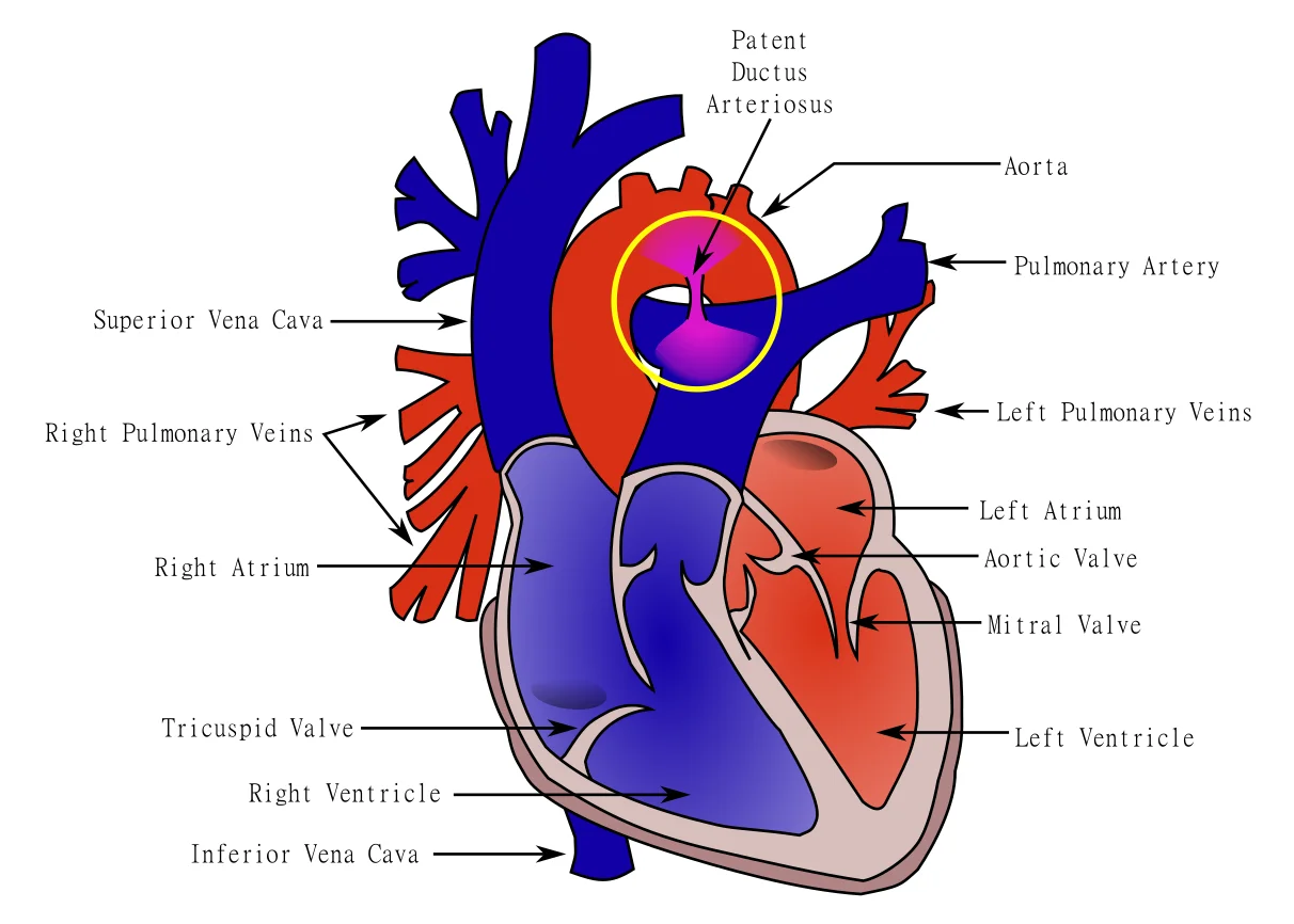
Long description
Labelled heart diagram. A circled channel links the underside of the aortic arch to the main pulmonary artery, letting aortic blood flow into the pulmonary circulation.

The ductus arteriosus normally closes within hours to days after birth. When it persists, it creates a left-to-right shunt from aorta to pulmonary artery, volume-loading the left ventricle.
- Examination. A continuous machinery murmur, loudest at the left infraclavicular area and second left intercostal space; bounding peripheral pulses with a wide pulse pressure; a left ventricular heave.
- Who needs closure. Very small and small ducts should be closed even when not haemodynamically significant; medium and large ducts are closed to avoid left ventricular volume overload, heart failure, pulmonary vascular disease and infective endocarditis. Closure is avoided once pulmonary vascular disease (Eisenmenger physiology) is established. [1]
- Timing. Closure can be undertaken at any age; asymptomatic infants are usually treated at 6 to 12 months, and large ducts before 18 months. Very small or silent ducts can be closed later. [1]
- Method. Term infants and children: catheter closure with coils or occluder devices, or surgical ligation/division. Premature infants: medical closure first, as below.
Clinical pearlCongenital rubella and the ductus
In congenital rubella, patent ductus arteriosus (often with peripheral pulmonary artery stenosis) is the classic cardiac lesion. [8] A continuous machinery murmur in a small-for-dates infant with a purpuric rash or cataracts should make you think of rubella.
Figure 4Chest radiograph in coarctation of the aorta: figure 3 sign and rib notching
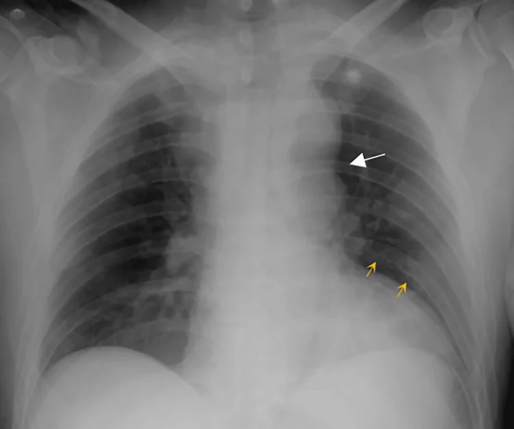
Long description
A posteroanterior chest radiograph of an adult with a focal coarctation of the aorta. At the aortic knob a white arrow marks the indentation of the left mediastinal contour with dilatation above and below it, the figure 3 sign. Two yellow arrows mark scalloped notches along the lower edges of the ribs on the left, produced by enlarged intercostal collateral arteries.
Figure 5Coarctation of the aorta
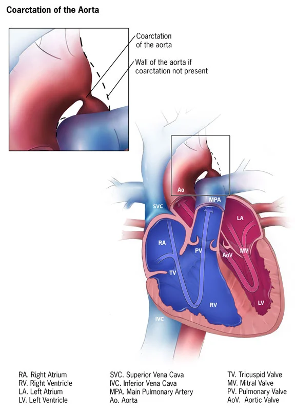
Long description
Labelled heart diagram with a magnified inset of the aortic arch. The aorta narrows beyond the origin of the left subclavian artery; a dashed line marks where the wall would lie without the coarctation.

These are the left-sided obstructive lesions. They share pressure overload and the risk of sudden deterioration in a newborn, and both can be associated with a bicuspid aortic valve.
Coarctation of the aorta is a narrowing of the aortic arch, usually near the ductus arteriosus. It accounts for 5 to 8% of congenital heart disease. [1]
- Presentation. Depends on the age. A critical neonatal coarctation presents when the ductus closes, with shock, absent or weak femoral pulses, and a systolic pressure gradient between arms and legs. Older children are found with hypertension and a radio-femoral delay.
- Examination. Systolic ejection murmur in the left interscapular area, radiating to the back; a systolic thrill between the scapulae; diminished or delayed femoral pulses; upper-limb hypertension.
- Thresholds. Significant coarctation is defined by significant hypertension and/or heart failure with a peak-to-peak systolic gradient greater than 20 mmHg. [1] Current guidance uses a gradient threshold of 30 mmHg for intervention in coarctation. [2]
- Management. Neonates in shock: prostaglandin E1 to reopen the duct, followed by surgical or transcatheter relief. In stable older children: surgery in newborns and young infants, balloon angioplasty in children, and stent implantation in adolescents and adults. [2]
- Complications. Persistent or late hypertension, recoarctation, aortic aneurysm or dissection, cerebral (circle of Willis) aneurysm, and coronary disease.
Aortic stenosis (valvar) is obstruction at the aortic valve, accounting for 5 to 6% of congenital heart disease. Subvalvar and supravalvar forms also exist.
- Presentation. Often asymptomatic in childhood; severe disease causes exertional dyspnoea, angina, syncope or sudden death.
- Examination. An ejection systolic murmur at the right upper sternal border radiating to the neck, with an ejection click; a slow-rising pulse; a left ventricular heave.
- Thresholds. Intervention is considered at a peak-to-peak gradient greater than 50 mmHg with symptoms or electrocardiographic ST-T changes, or a peak gradient greater than 70 mmHg irrespective of symptoms. [1] A common modern criterion for intervention in aortic stenosis is a peak gradient of 50 mmHg. [2]
- Management. Balloon valvuloplasty is the usual first intervention in children; surgical valvotomy or replacement is reserved for failed or unsuitable anatomy.
- Turner association. Coarctation of the aorta occurs in about 7.4% of patients with a 45,X karyotype versus 2.1% of those with a mosaic or structural karyotype; bicuspid aortic valve occurs in about 2.7 versus 2.9%. [7]
| Lesion | Key associations | Characteristic murmur | Principal features | Major complications | Definitive treatment |
|---|---|---|---|---|---|
| VSD | Down syndrome (often with AVSD); Turner syndrome | Harsh holosystolic at left lower sternal border, often with thrill | Small: asymptomatic; large: heart failure, poor growth | Pulmonary hypertension, Eisenmenger syndrome, aortic regurgitation, endocarditis | Observation if small; surgical or device closure by 6 to 12 months if moderate or large |
| ASD (secundum) | Turner syndrome; Down syndrome | Wide fixed split second sound, systolic ejection murmur left upper sternal border | Usually asymptomatic; right ventricular volume overload if large | Late pulmonary hypertension, arrhythmia, paradoxical embolism | Device closure when rims adequate and Qp:Qs greater than 1.5, usually at 4 to 5 years; surgery otherwise [1] |
| AVSD | Down syndrome (most common defect in this syndrome) | Holosystolic regurgitant murmur at apex plus ventricular component, low diastolic rumble | Early heart failure, poor growth, cardiomegaly | Premature pulmonary vascular disease, heart failure, post-repair valve regurgitation | Surgical repair, usually at 6 to 12 months; banding first in tiny infants [1] |
| PDA | Prematurity; congenital rubella; Turner syndrome | Continuous machinery murmur, loudest left infraclavicular | Bounding pulses, wide pulse pressure, heart failure if large | Pulmonary vascular disease or Eisenmenger syndrome, endocarditis | Term: catheter or surgical closure; premature: indomethacin or ibuprofen first |
| Coarctation of the aorta | Turner syndrome (45,X); bicuspid aortic valve | Systolic ejection murmur between the scapulae, radiating to the back | Neonatal shock when duct closes, or hypertension and radio-femoral delay later | Hypertension, recoarctation, aortic aneurysm or dissection | Neonatal: prostaglandin E1 then surgery or angioplasty; older: surgery, balloon or stent |
| Aortic stenosis | Bicuspid aortic valve | Ejection systolic at right upper sternal border, ejection click, radiating to neck | Exertional dyspnoea, angina, syncope | Sudden death, heart failure, endocarditis | Balloon valvuloplasty first; surgery when anatomy dictates |
Heart failure is the common presentation of large left-to-right shunts and is also how coarctation and severe aortic stenosis declare themselves in the newborn.
Recognition. Tachypnoea with feeding, poor suck and sweating during feeds, poor weight gain, recurrent chest infections, hepatomegaly and a gallop rhythm. In a small infant, watch the feeds: a baby who cannot finish a bottle without breathlessness, or who sweats, has heart failure until proven otherwise. Cyanosis is not part of the picture in pure acyanotic disease unless pulmonary pressures have already risen. [4]
Pathophysiology. Reduced cardiac output activates the renin-angiotensin-aldosterone system and sympathetic nervous system, producing vasoconstriction and sodium and water retention. Initially compensatory, this eventually produces unfavourable myocardial remodelling. [4]
- Diuretics (loop diuretic, usually furosemide) for congestion; add a thiazide if response is slow; monitor electrolytes and renal function, and avoid over-diuresis when physiology depends on preload.
- Afterload reduction with an ACE inhibitor once the child is beyond the neonatal period (ACE inhibitors should be avoided in neonates); uptitrate carefully and watch for hypotension and renal dysfunction.
- Digoxin as an adjunct for symptomatic systolic heart failure once weight and feeding are stable.
- Supportive: optimise nutrition (calorie-dense feeds, nasogastric or gastrostomy feeding if needed), maintain adequate haemoglobin, correct anaemia (consider transfusion when haemoglobin is under 100 g/L in acute symptomatic failure), and give oxygen if hypoxic. [4]
- Inotropes such as milrinone for severe low cardiac output in critical disease, and surgical or transcatheter relief of the lesion as the definitive treatment.
- More advanced, stage-based therapy (beta-blockers, mineralocorticoid antagonists, SGLT2 inhibitors, devices) follows established paediatric heart failure protocols. [4]
Two drugs recur in the management of acyanotic lesions, for opposite reasons.
Prostaglandin E1 (alprostadil). Its role in acyanotic disease is to keep the ductus arteriosus open, not to close it. In critical coarctation or severe left-sided obstruction, systemic perfusion depends on ductal flow until surgery. [5] [3]
- Indication: palliative, not definitive, maintenance of ductal patency in neonates whose defect depends on the duct, including severe coarctation of the aorta. [5]
- Standard label dosing: begin at 0.05 to 0.1 microg/kg/min intravenously; the recommended starting dose is 0.1 microg/kg/min. After response, reduce to 0.05, then 0.025, then 0.01 microg/kg/min. Doses up to 0.4 microg/kg/min may be used if response is inadequate, although higher rates are generally no more effective. [5]
- Some neonatal protocols use a lower prophylactic infusion of 5 to 10 ng/kg/min to keep an already open duct patent. [3]
- Warnings: apnoea occurs in about 10 to 12% of neonates, most often in those weighing under 2 kg and usually within the first hour of infusion; fever in about 14%, flushing in about 10%, bradycardia in about 7%, hypotension in about 4%. [5] Infants treated for more than 120 hours should be closely monitored, and gastric outlet obstruction (antral hyperplasia) is a known hazard. [5]
Indomethacin. This non-steroidal anti-inflammatory drug closes a haemodynamically significant patent ductus arteriosus in premature infants by inhibiting prostaglandin synthesis. [6]
- Indication: premature infants weighing 500 to 1750 g with a haemodynamically significant patent ductus that has not closed after 48 hours of usual medical management (fluid restriction, diuretics, respiratory support, and so on), with clear clinical evidence such as respiratory distress, continuous murmur, hyperactive precordium, cardiomegaly or pulmonary plethora. [6]
- Dosing by age at first dose: under 48 hours: 0.2, then 0.1, then 0.1 mg/kg; 2 to 7 days: 0.2, 0.2, 0.2 mg/kg; over 7 days: 0.2, 0.25, 0.25 mg/kg, given intravenously at 12 to 24 hour intervals. [6]
- If anuria or marked oliguria (urine output less than 0.6 mL/kg/h) is present at the time of the second or third dose, do not give further doses until renal function normalises. [6]
- After two courses without response, surgical closure may be needed. [6]
- Paediatric closure rates in preterm infants weighing 1750 g or less are reported at about 75 to 80% with indomethacin versus 25 to 30% with placebo; late reopening of the ductus occurs in about 26% of treated infants, but 70% of those close again without surgery or further drug. [6]
- Contraindication: congenital heart defects in whom ductal patency is necessary for adequate blood flow, such as pulmonary atresia, severe tetralogy of Fallot and severe coarctation of the aorta. [6]
Exam trapDo not close what keeps them alive
In duct-dependent left-sided lesions (severe coarctation, critical aortic stenosis, hypoplastic left heart), giving indomethacin to close the ductus, or leaving the duct to close spontaneously, can be fatal. These babies need the duct open, with prostaglandin E1, until specialist care is reached. [6] [5]
Figure 6Contrast CT of a patent ductus arteriosus
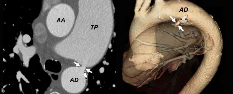
Long description
Two contrast-enhanced CT images from an elderly woman with a patent ductus arteriosus. The left image is an axial slice: the ascending aorta (AA) and a large pulmonary trunk (TP) lie above the descending aorta (AD), and two white arrows mark the 6 mm ductus joining the pulmonary trunk to the descending aorta, with small white calcifications beside it (asterisks). The right image is a 3D volume-rendered view of the same channel with arrows and asterisks.
Echocardiography is the diagnostic backbone. Transthoracic echocardiography defines cardiac anatomy, the presence, site and size of a shunt, valvar structure, ventricular function and pulmonary artery pressures. Transoesophageal imaging adds detail when a defect has poor acoustic windows, and is particularly useful for secundum atrial septal defects and patent ductus arteriosus. [1]
Colour and spectral Doppler answer the clinical question: they measure shunt velocity, estimate pulmonary artery systolic pressure from the tricuspid regurgitant jet, and quantify the Qp:Qs. Other tests play supporting roles. [1]
- Electrocardiogram may show right ventricular hypertrophy in a significant shunt, or left ventricular hypertrophy in obstruction; a normal trace never excludes disease.
- Chest radiograph shows cardiomegaly and plethoric lung fields in a large shunt, or pulmonary oedema and a narrow mediastinum in critical obstruction.
- Pulse oximetry in both hands and a foot, especially in the newborn, is an established screening tool for critical disease.
- Cardiac magnetic resonance or computed tomography is reserved for complex anatomy, anomalous pulmonary venous drainage, or pre-operative planning; catheterisation is used only when non-invasive data are insufficient or intervention is planned.
Practical thresholds that guide decisions in acyanotic disease are summarised below. They are operator- and guideline-dependent and should be read with local echocardiography and cardiology input. [1] [2]
- Ventricular septal defect. Moderate shunt when Qp:Qs is greater than 2:1; repair when Qp:Qs exceeds 1.5:1 with pulmonary vascular resistance still acceptable. [1]
- Pulmonary vascular resistance. Elevated pulmonary resistance with a pulmonary-to-systemic resistance ratio less than 0.35, and a pulmonary vascular resistance index less than 6 Wood units, identify shunts still feasible for closure. [1]
- Atrial septal defect. Closure indicated with right ventricular volume overload and/or Qp:Qs greater than 1.5:1. [1]
- Coarctation. Significant when there is hypertension and/or heart failure with a peak-to-peak systolic gradient greater than 20 mmHg, with intervention often quoted at a gradient of 30 mmHg. [1] [2]
- Aortic stenosis. Intervention at a peak-to-peak gradient greater than 50 mmHg with symptoms or electrocardiographic ST-T changes, or a peak gradient greater than 70 mmHg irrespective of symptoms; a working modern cut-off is a 50 mmHg peak gradient. [1] [2]
- Timing. Ventricular septal defect repair before 6 to 12 months (never later than 18 months); atrioventricular septal defect repair at 6 to 12 months; atrial septal defect electively at 4 to 5 years; large patent ductus arteriosus before 18 months. [1]
- Critical arch obstruction or aortic stenosis in a collapsed newborn: keep the duct open with prostaglandin E1, correct acidosis and perfusion, and transfer urgently to a centre that can perform neonatal cardiac surgery or intervention. [3] [5]
- Heart failure from a large shunt: diuretics, nutritional support, afterload reduction and close monitoring; treat anaemia and infection.
- An infant who is failing despite maximal medical therapy should be referred early for surgical closure rather than waiting for pulmonary vascular change.
- Left-to-right shunts: close the defect (surgical for ventricular and atrioventricular defects; device or surgery for atrial defects; device, coil or surgery for patent ductus) before irreversible pulmonary vascular disease develops.
- Coarctation: surgery in neonates and young infants; balloon angioplasty in children; stenting in adolescents and adults. [2]
- Aortic stenosis: balloon valvuloplasty first, surgery for residual or unsuitable anatomy.
- Note regional inconsistency: some lesions are managed very differently by experienced centres, and recommendations from European and North American guideline frameworks do not always agree. [11]
- Nutritional support is not optional: calorie-dense feeds, nasogastric feeding and, when necessary, gastrostomy feeding can decide whether an infant reaches surgery in good condition.
- Activity should not be unnecessarily restricted; most children are encouraged to exercise to tolerance.
- Family education about warning signs (poor feeding, sweating, breathlessness, reduced alertness) is essential.
- Small defects with no haemodynamic load can be followed by the general paediatrician with periodic echocardiography.
- Refer urgently when a defect is large, when heart failure is not controlled medically, when pulmonary pressures are rising, or when the diagnosis is in doubt.
- Children with syndromic disease need ongoing cardiac surveillance: Down syndrome infants should have their defect closed early, and patients with Turner syndrome need repeated aortic imaging because of the risk of progressive aortic dilatation. [1] [7]
- Pulmonary hypertension and Eisenmenger syndrome from long-standing uncorrected shunts, the most serious late complication. [9]
- Heart failure in infancy or childhood from a large left-to-right shunt or a severe obstruction.
- Aortic valve prolapse and regurgitation with ventricular septal defect.
- Endocarditis on any structural lesion, especially after repair or on a patent ductus.
- Arrhythmia, particularly atrial flutter or fibrillation after atrial septal repair, and after atrioventricular repair.
- Hypertension and recoarctation, with their vascular complications, after coarctation repair.
- Paradoxical embolism and stroke when a right-to-left shunt co-exists.
Outcome is excellent when the correct lesion is identified and treated before pulmonary vascular disease becomes irreversible. Most acyanotic defects, including the common small ventricular septal defect, carry a normal or near-normal life expectancy. [1]
Eisenmenger syndrome, by contrast, is a serious, multi-organ disease with substantial morbidity and mortality in adults, and no proven disease-modifying therapy has been established for the unrepaired shunt forms. [9] Early detection and timely closure are therefore the strongest modifiable determinants of prognosis.
- Prenatal and newborn screening (fetal echocardiography where available, routine neonatal examination, pulse oximetry) catches critical coarctation and other obstructive lesions that collapse when the ductus closes, as well as large shunts. [3]
- Vaccination. Rubella vaccination before pregnancy, with the standard 12- to 15-month and 4- to 6-year schedule, prevents congenital rubella and its cardiac manifestations. [8]
- Syndromic surveillance. All infants with Down syndrome and with Turner syndrome should undergo echocardiography (and, for Turner syndrome, ongoing aortic imaging) regardless of examination findings. [1] [7]
- Premature infants. The ductus often behaves pathologically: close according to the criteria above, use indomethacin or ibuprofen first, and operate if drugs fail. [6]
- Down syndrome. Atrioventricular septal defect is the commonest lesion; repair early (before 6 to 12 months) because pulmonary vascular disease develops faster than in other infants. [1]
- Turner syndrome. Coarctation, bicuspid aortic valve and atrial and ventricular septal defects are all over-represented; cardiology and imaging follow-up continue into adult life. [7]
- Congenital rubella. Survivors may have silent peripheral pulmonary stenosis as well as a patent ductus, and they should not be followed as though the heart were normal. [8]
- Diagnosis. A careful examination (pulses, blood pressure in all four limbs, precordial impulse, murmur quality) plus a chest radiograph identifies many obstructive lesions. Where echocardiography exists, it is the single most decisive test.
- Heart failure. Diuretics, nutritional rehabilitation and, where available, digoxin keep an infant alive and growing while a referral is arranged.
- The newborn in shock. If a critical arch obstruction cannot be excluded, an infusion of prostaglandin E1 at a low dose can be started at any facility before transfer; it will not harm a baby whose disease is not duct-dependent and may be life-saving if it is. [3] [5]
- Access. Surgery, catheter closure and prostaglandin are not universally available; refer early and keep the child well nourished and infection-free while waiting.
Most childhood heart murmurs are innocent. Less than 1% are associated with congenital heart disease, so the task is to recognise the few that matter without generating unnecessary anxiety or echocardiograms. [10]
A child with a heart murmur
- Hear any murmurHistory, growth, family, and full cardiovascular examination first
- Question: Any red flag?
Yes: symptoms, poor growth, syncope, chest pain, cyanosis, abnormal pulses, or non-innocent murmur quality
- Echocardiogram and paediatric cardiology referral
- Treat (for example, heart failure therapy) and plan closure or relief as indicated
No
- Question: Does it sound innocent? (Still, pulmonary flow, supraclavicular, venous hum)
Yes
- Reassure, explain, document; no further tests
Uncertain or not classic
- Refer for echocardiography rather than repeat guessing
Red flags include a diastolic murmur, any murmur in a neonate or very young infant, a harsh murmur accompanied by symptoms or abnormal findings, an ejection click, abnormal second sound, displaced apex, hepatomegaly, poor growth, exertional symptoms, or any murmur whose character does not fit a classic innocent pattern. [10]
- Acyanotic congenital heart disease comprises the left-to-right shunts (VSD, ASD, AVSD, PDA) and the left-sided obstructions (coarctation, aortic stenosis); oxygen saturation is normal at rest.
- A large shunt stays silent before birth and in the early newborn, then floods the lungs as pulmonary resistance falls over 6 weeks to 3 months, producing heart failure.
- Most acyanotic defects need only observation; the decision to close or relieve is driven by shunt size, pulmonary resistance and symptoms.
- VSD is the commonest lesion (20 to 25% of congenital heart disease); perimembranous defects are about 80% of them.
- AVSD is the defect most associated with Down syndrome and must be repaired by 6 to 12 months, sooner in Down syndrome, because pulmonary vascular disease develops quickly. [1]
- Closure thresholds: Qp:Qs greater than 1.5 to 2:1 for shunts, RV volume overload for ASD, and a coarctation or aortic stenosis gradient in the tens of millimetres of mercury.
- Premature pulmonary vascular disease is avoidable only by early closure of a large, unrestricted shunt.
- Once Eisenmenger syndrome develops, closure is dangerous: treat the pulmonary vasculature and follow the patient.
- Heart failure in infants is recognised at the bedside by feeding, sweating, poor growth and hepatomegaly; treat with diuretics, nutrition and, when appropriate, afterload reduction and digoxin.
- Prostaglandin E1 keeps the duct open in duct-dependent left-sided lesions; indomethacin closes the duct in premature infants but is contraindicated when ductal patency is essential.
- Echocardiography is the diagnostic test of choice, and a structured murmur algorithm prevents both missed disease and unnecessary testing.
- Think of the syndromic associations: Down syndrome with AVSD, Turner syndrome with coarctation, and congenital rubella with patent ductus arteriosus.
- Where care is limited, keep the baby nourished and infection-free, start prostaglandin E1 when duct-dependent obstruction cannot be excluded, and refer early.
Answer each one in your head first, then open the answer.
Why does an infant with a large ventricular septal defect often look well for the first few weeks of life and then develop heart failure?
Show answerHide answer
The fetal pulmonary vascular resistance is high, so at birth little blood shunts left to right and the defect is haemodynamically silent. Over 6 weeks to 3 months pulmonary resistance falls to near-adult levels, the shunt opens and grows, and pulmonary overcirculation and left heart volume overload produce pulmonary congestion and heart failure. In Down syndrome this process is faster and pulmonary vascular disease can appear by 6 months.
A loud, harsh, holosystolic murmur is heard at the left lower sternal border in a 6-week-old. The baby is small and breathless with feeds. What is the most likely lesion, and what is the management?
Show answerHide answer
A large ventricular septal defect. Management combines anti-congestive medical therapy (diuretics, afterload reduction, nutritional support, correction of anaemia) while arranging definite closure before 6 to 12 months of age, never later than 18 months, because pulmonary vascular disease begins early.
A 3-month-old has a soft systolic ejection murmur at the left upper sternal border and wide fixed splitting of the second heart sound; growth is normal. What is the lesion?
Show answerHide answer
An ostium secundum atrial septal defect. The wide fixed split S2 and flow murmur reflect increased pulmonary flow. The defect should still be followed: closure is indicated when there is right ventricular volume overload or a Qp:Qs greater than 1.5, usually performed electively at 4 to 5 years.
A term newborn becomes shocked 48 hours after birth, with absent femoral pulses and markedly different blood pressures in the arms and legs. What has happened, and what must you do immediately?
Show answerHide answer
The ductus arteriosus has closed in a baby with coarctation of the aorta, unmasking severe obstruction. Start prostaglandin E1 to reopen the duct, resuscitate, and arrange urgent transfer for surgery or intervention. Do not give indomethacin or allow the duct to close.
A premature infant weighing 1200 g has a continuous murmur and echocardiographic evidence of a haemodynamically significant patent ductus. How do you treat, and when is surgery used?
Show answerHide answer
Give intravenous indomethacin in the age-appropriate dosing (1200 g and under-7-day schedule: 0.2, 0.2, 0.2 mg/kg at 12 to 24 hour intervals), after 48 hours of usual medical management has failed. Withhold later doses if anuria or oliguria under 0.6 mL/kg/h is present. If the duct fails to close after two courses, surgical closure is needed. Indomethacin is contraindicated if ductal patency is essential for circulation. [6]
Explain in two sentences why early closure of an atrioventricular septal defect matters, mentioning the syndrome most often associated with it.
Show answerHide answer
Complete atrioventricular septal defect is most often seen in Down syndrome and presents in early infancy with a large left-to-right shunt and heart failure. Pulmonary vascular obstructive disease can appear by 6 to 12 months, sooner in Down syndrome, so repair is performed in the first 6 to 12 months.
A 10-year-old with an unrepaired atrial septal defect now has central cyanosis and clubbing. What complication has developed, and why must the defect not be closed?
Show answerHide answer
Eisenmenger syndrome: chronic pulmonary overcirculation has raised pulmonary resistance until the shunt reversed right to left. Closure at this stage removes the pop-off for the right ventricle and can cause acute right heart failure, so treatment is directed at the pulmonary vasculature rather than at the defect.
Plain-text related topics for this page, not linked: congenital cyanotic heart disease, Eisenmenger syndrome, heart failure in children, prostaglandin E1 and alprostadil, indomethacin for patent ductus arteriosus, Down syndrome, Turner syndrome, congenital rubella syndrome, echocardiography, hypertension in children, paediatric cardiac surgery and catheter intervention.
- Rao PS. Management of Congenital Heart Disease: State of the Art; Part I—ACYANOTIC Heart Defects. Children (Basel) 2019;6(3):42. doi:10.3390/children6030042 (PMID 30857252; PMCID PMC6463202). Accessed .
- Rao PS. Recent Advances in the Diagnosis and Management of Congenital Heart Disease. Children (Basel) 2024;11(1):84. doi:10.3390/children11010084 (PMID 38255397; PMCID PMC10814956). Accessed .
- Khalil M, Jux C, Rueblinger L, Behrje J, Esmaeili A, Schranz D. Acute therapy of newborns with critical congenital heart disease. Transl Pediatr 2019;8(2):114–126. doi:10.21037/tp.2019.04.06 (PMID 31161078; PMCID PMC6514285). Accessed .
- Dipchand AI. Management of Pediatric Heart Failure. Korean Circ J 2024;54(12):794–810. doi:10.4070/kcj.2024.0320 (PMID 39733776; PMCID PMC11685346). Accessed .
- Pharmacia & Upjohn Company LLC (DailyMed). PROSTIN VR PEDIATRIC (alprostadil) injection, solution [label]. DailyMed label, setid 930f8c02-46a8-4cff-a484-660468ab23c3. Accessed .
- Fresenius Kabi USA, LLC (DailyMed). INDOMETHACIN for Injection [label]. DailyMed label, setid dd5caa3f-3c09-4034-bf43-2df80c61c07b. Accessed .
- Huang Y et al. Karyotype-specific cardiovascular and metabolic profiles in Turner syndrome: a retrospective echocardiographic study. Orphanet J Rare Dis 2026;21:315 (PMID 42323690; PMCID PMC13563905). Accessed .
- Gailani AE, Dehority W, Katz SE. Clinical progress note: Rubella. J Hosp Med 2026;21(2):179–182. doi:10.1002/jhm.70169 (PMID 40938300; PMCID PMC12865257). Accessed .
- Yoshida K et al. Protocol for an open-label, randomised, controlled trial ... the SuMILE trial. BMJ Open 2026;16:e113430. doi:10.1136/bmjopen-2025-113430 (PMID 41819579; PMCID PMC12983897). Accessed .
- Doshi AR. Innocent Heart Murmur. Cureus 2018;10:e3689. doi:10.7759/cureus.3689 (PMID 30761241; PMCID PMC6368429). Accessed .
- Meng X et al. Congenital heart disease: types, pathophysiology, diagnosis, and treatment options. MedComm 2024;5:e631. doi:10.1002/mco2.631 (PMID 38974713; PMCID PMC11224996). Accessed .
Figure credits
- Figure 1: Mariana Ruiz LadyofHats, via Wikimedia Commons, Public domain, Public domain.
- Figure 2: Manco Capac, via Wikimedia Commons, CC BY-SA 3.0, CC BY-SA 3.0.
- Figure 3: Original uploader was BrownCow., via Wikimedia Commons, Public domain, Public domain.
- Figure 4: Gutiérrez Rangel VY, Morelos Guzmán M, Areán Martínez CA, Sandoval Martínez V, Fraga Ramos MS. Cureus, via PubMed Central, CC BY 4.0, CC BY 4.0. resized.
- Figure 5: Centers for Disease Control and Prevention, via Wikimedia Commons, CC0, CC0.
- Figure 6: Mueller S, Plank F, Klimes K, Feuchtner G, Mair J. Wiener klinische Wochenschrift, via PubMed Central, CC BY 4.0, CC BY 4.0. cropped to the lower (CT) panels.
Educational content for medical students and doctors. It is not a substitute for clinical judgement or local guidelines.