Cyanotic congenital heart disease
The cyanotic heart lesions: right-to-left shunt physiology, bedside and radiograph clues, initial stabilisation, and definitive surgery.
- Cyanotic congenital heart disease means a structural heart defect in which deoxygenated blood reaches the arterial circulation, so the infant is blue from birth or early infancy.
- The cause is one of three physiological problems: obstruction to pulmonary blood flow, complete mixing of the two circulations, or parallel systemic and pulmonary circuits, as in transposition of the great arteries.
- In the newborn with central cyanosis, pulse oximetry, a chest radiograph and echocardiography separate a cardiac cause from lung disease, and a hyperoxia test raises the suspicion of a right-to-left shunt before imaging confirms it.
- Most of these lesions are duct-dependent in the first days of life. If one is suspected, start prostaglandin E1 0.05 to 0.1 mcg/kg/min and arrange urgent echocardiography.
- Definitive repair is offered early: tetralogy of Fallot at 6 to 12 months, arterial switch for transposition within the first week of life, urgent repair for obstructed total anomalous pulmonary venous return, and staged Fontan surgery for tricuspid atresia.
By the end of this page you should be able to:
- Explain what makes a congenital heart lesion "cyanotic" and contrast obstruction, mixing and parallel circulations.
- Name the five Ts and summarise each one's anatomy and key clinical clue.
- Recognise the hypercyanotic spell, explain why squatting or the knee-chest position relieves it, and manage one step by step.
- Interpret a neonatal chest radiograph for the boot-shaped heart of tetralogy of Fallot, the egg-on-a-string of transposition and the snowman heart of supracardiac total anomalous pulmonary venous return.
- Use the hyperoxia test and pre-ductal and post-ductal oxygen saturations to decide when a cyanosed infant is cardiac rather than pulmonary.
- Outline the acute stabilisation of a duct-dependent lesion, the role of prostaglandin E1 and balloon atrial septostomy, and the timing and nature of corrective surgery.
- Describe Eisenmenger syndrome and the associations with DiGeorge syndrome (22q11 deletion), maternal diabetes and lithium.
DefinitionCyanotic congenital heart disease
A group of structural heart defects present from birth in which arterial oxygen saturation is low, because deoxygenated blood enters the systemic arterial circulation through a right-to-left shunt, because the pulmonary and systemic venous returns mix completely, or because the systemic and pulmonary circuits run in parallel rather than in series.
The arterial desaturation is the primary physiological abnormality, and its severity sets everything else: cyanosis, the depth of erythrocytosis, the tendency to clubbing, and the risk of brain abscess or stroke from polycythaemia. Chronic cyanosis increases red cell production by stimulating erythropoietin from the kidneys, and if iron stores are inadequate a relative anaemia results. Maintaining an adequate haemoglobin, avoiding dehydration, and performing timely surgery prevent most of these complications.[1]
Physiologically, cyanotic lesions fall into three groups.[1]
- Obstruction to pulmonary blood flow, as in tetralogy of Fallot, tricuspid atresia and pulmonary atresia. Blood cannot reach the lungs easily, so a right-to-left shunt (through a ventricular septal defect or a patent foramen ovale) lets deoxygenated blood into the systemic circuit. Cyanosis worsens when the obstruction tightens.
- Complete mixing, as in total anomalous pulmonary venous return, truncus arteriosus and single-ventricle hearts. The two venous returns merge and the resulting mixed blood is distributed to both circuits. Cyanosis depends on how much blood actually reaches the lungs.
- Parallel circulation, as in transposition of the great arteries. The aorta arises from the right ventricle and the pulmonary artery from the left ventricle, so the systemic and pulmonary circuits are independent loops. Such a circulation is incompatible with life unless blood mixes through an atrial septal defect, ventricular septal defect or patent ductus arteriosus.
Ductal-dependent lesions need the arterial duct to stay open: right-heart obstructive lesions need it to supply the lungs, and left-heart or parallel-circuit lesions need it to supply the body or to allow mixing. Most cyanotic lesions declare themselves within the first days of life as the duct begins to close.[1]
Transposition of the great arteries is the most common cyanotic congenital heart disease in the newborn period. Beyond infancy the commonest cyanotic lesion is tetralogy of Fallot.[1] Truncus arteriosus accounts for about 1 in 11,000 live births.[2]
Common targets of newborn pulse-oximetry screening for critical congenital heart disease are hypoplastic left heart syndrome, pulmonary atresia, tetralogy of Fallot, total anomalous pulmonary venous return, transposition of the great arteries, tricuspid atresia and truncus arteriosus. Most cyanotic lesions are diagnosed before discharge from the maternity unit, but obstructed total anomalous pulmonary venous return, transposition and tetralogy of Fallot are the lesions most often missed. Echocardiography is the gold standard for diagnosis once a suspicion has been raised.
Most cyanotic lesions are sporadic and multifactorial, with a genetic contribution that is strongest for the conotruncal defects. The conotruncal anomalies share a developmental origin in the neural crest and the aortopulmonary septum, which is why they cluster with deletion of chromosome 22q11 and with each other.[5]
Known associations worth remembering:
- Maternal diabetes mellitus increases the risk of transposition of the great arteries, which is described as more common in infants of diabetic mothers. A cardiac phenotype resembling 22q11.2 deletion has also been described in the same setting.[2][5]
- Maternal lithium exposure in the first trimester, especially the first 8 weeks of gestation, is strongly associated with Ebstein anomaly of the tricuspid valve, a cyanotic lesion in which the tricuspid leaflets are displaced towards the apex.[2]
- Deletion of chromosome 22q11.2 (DiGeorge or velocardiofacial syndrome) is the most common chromosomal microdeletion, with a prevalence of about 1 in 3,000 to 1 in 6,000 live births, and roughly 75% of children with the deletion have congenital heart disease. The cardiac lesions are conotruncal: tetralogy of Fallot (with or without pulmonary atresia), truncus arteriosus, interrupted aortic arch type B and ventricular septal defect. Patients with these lesions show 22q11.2 deletions at differing rates: 16% of those with tetralogy of Fallot, 34% of those with truncus arteriosus and 52% of those with interrupted aortic arch type B.[5] [2]
- Conotruncal defects are also linked with maternal retinoic acid exposure and with single-gene disorders such as CHD7 mutation (CHARGE syndrome), although these are uncommon.[5]
The normal circulation is in series: deoxygenated blood goes to the lungs, picks up oxygen, returns to the heart and is pumped to the body. Every cyanotic lesion breaks one part of this chain, and the clinical picture follows directly.
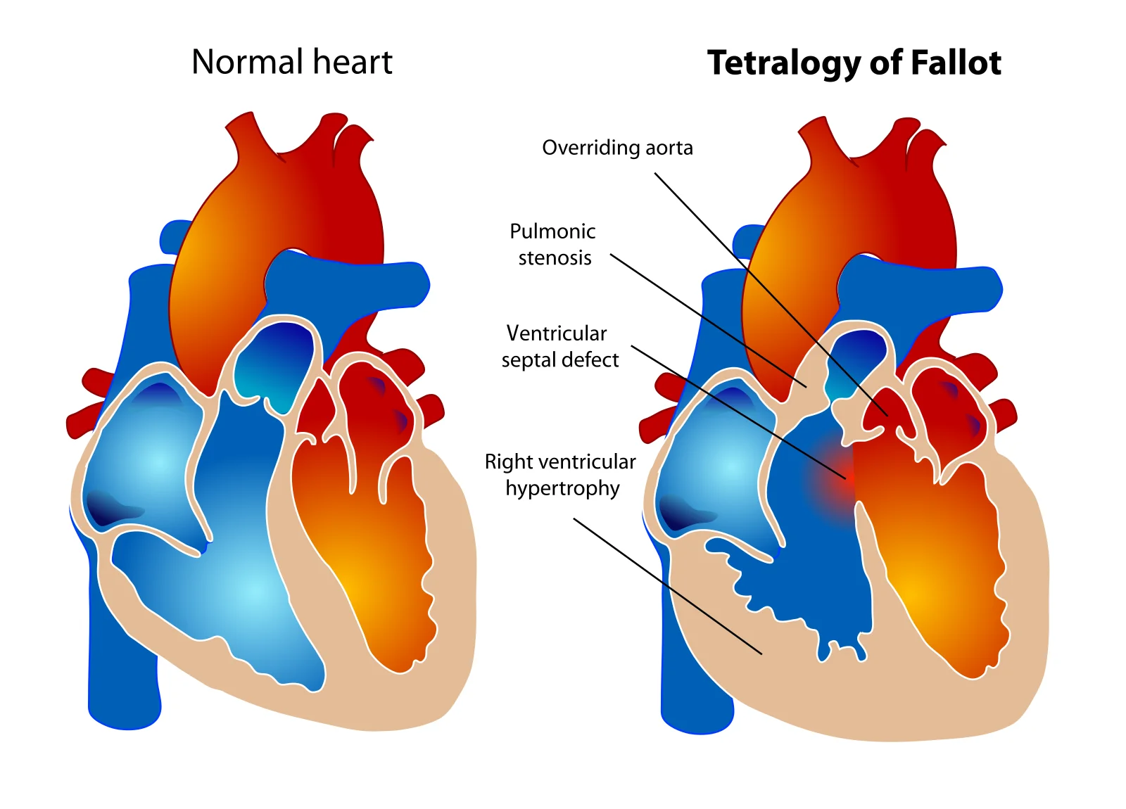
Tetralogy of Fallot combines a large ventricular septal defect with obstruction of the right ventricular outflow tract. The degree of cyanosis therefore depends on the severity of the obstruction. In "pink" or acyanotic tetralogy the obstruction is mild and pulmonary flow is preserved; in the severely obstructed form the child is deeply cyanotic. A right aortic arch is found in about a quarter of patients.[2] Hypercyanotic spells arise from a sudden increase in infundibular obstruction, most often in the morning, after awakening, crying, feeding or defaecation, usually in young infants. The mechanism is thought to be sudden catecholamine-driven spasm of the right ventricular infundibulum producing hyperpnoea, falling systemic vascular resistance and a larger right-to-left shunt.[1]
Transposition of the great arteries produces two independent loops. Without mixing, this is fatal, so the clinical severity depends entirely on the adequacy of the atrial septal defect, ventricular septal defect or ductus arteriosus. Babies with an intact ventricular septum are the sickest, with profound cyanosis within the first 24 hours.[1]
Truncus arteriosus has a single great vessel overriding a large ventricular septal defect. Pulmonary vascular resistance governs pulmonary blood flow, so these infants present with heart failure from pulmonary overcirculation once resistance falls, on top of mild cyanosis.[1]
Total anomalous pulmonary venous return returns all the oxygenated blood to the right atrium; survival depends on an atrial communication that lets it reach the left heart. Obstructed forms (most often the infracardiac type) present as critically ill neonates with severe pulmonary oedema and little cardiac enlargement.[2]
Tricuspid atresia has no opening between the right atrium and right ventricle, so systemic venous blood must cross the atrial septum. The amount of pulmonary blood flow, and therefore the cyanosis, depends on the associated defect and on whether pulmonary stenosis or atresia is present.[1]
Cyanosis is the defining symptom. In a newborn it is central (lips and tongue) rather than acral, and it does not improve with oxygen. Other clues:
- Poor feeding and sweating with feeds, especially when pulmonary overcirculation is present (truncus, total anomalous pulmonary venous return, transposition with a large ventricular septal defect).
- Hypercyanotic spells in tetralogy of Fallot: sudden deepening of cyanosis, hyperpnoea and distress, sometimes progressing to limpness or syncope, sometimes to convulsion or death.[1]
- Squatting after exertion, once the child is old enough to walk. Squatting increases systemic vascular resistance, which reduces the right-to-left shunt and improves pulmonary flow and oxygenation. The phenomenon is described less often now because most lesions are repaired early.
- Headache, dizziness or stroke from polycythaemia and hyperviscosity in long-standing cyanosis.
- Brain abscess in any child with a cyanotic lesion who presents with fever and focal neurological signs.
- Central cyanosis, with clubbing after a few months.
- Murmurs that reflect the lesion: an ejection systolic murmur at the upper left sternal edge in tetralogy of Fallot; often no murmur in transposition with an intact ventricular septum; a single loud second heart sound in truncus; a continuous murmur over the back in lesions with collateral flow.
- Diminished or absent femoral pulses suggest associated coarctation rather than the cyanotic lesion itself.
- Dysmorphic features and thymic absence point to DiGeorge syndrome.
- Saturation measurements at the right hand (pre-ductal) and a foot (post-ductal) are a standard bedside screen. A fall in the foot value indicates a right-to-left ductal shunt.
Not every blue newborn has a cyanotic heart defect. Pulmonary disease, persistent pulmonary hypertension of the newborn, central nervous system pathology, sepsis and methaemoglobinaemia all cause central cyanosis, and hypoxia from respiratory causes usually varies with time or with ventilator changes. The history, the chest radiograph and the hyperoxia test separate these. When a structural heart lesion is present, the differential among the cyanotic defects themselves rests mainly on the degree of pulmonary vascularity on the radiograph and on the echocardiographic anatomy.[2]
| Lesion | What is wrong | Key clinical or radiograph clue |
|---|---|---|
| Tetralogy of Fallot | Large ventricular septal defect with right ventricular outflow obstruction, overriding aorta, right ventricular hypertrophy | Boot-shaped heart and pulmonary oligemia; hypercyanotic spells; ejection systolic murmur[1][2] |
| Transposition of the great arteries | Aorta from the right ventricle, pulmonary artery from the left ventricle, in parallel | Profound cyanosis from day 1; egg-on-a-string chest radiograph; no response to oxygen[1][2] |
| Truncus arteriosus | Single great vessel overriding a ventricular septal defect supplying systemic, pulmonary and coronary flow | Early heart failure with increased vascularity; thymic atrophy and right aortic arch; DiGeorge association[1][2] |
| Total anomalous pulmonary venous return | All pulmonary veins drain to the systemic venous circulation or right atrium | Obstructed type: critically ill neonate with pulmonary oedema and a small heart; unobstructed supracardiac type: snowman mediastinum[1][2] |
| Tricuspid atresia | No tricuspid valve; right atrium drains across the atrial septum | Cyanosis from birth; single second sound; pulmonary vascularity varies with the associated defects[1] |
Measure pre-ductal (right hand) and post-ductal (foot) oxygen saturation in any cyanosed infant, together with an arterial blood gas on room air. A post-ductal value markedly lower than the pre-ductal value indicates a right-to-left ductal shunt.
Right axis deviation and right ventricular hypertrophy support tetralogy of Fallot or pulmonary stenosis; left axis deviation supports tricuspid atresia; the tracing is often normal in transposition. ECG alone does not diagnose the lesion.
The plain film is the first imaging step and it is most useful for two things: pulmonary vascularity and cardiac shape. Obstructive right-heart lesions (tetralogy of Fallot, tricuspid atresia, pulmonary atresia) produce decreased pulmonary vascularity; mixing and parallel-circuit lesions (transposition, truncus, total anomalous pulmonary venous return) produce normal to increased vascularity. Cardiac shape points to the lesion:
- Boot-shaped heart (coeur en sabot) in tetralogy of Fallot: concave main pulmonary artery segment with an upturned apex from right ventricular hypertrophy, pulmonary oligemia and often a right aortic arch.[2]
- Egg on a string in transposition of the great arteries: a globular heart with a narrow superior mediastinum created by the parallel aorta and pulmonary artery and by thymic atrophy, with increased pulmonary vascularity.[2]
- Snowman or figure-of-eight heart in supracardiac total anomalous pulmonary venous return: a dilated vertical vein and innominate vein form the "head" above the cardiac silhouette.[2]
- Box-shaped heart in Ebstein anomaly: massive right atrial enlargement.[2]
- A narrow pedicle with thymic atrophy and increased vascularity in truncus arteriosus, often with a right aortic arch.[2]
Transthoracic echocardiography is the gold standard: it defines the anatomy, the level of obstruction, the great-artery relationships, the venous drainage and the ductus, and it is done at the bedside. Transoesophageal views and CT or MRI are added when the anatomy is complex or before surgical planning.
A hyperoxia test differentiates a cardiac cause of cyanosis from a pulmonary one in an infant who is not simply desaturated from lung disease. An arterial blood gas is obtained on room air, the infant breathes 100% oxygen for 10 minutes, and the blood gas is repeated. In a respiratory cause the arterial partial pressure of oxygen rises above 150 mmHg. In a cyanotic congenital heart lesion with a right-to-left shunt the value does not rise significantly; instead the post-test saturation or partial pressure stays low. In practice pulse oximetry before and after 10 minutes of 100% oxygen is often used instead, with a post-test saturation below 95% (some sources use 85%) suggesting an intracardiac shunt. The result is not 100% specific, because favourable streaming in some cyanotic lesions can allow a larger rise, so an abnormal result should lead to echocardiography rather than reassurance.[3]
A teaching approach to central cyanosis in a newborn
- Central cyanosis under reviewPre-ductal and post-ductal pulse oximetry, arterial blood gas on room air, chest radiograph, ECG
- Question: Pre- and post-ductal saturation difference?
Post-ductal lower than pre-ductal
- Right-to-left ductal shunt likelyThink coarctation, interrupted aortic arch, hypoplastic left heart syndrome
- Start prostaglandin E1 and obtain echo
No significant difference
- Question: Hyperoxia test
PaO2 rises above 150 mmHg
- Pulmonary or other non-cardiac cause likelyInvestigate lung disease, PPHN, sepsis, methaemoglobinaemia
PaO2 does not rise significantly
- Cyanotic heart disease likelyStart prostaglandin E1 if duct-dependent and arrange urgent echocardiography
- Question: Lesion on echo?
TGA
- PGE1 plus balloon atrial septostomy if needed; arterial switch within about a week
Obstructed TAPVC
- Urgent surgical repair
Tetralogy of Fallot
- PGE1 if severe neonatal hypoxemia; surgical repair at 6 to 12 months
Tricuspid atresia
- PGE1 if pulmonary oligemia; staged Fontan palliation
Truncus arteriosus
- Medical stabilisation; repair with Rastelli-type procedure in the first week of life
Handle the baby gently, keep the temperature normal, correct acidosis and hypoglycaemia, and give modest supplemental oxygen. An inspired oxygen fraction of 0.3 to 0.4 is reasonable in cyanotic heart disease; 100% oxygen is unnecessary and, in some duct-dependent pulmonary lesions, can promote ductal closure by lowering pulmonary vascular resistance.[1]
If duct-dependent flow is suspected, start an intravenous infusion of prostaglandin E1 (PGE1, alprostadil) 0.05 to 0.1 mcg/kg/min without waiting for the echocardiogram, and keep two venous access routes open so the infusion is never interrupted. PGE1 reopens or maintains the ductus and is effective in most infants within 30 minutes to 2 hours. The lowest dose that achieves the response should be used: severe neonatal obstruction may need the higher end of the range, an almost closed duct often responds to 0.02 mcg/kg/min, and in some settings 0.01 mcg/kg/min is effective. Doses must then be reduced stepwise, to 0.02 to 0.025 mcg/kg/min, once the ductus is open, because higher doses add hypotension, apnoea and fever. Watch continuously for apnoea, especially when the dose is increased, and have airway support ready. Other adverse effects include vasodilation, tachycardia and, with prolonged infusion, gastric outlet obstruction or cortical hyperostosis.[1][3]
Correct metabolic acidosis with bicarbonate, transfuse for anaemia, and rehydrate. Keep the baby nil by mouth until the diagnosis and plan are settled.
Treat any prolonged spell as an emergency. Steps:[1]
- Place the baby in the knee-chest position (or hold an older child in a squat).
- Give humidified oxygen by face mask, although oxygen helps alveolar hypoxia less than it helps a tired parent feel useful; the real problem is pulmonary oligemia.
- Give morphine sulphate 0.1 mg/kg subcutaneously. [1]
- Leave the infant undisturbed to rest once examination and essential sampling are complete.
- Correct metabolic acidosis, anaemia by transfusion and dehydration by intravenous fluids.
- If the spell continues, raise systemic vascular resistance with phenylephrine, or give propranolol or esmolol intravenously. Oral propranolol may be used chronically once the spell aborts. [1]
- If all else fails, general anaesthesia, an emergency modified Blalock-Taussig shunt or urgent total correction is required. The governing principle is that the infant needs more pulmonary blood flow.
Figure 1Tetralogy of Fallot
Long description
Two heart diagrams side by side. The right-hand heart is labelled with an overriding aorta, pulmonic stenosis, a ventricular septal defect and a thickened right ventricle.

Tetralogy of Fallot. Most infants are minimally cyanotic or acyanotic at birth and do not need immediate surgery. The preferred approach is elective total correction between 6 and 12 months of age, closing the ventricular septal defect so the left ventricle ejects into the aorta and relieving the infundibular, valvar and pulmonary arterial obstruction, on cardiopulmonary bypass, with or without a transannular patch. If neonatal hypoxaemia is severe, PGE1 is used first and then a modified Blalock-Taussig shunt (a Gore-Tex interposition graft between the subclavian artery and the ipsilateral pulmonary artery) or, in suitable anatomy, balloon pulmonary valvuloplasty or right ventricular outflow tract stenting, to allow the pulmonary arteries and the child to grow before correction.[1]
Transposition of the great arteries. With an intact ventricular septum, PGE1 is started and, if oxygenation remains inadequate, a balloon atrial septostomy (Rashkind procedure) creates or enlarges the atrial communication. The arterial switch operation (Jatene procedure, with the LeCompte manoeuvre) is performed at around one week of age, earlier in some centres, so not every baby needs a septostomy. With a ventricular septal defect the arterial switch with closure is undertaken early; with a ventricular septal defect and pulmonary stenosis the Rastelli procedure (intraventricular baffle directing left ventricular blood to the aorta, plus right ventricle to pulmonary artery conduit) is performed at 1 to 2 years.[1]
Tricuspid atresia. Surgery is palliative and staged: first, the haemodynamic problem is addressed (a modified Blalock-Taussig shunt for pulmonary oligemia, pulmonary artery banding for pulmonary plethora, or balloon atrial septostomy for an obstructed foramen ovale). The second stage, a bidirectional Glenn anastomosis at about 6 months, sends superior vena caval blood directly to both pulmonary arteries. The third stage is a fenestrated Fontan (total cavopulmonary connection with an extracardiac conduit), which is the "corrective" step for a single ventricle, relying on low pulmonary vascular resistance and an unobstructed pathway. Desaturating patients need PGE1 first.[1]
Total anomalous pulmonary venous return. Unobstructed types are repaired electively in infancy by anastomosing the common pulmonary vein to the left atrium. Obstructed types are surgical emergencies.[1][2]
Truncus arteriosus. Repair in the first week of life: closure of the ventricular septal defect and placement of a valved conduit between the right ventricle and the pulmonary arteries (Rastelli-type repair), at about the time the falling pulmonary vascular resistance has produced heart failure.[1][2]
In transposition, balloon atrial septostomy is the classic palliative manoeuvre. A balloon-tipped catheter is passed from the femoral or umbilical vein into the left atrium, inflated and pulled sharply back across the atrial septum, tearing it to create a mixing hole. It is performed at the bedside or in the catheter laboratory, guided by fluoroscopy or echo, and it is highly effective. Blade septostomy is reserved for older infants in whom the septum has thickened.[1]
Eisenmenger syndrome is the end result of a large, unrepaired systemic-to-pulmonary shunt. Over years, pulmonary vascular resistance rises until the shunt reverses through the defect, producing cyanosis and pulmonary arterial hypertension. Once the syndrome is established, closing the defect is generally contraindicated, because the defect has become a necessary pop-off. Management therefore centres on targeted pulmonary arterial hypertension therapy (endothelin-receptor antagonists such as bosentan, phosphodiesterase type-5 inhibitors such as sildenafil or tadalafil, and prostacyclin or iloprost), plus diuretics, antiarrhythmics where indicated and anticoagulation in selected patients. Conventional measures such as supplemental oxygen have not shown a clear mortality benefit. Transplantation (heart-lung, or lung with repair) is the only curative option. Clinicians keep patients away from pregnancy, dehydration, isometric exercise, significant time at high altitude and dangerous hypoxia, and avoid peripheral vasodilators that can worsen the right-to-left shunt. Early surgical repair of the original defect, before pulmonary vascular change, is what prevents the syndrome.[6]
- Hypercyanotic spells, seen in tetralogy of Fallot and in other lesions with a large interventricular communication and right ventricular outflow obstruction.[1]
- Polycythaemia and hyperviscosity from chronic hypoxaemia, with headache, visual disturbance and stroke.
- Cerebral abscess, because right-to-left shunting bypasses the pulmonary filter.
- Paradoxical embolism and stroke, especially with an interatrial communication.
- Iron deficiency masked by polycythaemia (relative anaemia), which should be sought and corrected.
- Endocarditis in unrepaired or repaired lesions with residual shunts or patches.
- Pulmonary vascular disease and Eisenmenger syndrome from late repair.[6]
- Arrhythmia and heart failure long after repair (pulmonary regurgitation and right ventricular dilatation after tetralogy of Fallot repair, neoaortic root dilatation and coronary ostial problems after the arterial switch, and Fontan-associated liver disease, plastic bronchitis or protein-losing enteropathy after Fontan surgery).
Prognosis is now excellent in centres that offer early repair: long-term survival after tetralogy of Fallot repair is high, and arterial switch for transposition has transformed an inoperable neonatal condition into one with good medium-term outcomes. Long-term follow-up is lifelong, because residual haemodynamic lesions (pulmonary regurgitation, conduit stenosis, arrhythmia, ventricular dysfunction) appear decades later and often need reintervention. Eisenmenger syndrome, by contrast, remains a multisystem disorder with reduced life expectancy, and pregnancy in Eisenmenger syndrome is dangerous.
Pre-pregnancy counselling is essential. Women with repaired cyanotic lesions may tolerate pregnancy, depending on residual physiology and ventricular function, but women with cyanotic lesions or with Eisenmenger syndrome are at very high risk of maternal death and should be counselled accordingly. Delivery and anticoagulation plans belong to a cardio-obstetric team.
Maternal diabetes mellitus. Transposition of the great arteries is more common in infants of diabetic mothers, and a cardiac phenotype resembling 22q11.2 deletion has been described in the same setting.[2][5]
Maternal lithium. Lithium exposure in the first trimester, particularly in the first 8 weeks, is strongly associated with Ebstein anomaly, and women on lithium need a fetal echocardiogram and a balanced discussion of continuing treatment.[2]
DiGeorge and 22q11.2 deletion. Look for it in any child with a conotruncal defect, especially tetralogy of Fallot, truncus arteriosus, interrupted aortic arch or a ventricular septal defect, together with abnormal facies, thymic hypoplasia, cleft palate or hypocalcaemia. The acronym CATCH 22 captures the clinical spectrum: Cardiac defects, Abnormal facies, Thymic hypoplasia, Cleft palate, Hypocalcaemia, resulting from 22q11 deletion.[4] The deletion is the most common microdeletion, with prevalence about 1 in 3,000 to 1 in 6,000 live births; most are de novo (90 to 95%). Around 75% of affected children have cardiac defects and around 75% have immune deficiency from thymic hypoplasia; hypocalcaemia from hypoparathyroidism occurs in about 50%. Cardiovascular disease is the main cause of childhood mortality, accounting for about 87% of deaths in children with the deletion. Screening uses fluorescence in situ hybridisation or, more commonly now, chromosomal microarray. [5]
- Echocardiography anywhere in the hospital is the test that confirms the diagnosis and should be sought early; a portable scan at the bedside is enough to establish anatomy and ductal flow.
- Where echo is unavailable, the combination of central cyanosis, a chest radiograph (pulmonary vascularity and cardiac shape), and a hyperoxia test (PaO2 failing to rise significantly on 100% oxygen, whereas it rises above 150 mmHg in respiratory disease) separates cardiac from respiratory disease.
- Start prostaglandin E1 on clinical suspicion of a duct-dependent lesion while arranging transfer, because waiting for imaging can cost lives.
- Identify maternal diabetes and lithium exposure in the antenatal history, since they raise the pre-test probability of transposition and Ebstein anomaly.
- Newly diagnosed children with 22q11.2 deletion need calcium, infection and feeding assessment alongside the heart.
- In Eisenmenger syndrome, early referral to a centre with pulmonary arterial hypertension expertise and avoidance of pregnancy are the priorities.
- Cyanotic congenital heart disease is a structural heart defect in which deoxygenated blood reaches the arterial circulation, from obstruction to pulmonary flow, complete mixing, or parallel circulations.
- The five Ts are tetralogy of Fallot, transposition of the great arteries, truncus arteriosus, total anomalous pulmonary venous return and tricuspid atresia.
- Transposition is the commonest cyanotic lesion in the newborn; tetralogy of Fallot is the commonest beyond infancy.
- The intensity of cyanosis follows the mechanism: obstruction tightens in tetralogy, mixing is fixed in truncus and total anomalous pulmonary venous return, and transposition is fatal without mixing.
- Hypercyanotic spells are most common in young infants; knee-chest position, morphine and phenylephrine target the infundibular spasm and falling systemic vascular resistance.
- Squatting after exertion raises systemic vascular resistance and improves oxygenation.
- A boot-shaped heart, egg-on-a-string and snowman mediastinum are classic radiograph clues; echocardiography confirms the anatomy.
- In the hyperoxia test, a PaO2 that does not rise significantly on 100% oxygen, while it rises above 150 mmHg in respiratory disease, points to a cardiac cause. [3]
- Duct-dependent lesions need prostaglandin E1 0.05 to 0.1 mcg/kg/min started on suspicion, with stepwise reduction to 0.02 to 0.025 mcg/kg/min once the ductus is open.
- Balloon atrial septostomy creates mixing in transposition; the arterial switch is performed at about one week of age. [1]
- Tetralogy of Fallot is repaired electively at 6 to 12 months; truncus arteriosus and obstructed total anomalous pulmonary venous return are repaired in the first days of life; tricuspid atresia needs staged Fontan surgery.
- Eisenmenger syndrome is shunt reversal from chronic pulmonary hypertension; the defect should not be closed once it has developed.
- Maternal diabetes, maternal lithium and 22q11 deletion are the associations that change counselling and fetal imaging.
- Untreated cyanosis leads to polycythaemia, iron deficiency, cerebral abscess, stroke and endocarditis; early repair prevents most of these.
Answer each one in your head first, then open the answer.
A 4-day-old infant is deeply cyanosed (SpO2 72%) with no improvement on 100% oxygen. The chest radiograph shows a globular heart with a narrow superior mediastinum and plethoric lungs. What is the most likely lesion, what physiological problem does it create, and what is the first palliative step?
Show answerHide answer
Transposition of the great arteries. The aorta and pulmonary artery run in parallel, so systemic and pulmonary circulations are independent and survival depends on mixing. Give prostaglandin E1 0.05 to 0.1 mcg/kg/min and, if oxygenation remains inadequate, perform a balloon atrial septostomy; then an arterial switch operation at about one week of age.
A 3-month-old with known tetralogy of Fallot suddenly becomes more cyanosed and hyperpnoeic after a feed, then goes limp. Describe the physiology and the immediate management.
Show answerHide answer
This is a hypercyanotic spell from sudden right ventricular infundibular spasm with rising right-to-left shunt. Place the infant in the knee-chest position, give humidified oxygen and morphine 0.1 mg/kg subcutaneously, keep him undisturbed, correct acidosis, anaemia and dehydration, then raise systemic vascular resistance with phenylephrine or beta blockade; if necessary proceed to anaesthesia or surgery. The key is to increase pulmonary blood flow.
How does a hyperoxia test distinguish a cyanotic heart defect from lung disease, and why can it mislead?
Show answerHide answer
The infant breathes 100% oxygen for 10 minutes between two arterial blood gases. A respiratory cause usually raises the arterial PO2 above 150 mmHg; a right-to-left cardiac shunt does not raise it significantly, and a value staying below 100 mmHg or rising less than 30 mmHg points to a cardiac cause. It can mislead because favourable intracardiac streaming in some cyanotic lesions can allow a larger rise, so an abnormal clinical picture still needs echocardiography.
What chest-radiograph findings fit (a) tetralogy of Fallot and (b) supracardiac total anomalous pulmonary venous return, and what does each shape mean anatomically?
Show answerHide answer
(a) A boot-shaped heart: a concave main pulmonary artery segment with an upturned apex from right ventricular hypertrophy, plus pulmonary oligemia, often with a right aortic arch. (b) A snowman or figure-of-eight heart: a dilated vertical vein and innominate vein forming the "head" above the cardiac silhouette, with plethoric lungs. The shapes reflect the underlying anatomy rather than the severity.
Why does squatting relieve cyanosis in a toddler with tetralogy of Fallot, and why might a 10-year-old with an unrepaired ventricular septal defect no longer be blue for the same reason?
Show answerHide answer
Squatting raises systemic vascular resistance, which reduces the right-to-left shunt through the ventricular septal defect and increases pulmonary blood flow. An older child with a large left-to-right shunt and no outflow obstruction has already developed pulmonary vascular disease; cyanosis then returns if Eisenmenger physiology supervenes, not because the original right-to-left physiology recurred.
A pregnant woman in the first trimester is found to take lithium for bipolar disorder. What fetal heart problem is associated, what investigation is warranted, and how does the finding change management?
Show answerHide answer
First-trimester lithium exposure, particularly in the first 8 weeks, is strongly associated with Ebstein anomaly, an apical displacement of the tricuspid valve. A fetal echocardiogram is warranted. Management balances the maternal psychiatric need for treatment against the small, apparently dose-related fetal cardiac risk, within a shared cardio-obstetric plan rather than an automatic stop.
Name the clinical acronym for 22q11 deletion syndrome and list what each letter stands for.
Show answerHide answer
CATCH 22: Cardiac defects, Abnormal facies, Thymic hypoplasia, Cleft palate, Hypocalcaemia, resulting from deletion of chromosome 22q11. It reminds the clinician to look for all of these when one of them, especially a conotruncal heart defect, is found.
A 20-year-old with an unrepaired ventricular septal defect is cyanosed and has haemoptysis and clubbing. Why is the defect no longer closed, and what treatment remains?
Show answerHide answer
She has Eisenmenger syndrome: chronic left-to-right shunting raised pulmonary vascular resistance until the shunt reversed, so the defect now acts as a necessary vent for the right ventricle. Closing it would precipitate right heart failure. Treatment is targeted pulmonary arterial hypertension therapy, avoidance of pregnancy, dehydration, high altitude and isometric exercise, and consideration of transplantation.
Read next: heart failure in children for the pulmonary overcirculation phase of truncus and total anomalous pulmonary venous return; pulmonary arterial hypertension and Eisenmenger syndrome for the vascular complications of late repair; and conotruncal anomalies and 22q11.2 deletion for the genetics and extracardiac follow-up. Within paediatric cardiology see also balloon atrial septostomy, Blalock-Taussig shunt and Fontan circulation for the procedures these children undergo. Low-resource readers should focus on neonatal resuscitation and transfer of the sick newborn.
- Rao PS. Management of Congenital Heart Disease: State of the Art—Part II—Cyanotic Heart Defects. Children 2019;6(4):54. doi:10.3390/children6040054. Accessed .
- Bhat V, Belaval V, Gadabanahalli K, Raj V, Shah S. Illustrated Imaging Essay on Congenital Heart Diseases: Multimodality Approach Part III: Cyanotic Heart Diseases and Complex Congenital Anomalies. J Clin Diagn Res 2016;10(7):TE01–TE10. doi:10.7860/JCDR/2016/21443.8210. Accessed .
- Fern NM. The Hyperoxia Test for the Cyanotic Infant. UMEM Educational Pearls, University of Maryland School of Medicine 2019. Accessed .
- Wilson DI, Burn J, Scambler P, Goodship J. DiGeorge syndrome: part of CATCH 22. J Med Genet 1993;30(10):852–856. doi:10.1136/jmg.30.10.852. Accessed .
- McDonald-McGinn DM, Sullivan KE, Marino B, et al. 22q11.2 deletion syndrome. Nat Rev Dis Primers 2015;1:15071. doi:10.1038/nrdp.2015.71. Accessed .
- Kim M, Chung W-J. Current therapy of Eisenmenger syndrome. J Thorac Dis 2016;8(11):3009–3011. doi:10.21037/jtd.2016.11.27. Accessed .
Figure credits
Educational content for medical students and doctors. It is not a substitute for clinical judgement or local guidelines.