Pyloric stenosis
Infantile hypertrophic pyloric stenosis: gastric outlet obstruction, projectile vomiting, metabolic alkalosis, ultrasound diagnosis and pyloromyotomy.
- Pyloric stenosis (infantile hypertrophic pyloric stenosis) is a thickening of the muscular wall of the pylorus that narrows and lengthens the outlet of the stomach, producing gastric outlet obstruction in young infants.
- It most often appears between 2 and 8 weeks of age, mainly in first-born boys, as progressively forceful, non-bilious vomiting shortly after feeds; the infant remains hungry and eager to feed.
- Dehydration and loss of gastric acid lead to a hypochloraemic, hypokalaemic metabolic alkalosis.
- Palpation may detect a small, firm, olive-shaped mass in the epigastrium or right upper quadrant; ultrasound confirms the diagnosis and rules out other causes.
- After fluid and electrolyte resuscitation, a pyloromyotomy (splitting the hypertrophied muscle without opening the mucosa) is curative.
By the end of this page you should be able to:
- Recognise infantile hypertrophic pyloric stenosis from its age of onset, pattern of vomiting and physical findings.
- Explain how a thickened pylorus produces vomiting and hypochloraemic hypokalaemic metabolic alkalosis.
- Interpret an abdominal ultrasound for pyloric stenosis and state why published measurement cut-offs differ.
- Resuscitate an affected infant before surgery and outline what pyloromyotomy involves.
- Distinguish pyloric stenosis from the other common causes of vomiting in early infancy.
- Counsel parents about prognosis, feeding after surgery and low-resource alternatives to ultrasound.
DefinitionInfantile hypertrophic pyloric stenosis (IHPS)
A condition of young infants in which the circular (and to a lesser degree the longitudinal) muscle of the pylorus hypertrophies and thickens, lengthening and narrowing the pyloric canal so that the stomach cannot empty normally: gastric outlet obstruction. It is not a congenital malformation in the usual sense - the pylorus is present and positioned normally - but the hypertrophy develops in early infancy.
It is the commonest surgical cause of early-infancy vomiting. The commoner functional problem it is confused with is pylorospasm, in which the pyloric muscle is not hypertrophied and vomiting usually settles.
Incidence varies by population and era: the index series quotes about 2 to 4 per 1,000 live births, with higher rates in temperate than in equatorial countries (suggesting that environmental factors play a part); Medscape 2024 quotes 1.5 to 4 per 1,000 live births among White infants (lower in Black, Asian and Hispanic infants), and Children's Hospital of Philadelphia quotes 2 to 3 per 1,000.[2][5][8] One regional population study found it in roughly 9 to 14 per 10,000 births before 2009.[4]
- Sex. Boys are affected far more often than girls. Reported male-to-female ratios range from about 2:1 to 6:1 depending on the series; Medscape 2024 quotes 2:1 to 5:1, Bašković 4:1 to 6:1, and Feng 2.5:1 to 5.5:1.[1][2][5]
- Birth order. It is a classic disorder of first-born infants, especially first-born boys, although the proportion reported as firstborn varies between series (76.3% in one Chinese hospital series) and other sources only confirm first-born male infants as the highest-risk group.[1][5][7]
- Age at presentation. Vomiting usually begins between 2 and 8 weeks of age, and most infants are diagnosed between 2 and 12 weeks after birth, with a median around the 40th day of life.[6][8][2][7] Occasional late presentation occurs up to 12 weeks of age or beyond.[6] [1]
- Ethnicity and geography. The disorder is most common in infants of Northern European descent and less common in infants of Afro-Caribbean and Asian descent.[1]
- Family history and genetics. A minority of cases run in families; one series found a positive parental history in about 2% of cases. Coexisting congenital anomalies occur in about 20.6% of cases.[1][2]
The exact cause is unknown. IHPS is generally regarded as multifactorial, with both genetic and environmental contributions.[1][2]
- Bottle feeding. Bottle feeding after birth has been associated with an increased risk of pyloric stenosis in a large population-based study; the association appears stronger in older and multiparous mothers. The same trend is compatible with the observed rise in breastfeeding and fall in incidence in recent decades.[4]
- Prematurity and birth weight. Prematurity is found in a small proportion of cases. Birth weight overlaps broadly with unaffected infants.[1]
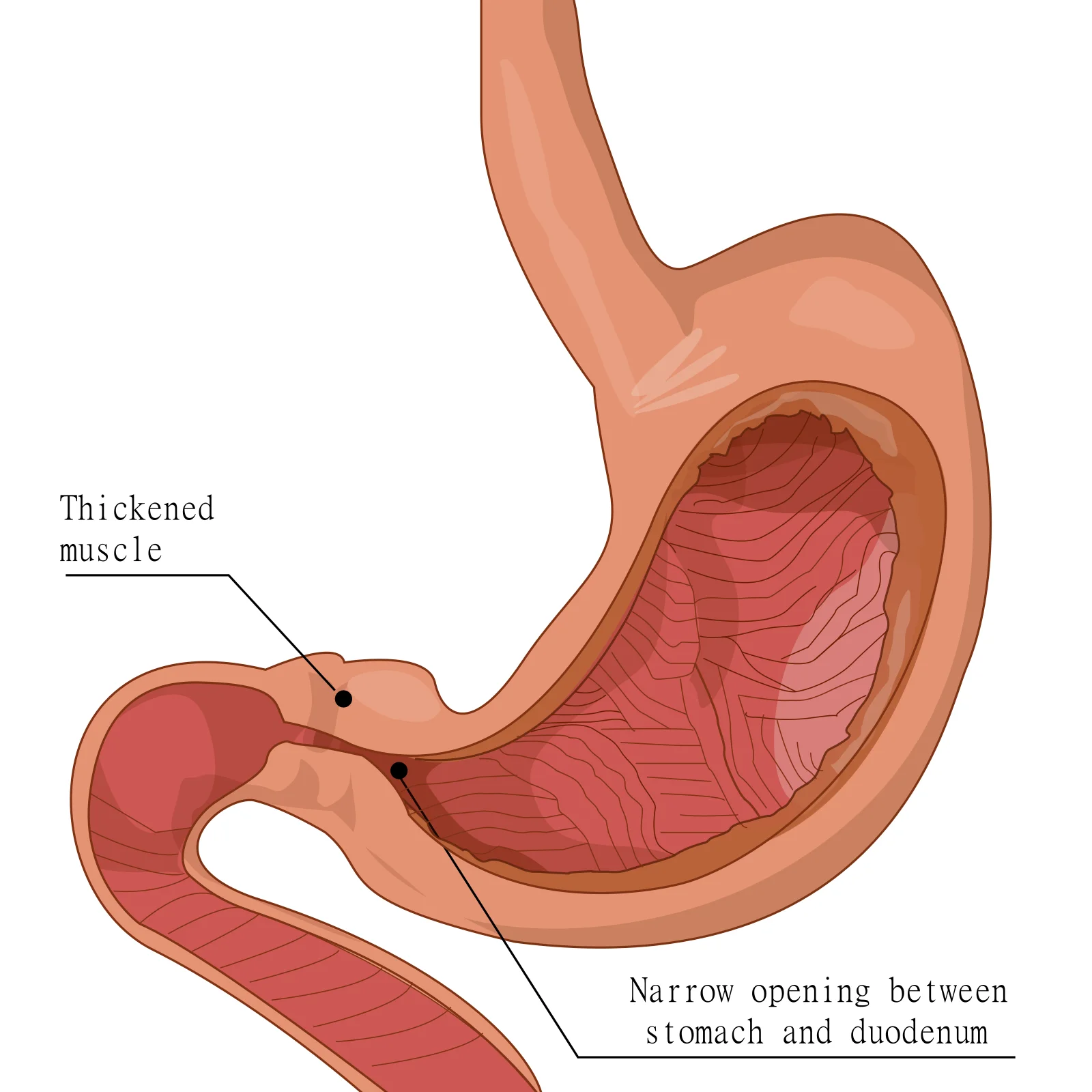
Figure 1Pyloric stenosis
Long description
Diagram of infantile hypertrophic pyloric stenosis: a thickened pylorus narrowing the outlet of the stomach.

The pylorus is the muscular gateway between the stomach and the duodenum. In IHPS the circular muscle (and to a lesser extent the longitudinal muscle) hypertrophies, so the canal becomes long, thick and rigid. Food can enter the stomach, but emptying into the duodenum is obstructed, especially after a meal.
- The stomach generates powerful peristaltic waves to push past the narrowed outlet, and the infant vomits non-bilious (gastric) contents with considerable force.
- Because the blockage is at the pylorus, before the duodenum, the vomit never contains bile.
- Repeated vomiting of gastric acid (hydrochloric acid) removes hydrogen and chloride ions from the body, producing hypochloraemic metabolic alkalosis. Chloride loss exceeds the kidney's ability to compensate, and potassium is lost with the vomitus and in the urine; the result is hypokalaemia.
- Dehydration follows, and the kidney conserves water and sodium; in compensation the kidney excretes acid urine in the presence of alkalosis, so-called paradoxical aciduria.
The degree of alkalosis tends to be worse when vomiting has lasted longer than about three days.[1] [2]
The history and the baby's general condition point strongly to the diagnosis.
- Projectile vomiting of stomach contents, usually 10 to 30 minutes after a feed, increasing in frequency and force over days. The vomit is non-bilious and may be blood-streaked if the mucosa is irritated.[1]
- Persistent hunger: the infant wants to feed again immediately after vomiting, and takes feeds readily (the "hungry vomiter").
- Constipation or scanty stools, because little milk reaches the intestine.
- Failure to gain weight or weight loss, irritability and lethargy as dehydration develops.
- Dehydration: sunken fontanelle, dry mucous membranes, poor skin turgor, reduced urine; in severe cases shock.
- Visible gastric peristalsis: a wave moves from left to right across the upper abdomen during or after a feed.
- The olive: a small, firm, mobile, cylindrical mass palpable in the epigastrium or right upper quadrant, best felt after a vomit or with the infant relaxed. It is felt in many but not all cases and is harder to feel in early, mild disease.
- Metabolic alkalosis on blood testing: high pH, high bicarbonate, low serum chloride and often low potassium.
A vignette: a 3-week-old first-born boy who has been feeding well, now vomits forcefully after every feed and then cries for more. His weight gain has faltered. A wave is visible crossing the abdomen after feeds, and a firm elongated mass is felt just to the right of the midline below the liver.
In large series most affected infants have a raised pH and bicarbonate and lowered chloride; hypokalaemia occurs in roughly 40% of infants in older presentation series and less often in sonography-era cohorts, hypochloraemia in about 63%, and an acid-base abnormality overall in one series (84.9%, 203/239), whereas many contemporary infants have normal or mildly abnormal electrolytes.[1][2] Severity worsens with longer symptom duration.[1][2]
The key distinction is between gastric outlet obstruction at the pylorus (non-bilious projectile vomiting in a 2 to 8 week old) and everything else. The table separates the usual alternatives by the feature that points away from pyloric stenosis.
| Condition | Typical age | Feature that separates it from pyloric stenosis |
|---|---|---|
| Gastro-oesophageal reflux (physiological) | Any age | Small, effortless possets; infant well, gaining weight; no olive mass |
| Pylorospasm or an evolving pyloric stenosis | Early infancy | Same picture, but ultrasound shows a normal muscle thickness |
| Milk protein (cow's milk or soya) enteropathy | Early infancy | Diarrhoea or blood/mucus in the stool; eczema or atopy; improves with elimination diet |
| Malrotation with midgut volvulus | Any age | Bilious vomiting; sudden onset; painful, distended abdomen - surgical emergency |
| Duodenal atresia or stenosis | From first feeds | Bilious vomiting from birth; double-bubble on abdominal X-ray |
| Oesophageal atresia / tracheo-oesophageal fistula | First feeds | Coughing, choking and cyanosis with feeds; inability to pass a nasogastric tube |
| Intussusception | Older infant | Episodic screaming, pallor and drawing up of legs; redcurrant-jelly stool; sausage-shaped mass |
| Viral gastroenteritis | Any age | Diarrhoea and fever; contacts ill; no progressive projectile vomiting |
| Increased intracranial pressure (e.g. hydrocephalus, meningitis) | Any age | Vomiting not tied to feeds; bulging fontanelle, altered tone or irritability |
| Systemic infection or metabolic disease | Neonatal period | Lethargy, poor feeding, temperature disturbance; often no focus |
| Congenital adrenal hyperplasia | Neonatal period | Vomiting, poor weight gain, dehydration, virilisation; hyponatraemia with hyperkalaemia |
Approach to an infant presenting with vomiting
- Vomiting infantHistory of age, onset, feeding, vomitus (bilious/non-bilious), growth; examine for dehydration, olive mass, peristalsis
- Question: Any red flag? (bilious vomiting, sick/unwell infant, abdominal distension, shock, onset at birth)
Yes
- Treat as emergencyResuscitate, imaging per surgical team; exclude malrotation/volvulus, atresia, sepsis
No
- Age 2-8 weeks, projectile non-bilious vomiting, hungry babyHypertrophic pyloric stenosis likely
- Examine the abdomen after a feedVisible peristalsis and an olive-shaped epigastric/right-upper-quadrant mass
- Blood testsUrea and electrolytes, venous blood gas: look for hypochloraemic hypokalaemic metabolic alkalosis
- Abdominal ultrasoundPyloric muscle thickness and canal length; compare with the criteria your department uses (see Investigations)
- Question: Ultrasound supportive of HPS?
Yes
- Resuscitate, then pyloromyotomyBoluses of saline, then infusion; correct alkalosis before anaesthesia
No or equivocal
- Re-image and reconsiderRepeat ultrasound, upper GI contrast study, and broaden the differential
Examination is best done with the infant relaxed, and rocking or offering a dummy between feeds. A test feed can make peristalsis visible. If a mass is felt, do not keep re-examining - confirm with ultrasound.
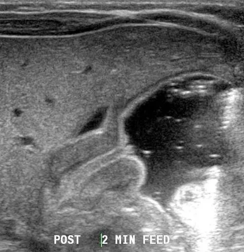
Figure 2PyloricStenosisUS
Long description
Abdominal ultrasound of infantile hypertrophic pyloric stenosis in a 6-week-old, showing a thickened pyloric muscle.

Ultrasound is non-invasive; reported sensitivity ranges from about 94% to 99.5% and specificity from about 98% to 100% depending on the muscle-thickness cut-off used and the setting, with a pooled sensitivity of 97.6% and pooled specificity of 98.8% at a cut-off of 3 mm.[2] Typical findings are a thickened, elongated pylorus with a non-relaxing canal; if the infant drinks water the canal can be seen to stay closed. One hospital series found ultrasound to be diagnostic in 98.2% of cases.[1] [3]
Published measurement criteria differ, and clinicians should know which cut-offs their department applies:
- Traditional criteria: pyloric muscle thicker than 3 mm and pyloric canal longer than 15 mm.[2]
- Contemporary data: a surgically confirmed series found pyloric lengths as low as 10 mm and muscle thickness as small as 2.0 mm, and argues that a canal length greater than 10 mm is reliable while muscle thickness greater than 2.2 mm is supportive.[3]
- Series data: in another large series muscle thickness ranged from 3 to 8 mm and canal length from 11 to 38 mm.[1]
- The overall pyloric diameter and transverse measurements are not reliable discriminators on their own.[3]
No ultrasound criterion on its own overrides a strong clinical picture or an equivocal scan: if doubt remains, repeat the scan after a short interval or use an upper gastrointestinal contrast study.
- Urea and electrolytes and venous or capillary blood gas: expect hypochloraemic hypokalaemic metabolic alkalosis.
- Glucose (infants may be hypoglycaemic after prolonged vomiting).
- Full blood count if anaemia or infection is suspected.
- Conjugated bilirubin is often transiently raised in jaundiced infants with pyloric stenosis and resolves after treatment.
An upper gastrointestinal contrast study (barium or water-soluble contrast meal) shows a string sign or a double-track sign through an elongated pyloric canal and delayed gastric emptying. It is less favoured where ultrasound is readily available because of radiation and aspiration risk.
There is no single agreed numeric threshold worldwide: sources quote muscle thickness cut-offs of 2.2 to 3 mm and canal length cut-offs of 10 to 15 mm.[2][3] In practice the diagnosis rests on a consistent clinical picture plus an ultrasound morphology that is clearly abnormal for the child's age, with the clinical team integrating the two. For this reason the integrity of the technique (a relaxed, fasted infant, water to fill the stomach, measurements taken at right angles to the beam) matters as much as any number.[3]
Severity is judged clinically (degree of dehydration, biochemical disturbance and weight loss) rather than by a staging score.
The operation itself is straightforward; the risk is operating on an unresuscitated infant. Uncorrected alkalosis can impair respiratory function in neonates in the perioperative period.[2]
- Volume. Many infants need an intravenous bolus of normal (0.9%) saline 20 mL/kg, once or repeatedly, then an infusion at 4 to 6 mL/kg/h until biochemical abnormalities are corrected.[2]
- Electrolytes. Replace potassium once urine output is adequate and ensure chloride is restored; the goal of saline resuscitation is in part to give a relative excess of chloride, which allows the kidney to excrete bicarbonate and correct the alkalosis.
- Monitoring. Repeat urea, electrolytes and acid-base status until they normalise.
- Timing. Correction usually takes 12 to 48 hours; surgery is performed once electrolytes and acid-base balance are satisfactory.[2]
The standard operation is the Ramstedt pyloromyotomy: the hypertrophied muscle of the pylorus is incised along its full length and gently split apart, but the mucosa is left intact so the lumen opens.
- The incision extends from the prepyloric vein at the duodenal junction to the circular fibres of the distal antrum, proximal to the pylorus.
- The muscle is spread until the intact mucosa bulges into the incision.
- Surgery may be open (through an upper mini-incision or circumumbilical approach) or laparoscopic.
- Laparoscopic pyloromyotomy is an efficient and reliable alternative: hospital stay and recovery are shorter, but the risk of incomplete pyloromyotomy or mucosal perforation is higher in the learning curve.[2]
Feeding is restarted once the infant is awake and not vomiting, usually beginning with small frequent feeds and advancing as tolerated. Postoperative vomiting is common initially and usually settles. Discharge is typically once full feeds are tolerated without vomiting; exact pathways are set by the surgical team.
All suspected or confirmed cases should be discussed with a paediatric surgeon early, because resuscitation and surgery are time-critical and electrolyte correction takes a day or more. Any infant with bilious vomiting, shock, or a mass not typical of pyloric stenosis needs same-day paediatric or surgical review.
- Preoperative: dehydration, hypochloraemic hypokalaemic metabolic alkalosis, malnutrition and aspiration risk.
- Perioperative: mucosal perforation (if the mucosa is breached, it is repaired and the infant is managed accordingly), incomplete pyloromyotomy (recurrent vomiting), wound infection, ileus.
- Postoperative: postoperative vomiting is common initially; surgical-site infection or bleeding are uncommon.
With prompt resuscitation and pyloromyotomy, outcomes are excellent: the obstruction is relieved permanently and most infants feed normally and grow well afterwards. Regrowth of the muscle and recurrent stenosis are rare. Infants who presented late with severe alkalosis may take longer to recover feeding.
There is no established screening test and no proven preventive measure for the general population. Prenatal macrolide use should be avoided where possible, and feeding and medication choices in early infancy should follow standard guidance. There is no routine postoperative surveillance beyond normal growth follow-up.
- Premature infants may present earlier and are at higher risk of postoperative apnoea; anaesthetic and surgical teams familiar with neonates are important.
- Children with other congenital anomalies (6% to 20% of cases, up to 20.6% in one series, commonly cardiac defects) need these addressed alongside the pyloromyotomy.[1][5]
- Pregnancy/maternal: the condition is a disease of infants; maternal pregnancy is not affected, but recurrence risk in later siblings is raised, so parents should be told of the possibility.
Ultrasound and a paediatric surgeon are not available everywhere, but the diagnosis is clinical in many cases.
- Clinical diagnosis: in an infant of 2 to 8 weeks with forceful non-bilious vomiting, a visible peristaltic wave and a palpable olive-shaped epigastric mass is highly suggestive; basic blood tests show the characteristic hypochloraemic hypokalaemic metabolic alkalosis.
- Where ultrasound is scarce: a test feed and careful palpation (after the infant vomits and relaxes) can establish the diagnosis; upper gastrointestinal contrast studies are an alternative where imaging exists.
- Resuscitation is cheap and essential: correct dehydration and electrolytes with saline and potassium before surgery even if the definitive operation must wait for transfer.
- Surgery: if cardiopulmonary or surgical facilities are unavailable, arrange early referral to a centre that can perform pyloromyotomy; in the interim keep the infant nil-by-mouth with nasogastric decompression if there is persistent vomiting.
East Africa
In East African practice delayed presentation is common; by the time of diagnosis many infants arrive severely dehydrated with marked electrolyte disturbance. Empirical resuscitation with saline and potassium and careful re-feeding after pyloromyotomy are emphasised.
- Infantile hypertrophic pyloric stenosis is gastric outlet obstruction from hypertrophy of the pyloric muscle, not a fixed malformation.
- It typically presents at 2 to 8 weeks (most diagnosed by 12 weeks), mainly in first-born boys, with progressive projectile non-bilious vomiting and a persistently hungry infant.
- Loss of gastric acid produces hypochloraemic hypokalaemic metabolic alkalosis, worsened by prolonged vomiting.
- Look for visible gastric peristalsis and a palpable olive-shaped mass in the epigastrium or right upper quadrant.
- Abdominal ultrasound is the diagnostic test of choice; published muscle and canal-length cut-offs vary, so apply your department's criteria and good technique.
- Resuscitate fully with saline (20 mL/kg bolus, then 4 to 6 mL/kg/h) and correct electrolytes before surgery (usually 12 to 48 hours).
- Pyloromyotomy (open or laparoscopic, preserving the mucosa) is curative.
- Prognosis after surgery is excellent; recurrence of stenosis is rare.
- In low-resource settings the diagnosis can often be made clinically and the infant stabilised with saline while arranging referral.
Answer each one in your head first, then open the answer.
Which infants are most at risk of pyloric stenosis, and at what age does it usually present?
Show answerHide answer
First-born boys presenting between 2 and 8 weeks of age (most diagnosed by about 12 weeks). Male-to-female ratios of about 2:1 to 6:1 are reported; first-born male infants are at highest risk.
A 4-week-old boy has forceful vomiting after every feed but begs to feed again. Why is the vomit not bile-stained, and why does the baby stay hungry?
Show answerHide answer
The obstruction is at the pylorus, proximal to the duodenum where bile enters, so only gastric contents are expelled. Because the blockage is mechanical and intermittent, the stomach's hungry sensation returns immediately, hence the eager re-feeding.
What is the characteristic acid-base and electrolyte pattern, and what two factors produce it?
Show answerHide answer
Hypochloraemic hypokalaemic metabolic alkalosis. Loss of hydrochloric acid in the vomitus removes hydrogen and chloride; renal water conservation and potassium loss compound the hypokalaemia. Paradoxical aciduria occurs because the kidney conserves sodium and water at the expense of acid excretion.
On ultrasound what are the published cut-offs you may see quoted, and how do centres differ?
Show answerHide answer
Traditional criteria quote pyloric muscle greater than 3 mm and canal length greater than 15 mm; contemporary data argue that a canal length greater than 10 mm and muscle greater than 2.2 mm can be diagnostic, and a large series reports thickness of 3 to 8 mm and length of 11 to 38 mm. Apply your own department's criteria and good technique. [2]
Before pyloromyotomy, what resuscitation is required and why can surgery not proceed immediately?
Show answerHide answer
Intravenous normal saline 20 mL/kg boluses plus maintenance 4 to 6 mL/kg/h with potassium replacement, usually over 12 to 48 hours, until urea, electrolytes and acid-base balance normalise. Uncorrected alkalosis can impair neonatal respiratory function in the perioperative period.
A 6-week-old vomits after feeds. What finding at the bedside or on imaging would most strongly point to midgut volvulus rather than pyloric stenosis?
Show answerHide answer
Bilious vomiting. Pyloric stenosis vomits are non-bilious; bilious, sudden or severe vomiting in an infant means treat as a surgical emergency and exclude malrotation with volvulus.
- Gastro-oesophageal reflux disease"""
- Malrotation and midgut volvulus
- Vomiting in the infant (presentation approach)
- Neonatal intestinal obstruction
- Dehydration and fluid therapy
- Congenital adrenal hyperplasia
- Feng Z, Nie Y, Zhang Y, et al. The Clinical Features of Infantile Hypertrophic Pyloric Stenosis in Chinese Han Population: Analysis from 1998 to 2010. PLoS One 2014;9(2):e88925. Accessed .
- Bašković M, Sinjeri D. Diagnostic Accuracy of Acid-Base Status in Infants with Hypertrophic Pyloric Stenosis. Children (Basel) 2022;9(12):1815. Accessed .
- Piotto L, Gent R, Taranath A, Bibbo G, Goh DW. Ultrasound diagnosis of hypertrophic pyloric stenosis - Time to change the criteria. Australas J Ultrasound Med 2022;25(3):116-126. Accessed .
- Rosen NG, Stetson AE. Infantile Hypertrophic Pyloric Stenosis. Medscape. Updated 2024-02-07. Accessed .
- Johns Hopkins All Children's Hospital. Hypertrophic pyloric stenosis clinical pathway. Clinical pathway, revised 2026-03-24. Accessed .
- BMJ Best Practice. Pyloric stenosis. Topic updated 2024-01-05. Accessed .
- Children's Hospital of Philadelphia. Pyloric stenosis. Patient information. Accessed .
- van den Bunder FA, Derikx JP, Kiblawi R, van Rijn RR, Dingemann J. Diagnostic accuracy of palpation and ultrasonography for diagnosing infantile hypertrophic pyloric stenosis: a systematic review and meta-analysis. Br J Radiol 2022;95(1139):20211251. Accessed .
Figure credits
Educational content for medical students and doctors. It is not a substitute for clinical judgement or local guidelines.