Intussusception
Telescoping of the intestine in infants: causes, recognition, ultrasound and enema reduction, surgery, and recurrence.
- Intussusception is a telescoping of one part of the intestine into the next, usually the ileum sliding into the colon; it is the commonest cause of bowel obstruction in infants and toddlers.
- It is most common between about 3 and 36 months of age, with a peak in later infancy, and it is more common in boys.[1][3]
- The usual picture is sudden, colicky abdominal pain that comes and goes every 15 to 20 minutes, with vomiting; "currant-jelly" stool and a sausage-shaped mass are later or less common signs.[2][3]
- Ultrasound is the investigation of choice and shows the target sign; if the child is stable with no perforation or peritonitis, an air or liquid enema reduces the bowel and treats the problem in about 80% or more of cases.[1][2]
- Surgery is needed for perforation, peritonitis, shock, failed enema reduction, or a structural lead point; recurrence after successful enema reduction is about 9 to 15%.[1]
By the end of this page you should be able to:
- Explain what an intussusception is, name the usual type and the usual age group, and describe the common triggers.
- Recognise the presentation, including early and atypical forms, and know which features are late.
- Use ultrasound sensibly, explain the target sign, and decide which children can go straight to enema reduction.
- Carry out or arrange the standard enema reduction safely, state the pressure limit and contraindications, and know when to stop and operate.
- List the common lead points and the clinical situations in which to look for one.
- Counsel a family about recurrence and about the very small risks and large benefits of rotavirus vaccination.
DefinitionIntussusception
Intussusception is the telescoping (invagination) of a proximal segment of intestine into the lumen of an adjacent distal segment. The part that slides in is the intussusceptum; the part that receives it is the intussuscipiens. The commonest type is ileocolic, in which terminal ileum telescopes into the caecum and colon, and it makes up more than 80% of cases.[1]
The other types are ileoileocolic, small-bowel (enteroenteric or jejunoileal) and, rarely, colocolic intussusception.[1] Small-bowel intussusceptions are usually short and transient and often settle with observation alone; this page focuses on the ileocolic type, which is the one that presents as an acute abdomen in infants.[1]
About 75% of cases have no single identified cause and are called idiopathic; the usual mechanism is thought to be hypertrophy (hyperplasia) of lymphoid tissue (Peyer patches) in the terminal ileum, often after a viral or, less often, bacterial infection.[3][4] The rest have a pathologic lead point, more often in very young infants, in older children, and after repeated episodes.[2]
Intussusception is a disease of infants and toddlers. It typically occurs in children aged 3 to 36 months, with incidence highest in the first to third years of life; about 10% of cases occur in children older than 5 years.[1][3] One well-quoted description gives onset at about 5 months of age, peaking between 4 and 9 months and gradually declining by 18 months.[4]
- Numbers. About 2,000 infants in the United States experience intussusception in the first year of life.[4] Incidence estimates vary by country and study; examples include about 196.7 cases per 100,000 children under 2 years in a Korean nationwide survey, an overall rate of 28.3 per 100,000 person-years in Korea, and 56.1 per 100,000 per year in children under 1 year in New Zealand before rotavirus vaccination, close to a worldwide average estimate of about 74 per 100,000.[1][3]
- Sex. Boys are affected more often. Ratios of about 3 to 1 (male to female) are reported in one review, and about 3 to 2 in another.[3][4]
- Age pattern differs by region. In a prospective Indian surveillance series the median age was 8 months (interquartile range 5 to 13 months), about three-quarters of cases were in infants, and 65.7% were male.[6]
- Season. Many series describe peaks that coincide with the season of viral enteritis; other series find no clear seasonal pattern.[1]
Where intussusception occurs outside the usual age range, a pathologic lead point is likely, so age outside 3 months to 5 years should raise the search for a cause.[1][2]
In about 75% of children the cause is unknown (idiopathic).[3] Known triggers fall into three groups.
Infections. Approximately 30% of children have a recent viral illness.[2] A strong association with adenovirus infection is described in multiple populations, with enteric and non-enteric adenovirus in roughly 30 to 40% of cases; infection probably causes hypertrophy of lymphoid tissue in the bowel wall, which then acts as a lead point.[1] Bacterial enteritis with Salmonella, Escherichia coli, Shigella and Campylobacter species has also been reported.[3] Other associated viruses include rotavirus and human herpesvirus 6.[3]
Systemic or anatomical conditions that thicken or distort the bowel wall or alter motility. Examples are cystic fibrosis, immunoglobulin A vasculitis (IgAV, formerly Henoch-Schonlein purpura) when purpura involve the bowel wall, haemolytic-uraemic syndrome, and (in older lists) coeliac disease.[2][3]
Focal lead points (see table below) such as Meckel diverticulum, intestinal polyps, duplication cysts, lymphoma and, in adults, a neoplasm.[1][2]
Approximately 25% of children, typically very young infants and older children, have a lead point.[2]
| Lead point | Notes |
|---|---|
| Meckel diverticulum | The most common focal lead point in children in most series.[1] |
| Intestinal polyps | Including polyps from Peutz-Jeghers syndrome; may also be solitary.[2] |
| Duplication cyst | Often small-bowel or ileocaecal.[1] |
| Lymphoma or other tumour | Especially in older children; small-bowel lymphoma in particular.[1][2] |
| IgAV / Henoch-Schonlein purpura | A small-bowel wall haematoma acts as the lead point, often after the acute abdominal pain of the vasculitis has settled.[1][2] |
| Cystic fibrosis | Thick secretions and altered motility; also a risk factor rather than a focal mass.[2] |
| Inverted appendiceal stump, vascular malformation, parasite, foreign body | Less common; usually found at surgery.[1][3] |
| Reactive lymphoid hyperplasia | A diffuse lead point rather than a focal one; the common idiopathic mechanism.[1] |
Lead points are found in a minority of cases overall (a pooled estimate of 4%, with reported ranges of 0.3 to 20%), and the proportion rises with age, being much higher in children older than the typical age range.[1] Meckel diverticulum is the most common pathologic lead point, followed by polyps and then duplication cysts or IgAV.[1]
Figure 1Intussusception EN
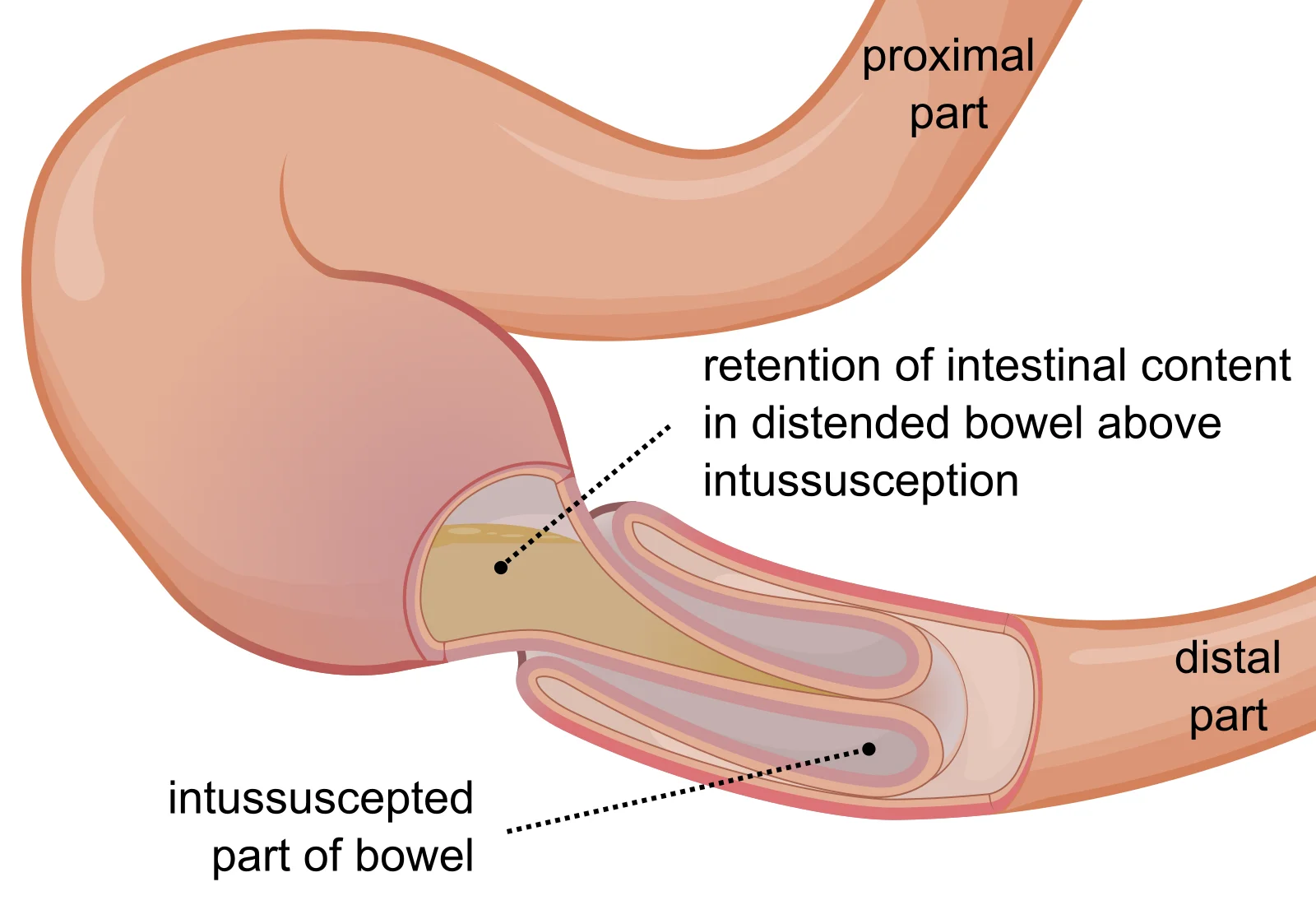
Long description
Diagram of bowel intussusception with one segment of intestine telescoped into the next.

Ileocolic intussusception is driven by peristalsis. A segment of terminal ileum, sometimes carrying a lead point, is pulled forward into the caecum and then further along the colon. The mesentery is dragged in with it, so veins are compressed before arteries, and the bowel wall becomes oedematous, congested and, with time, ischaemic. If untreated, gangrene and perforation follow. The illness can deteriorate from well-appearing intervals to shock within hours, which is the reason for treating it urgently.[1][2]
The "currant-jelly" stool is a late consequence of this chain: as the mucosa sloughs from ischaemia and mixes with blood and mucus, the stool takes on the appearance of red currant jelly. Waiting for it before suspecting intussusception is a dangerous error, because it is a late sign and by then bowel is already threatened.[2][4]
The classic story is sudden, severe, colicky abdominal pain in a previously well infant or toddler, coming in waves every 15 to 20 minutes, with the child inconsolable and drawing the legs up towards the abdomen. Between waves the child may look relatively well, which can be misleading.[3][2]
- Vomiting. Vomiting usually follows the first pain; it is non-bilious early and becomes bilious as obstruction develops.[2]
- Stool. In nearly half of cases the stool is grossly bloody. "Currant jelly" stool is a mixture of blood and mucus and is a late sign.[3][2]
- Lethargy/apathetic presentation. Up to about 20% of young infants show no obvious pain; they appear lethargic, floppy or "drugged", and intussusception may be missed until a mass is felt or bloody stool appears.[3]
- Incomplete triad. The textbook triad of abdominal pain, vomiting and bloody stool is found in only a minority: an Indian surveillance series reported the classic triad in 34.8% of cases.[6] In a Korean review, vomiting, irritability, lethargy or bloody stools were more common in younger children, while older children more often presented with abdominal pain alone.[1]
- Onset timing. Episodes typically last only minutes and recur at 15 to 20 minute intervals, increasing in frequency and severity if untreated.[3][2]
- General. The child may be pale, sweaty and tachycardic if dehydrated or shocked. Check hydration carefully and weigh the child's overall appearance, not only the abdomen.[2]
- Abdomen. A sausage-shaped mass is sometimes felt, most often in the right upper or central abdomen; there may be emptiness in the right lower quadrant (the dance sign). Bowel sounds are variable.[2]
- Rectal examination. May reveal occult blood or mucus; occasionally the intussusceptum is felt.[3]
- Peritonitis. Tenderness, guarding and rigidity appear if the bowel has perforated and mean the child has moved from enema to emergency surgery.[2]
- Vital signs. Tachycardia, diaphoresis and pallor point to shock.[2]
The main alternatives in an infant or toddler with vomiting and abdominal pain or blood in the stool:
| Alternative | Feature that points to it |
|---|---|
| Viral gastroenteritis | Vomiting and diarrhoea without a mass; no waves of colicky pain; ultrasound does not show a target sign.[1] |
| Appendicitis / appendicular mass | Focal right-lower-quadrant pain and tenderness; older child on average.[2] |
| Malrotation with volvulus | Sudden bilious vomiting in a younger infant; emergency; managed differently.[2] |
| Hirschsprung disease enterocolitis | Distended abdomen, explosive stool, neonatal/early history of delayed meconium. |
| Meckel diverticulitis or bleeding | Rectal bleeding without the waves of colicky pain; may itself act as a lead point.[2] |
| Necrotising enterocolitis | Preterm or sick neonate; pneumatosis on X-ray. |
| IgAV (Henoch-Schonlein purpura) with bowel involvement | Purpura on lower limbs and a colicky abdomen; intussusception is a complication rather than the first diagnosis.[2] |
Figure 2Intussusception on ultrasound
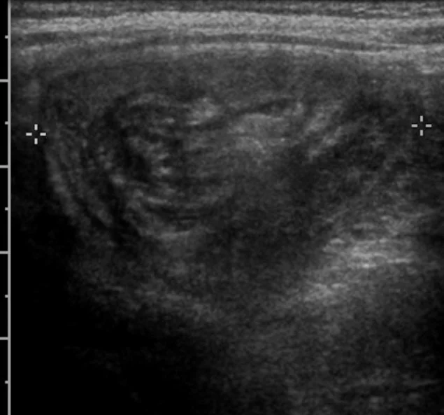
Long description
Ultrasound with the target sign of intussusception in a 3-year-old boy.

Ultrasound is the investigation of choice: it is safe, quick, widely available, and does not use radiation. Sensitivity is 97.9 to 98.5% and specificity 97.8 to 100%, with a negative predictive value of 99.7%, so a negative study by an experienced sonographer effectively rules out intussusception.[1] It is also better than fluoroscopy at showing lead points and at offering alternative diagnoses such as appendicitis.[1]
The classic finding is the target sign on the transverse view (concentric rings, a "doughnut" or "bull's eye") and the pseudokidney sign on the longitudinal view, representing the intussusceptum with its mesenteric fat inside the intussuscipiens. Color Doppler may show absent flow in the intussusceptum, suggesting ischaemia.[1]
Ultrasound can also be used for hydrostatic (saline) reduction under live guidance, combining diagnosis with treatment, with comparable success rates and no radiation. Point-of-care ultrasound by a trained paediatric emergency physician can screen effectively, but accuracy depends on training.[1]
Plane abdominal films are not good at confirming intussusception (sensitivity is only about 40% including equivocal cases), but they are useful for one job: ruling out pneumoperitoneum or high-grade obstruction before an enema is attempted.[1] Free air on the film means operate, not reduce.
CT is not first line in children because of the radiation load, but it is appropriate when the presentation is atypical, the child is older than the usual range, or a focal mass or systemic disease (for example Peutz-Jeghers syndrome) is suspected.[1]
There is no single blood test or score that diagnoses intussusception; diagnosis is confirmed by imaging, usually ultrasound. Several things raise (or support) the diagnosis and guide urgency:
- Age outside 3 months to 5 years makes a pathologic lead point likely.[1][2]
- Lethargy or an "apathetic" presentation is a recognised but atypical form and delays diagnosis.[2][3]
- Bloody stool, vomiting with pain, or an abdominal mass in a previously well infant is intussusception until proved otherwise.[3]
- Pallor, tachycardia, guarding signal ischaemia or perforation and shift management to surgery.[2]
The Brighton collaboration case definition (used in vaccine surveillance) separates definite, probable and possible cases; in practice, a child who meets clinical criteria plus ultrasound confirmation is a definite case.[6]
Ill children with signs of peritonitis, shock or suspected perforation need fluid resuscitation, broad-spectrum antibiotics (for example ampicillin with gentamicin and clindamycin, or metronidazole with cefotaxime or piperacillin/tazobactam), nasogastric decompression, and urgent surgery.[2] Clinically stable children with no contraindication proceed to imaging and enema reduction.
- Nothing by mouth, intravenous access, analgesia and antiemetics as needed.[5]
- Antibiotics are not needed before a planned enema reduction: prophylactic antibiotics do not appear to reduce post-reduction complications.[5]
- Surgeon availability. Centres performing reduction need surgical capability available; a physician capable of decompressing a tension pneumoperitoneum should be present.[5]
For a stable child with ileocolic intussusception and no perforation or peritonitis, pneumatic (air) or hydrostatic (liquid) enema is the treatment of choice, and it both confirms and treats the condition. [1]
- Technique. A soft catheter is placed in the rectum and air or liquid is run in under imaging. A manometer monitors pressure; the recommended upper limit of intraluminal pressure is 120 mmHg. Signs of success are disappearance of the soft-tissue mass and rapid reflux of air (or saline) into the distal small bowel.[1] The generally quoted practical rule is up to about 3 attempts of about 3 minutes each in the same position, with partial reduction between attempts.[1]
- Air versus liquid. Air under fluoroscopy is the most widely used medium: it is faster, cheaper, clearer and exposes the child to less radiation. A meta-analysis of 32,451 children reported higher success with air than with liquid (82.7% versus 69.6%), with similar perforation rates (0.39% versus 0.43%).[1] Hydrostatic (saline) reduction under ultrasound avoids radiation and is increasingly used, with comparable success rates of 73 to 86% and a perforation rate around 1% in experienced hands.[1]
- Overall success. The pooled success rate of non-operative reduction is about 82%, and in excess of 80% with an air enema.[1][2]
- Sedation for comfort is used in many units, but evidence is mixed about whether it improves success; risks and benefits depend on local practice and the child's state.[1]
- Delayed repeat enema (DRE). If the first attempt only partially reduces the bowel and the child stays stable without peritonitis, a second or third attempt after an interval (anything from about 15 minutes to a few hours, most often 30 minutes to 4 hours in practice) can rescue about half of the cases that would otherwise go to surgery, with a reported success rate near 50%.[1][5]
- Contraindications to enema reduction are pneumoperitoneum on the radiograph, peritonitis, or shock; these children need surgery.[1]
- Complication. Bowel perforation occurs in roughly 0.4% of air and liquid enemas. Air enema perforation can cause tension pneumoperitoneum, requiring emergency needle decompression.[1]
Suspected intussusception in a child
- Suspected intussusceptionInfant or toddler with colicky pain waves, vomiting, blood in stool, or an abdominal mass
- Assess stabilityAirway, breathing, circulation; signs of peritonitis, shock or perforation?
- Question: Unstable, peritonitis, or free air on X-ray?
Yes
- Resuscitate and operateFluids, antibiotics, nasogastric decompression; surgical reduction
No
- Abdominal ultrasoundConfirms or excludes intussusception; assesses lead points and perfusion
- Question: Intussusception confirmed?
No
- Reconsider diagnosisInvestigate alternatives; repeat ultrasound if doubt persists
Yes
- Enema reductionAir or saline under fluoroscopy/ultrasound; pressure under 120 mmHg; up to about 3 attempts
- Question: Reduced?
Yes
- Observe 4 hoursOral trial when pain-free; discharge if well, well counselled, and able to return
Partial / no
- Delayed repeat enema or surgeryDRE if stable and no peritonitis; operate if not reducible or lead point suspected
Children are usually observed for about 4 hours after a successful reduction; feeding is reintroduced gradually. Discharging from the emergency department after observation appears safe, with recurrence rates similar to inpatient observation, although very young children and families who cannot readily return may be admitted.[5]
Surgery is required when:
- There is perforation, peritonitis, shock or failed non-operative reduction;
- An enema is unavailable locally, or timely transfer is not possible;
- A pathologic lead point is suspected or confirmed; or
- The child is too unwell for an attempted enema.[1][5]
At operation the surgeon reduces the intussusception by gentle manual pressure (the Hutchinson manoeuvre) with as little handling as possible to avoid crushing the bowel, and resects any necrotic segment or the lead point if it cannot be preserved. For ileocolic disease, laparoscopic reduction is increasingly used and has equivalent complication rates to open surgery, with a significantly shorter hospital stay in most comparative series; the choice follows local expertise.[5] Delay matters: success of enema reduction and survival both fall with time, and one surgical review notes operative reduction becomes more likely after longer symptom duration.[6]
- Anticoagulation or bowel-sterilising drugs are not part of standard care; antibiotics (prophylactically) add nothing before reduction.[5]
- Dexamethasone used as an adjuvant around reduction may reduce recurrence in some small studies, but the evidence is limited.[4]
- Observation after a successful reduction of about 4 hours is supported by current systematic reviews.[5]
- Bowel ischaemia, necrosis and perforation if the intussusception is left untreated, leading to peritonitis and sepsis; untreated disease can be fatal.[2]
- Mortality. Outcome is excellent if treated promptly; a prospective Indian series recorded 1.8% mortality, with most deaths in children who presented late.[6]
- Enema-related perforation, roughly 0.4%, occasionally with tension pneumoperitoneum.[1]
- Recurrence, 9 to 15% after successful enema reduction.[1]
- Pathologic lead point discovered only at operation in some children.[1]
With prompt reduction the prognosis is excellent. Delay increases the need for surgery, the need for bowel resection, and the rare risk of short bowel. Recurrent intussusception is commonest in the first days after reduction (about half of recurrences) and can also occur months or years later.[1] Older age at first presentation (over 1 year) is associated with higher recurrence, and multiple recurrences (reported in 14 to 19% of children with more than one episode) raise the chance of an underlying lead point, although most recurrences are still idiopathic.[1]
There is no drug that prevents idiopathic intussusception, but early diagnosis on a high index of suspicion prevents complications.
Rotavirus vaccination deserves a careful explanation to families, because a rotavirus vaccine has been associated with intussusception.
- The first rotavirus vaccine used in the United States (RRV-TV, RotaShield) was withdrawn after a roughly 22-fold increase in intussusception among vaccinated infants (1 to 2 excess cases per 10,000 recipients).[2]
- Currently licensed rotavirus vaccines carry a much smaller risk, estimated at 1 to 2 excess cases per 100,000 recipients, confined to the first 7 days after the first dose, and at least one large study found no increase beyond background rates.[2][3] Another estimate from some countries is 1 to 6 excess cases per 100,000 vaccinated infants.[3]
- In large surveillance from India, 12 children received rotavirus vaccine within 1 to 21 days before intussusception, with no increase in case load or case ratio after vaccine introduction in the sites studied.[6]
- The benefits of rotavirus vaccination continue to outweigh the risks of harm; the vaccine is not given to children with a history of intussusception or an uncorrected congenital gastrointestinal malformation.[3]
- Rotavirus vaccination is usually completed before the peak age for intussusception, which is another reason to vaccinate on schedule.[3]
- Counselling tip: keep the absolute risk small and the benefit large in the family's mind; serious gastroenteritis and hospitalisation are far more common than intussusception, which is why programmes continue to recommend vaccination. Where sources differ on the size of the excess risk, say so plainly and quote both figures rather than choosing one.[3]
- Neonates and preterm infants. Intussusception is rare and easily confused with necrotising enterocolitis; it tends to be small-bowel (91% of preterm cases) and usually has no pathologic lead point.[1]
- Infants under 3 months. A lead point (including Meckel diverticulum) is more likely; a high index of suspicion is needed.[2]
- Older children (over 3 to 5 years). Lead points are much more common; search for a structural cause and malignancy if adult.[1][2]
- Adults. Intussusception is linked to neoplasia and accounts for about 1% of bowel obstructions; surgery, not enema, is the standard treatment.[4][5]
- Recurrent cases. Each recurrence raises the chance of a lead point, but enema reduction remains effective and safe when the child is stable.[1]
- Pregnancy. Not specifically addressed here; manage with obstetric, paediatric surgical and imaging input.
- Ultrasound, even a portable machine, is the single most useful tool; learn to look for the target sign in any infant with unexplained colic or bloody stool. Pneumoperitoneum on X-ray is the reason not to attempt an enema.[1]
- Where non-operative reduction is unavailable, children with intussusception are usually managed surgically.[1]
- Late presentation is the main driver of poor outcome; educating families and primary care to seek help early is the highest-value intervention.[6]
- Pre-reduction antibiotics are unnecessary in stable children, which reduces cost and side effects.[5]
- Send every resected bowel segment for histology, so that lead points are not missed.
- Intussusception is telescoping of intestine into the adjacent segment; the common type is ileocolic and is the commonest cause of bowel obstruction in infants and toddlers.
- Peak age is 3 to 36 months (onset about 5 months, peak 4 to 9 months); about 10% occur after age 5.[1][3][4]
- Boys are affected more often (ratios roughly 3 to 1 or 3 to 2 depending on the series).[3][4]
- About 75% are idiopathic and follow viral illness; 25% have a pathologic lead point, more often at the extremes of age.[2][3]
- Common lead points: Meckel diverticulum, intestinal polyps, duplication cysts, lymphoma, IgAV (Henoch-Schonlein purpura).[1][2]
- Colicky pain in waves every 15 to 20 minutes, vomiting, drawing up of the legs; currant-jelly stool and a sausage mass are late or inconstant signs.[2][3]
- Up to about 20% of infants present lethargically without obvious colic.[3]
- Ultrasound is the investigation of choice (sensitivity 97.9 to 98.5%, specificity 97.8 to 100%, negative predictive value 99.7%); look for the target sign.[1]
- Air or liquid enema under imaging is first-line treatment for stable children without perforation or peritonitis; success is about 82% (air 82.7% versus liquid 69.6%).[1][2]
- Enema pressure should not exceed 120 mmHg; contraindications are free air, peritonitis, or shock.[1]
- Delayed repeat enema rescues about half of first failures; observe for about 4 hours after a successful reduction.[1][5]
- Surgery is needed for contraindications, failure, a suspected lead point, or unavailability of enema.[1][5]
- Recurrence after successful enema reduction is about 9 to 15%; older age at first presentation and multiple recurrences raise the chance of a lead point.[1]
- Rotavirus vaccination carries, at most, a tiny absolute risk (1 to 2, or up to 1 to 6, excess cases per 100,000 recipients) and its benefits outweigh the risks.[2][3]
Answer each one in your head first, then open the answer.
A 9-month-old boy is brought in with episodic, severe abdominal pain every 20 minutes, drawing his legs up, then settling between episodes. He vomits once. There is no blood in the stool. What is the most likely diagnosis and the test of choice?
Show answerHide answer
Intussusception, most likely ileocolic. The absence of bloody stool does not exclude it, because currant-jelly stool is a late sign. The test of choice is abdominal ultrasound, looking for the target sign; a stable child with no perforation or peritonitis can proceed to enema reduction.[1]
An enema reduction for ileocolic intussusception is about to start. What pressure should not be exceeded, and which three findings would stop the team and send the child to surgery?
Show answerHide answer
The intraluminal pressure should stay at or under 120 mmHg. Pneumoperitoneum on the radiograph, peritonitis, or shock are contraindications to enema reduction and mean the child needs surgery.[1]
A child's first enema only partially reduces the intussusception and the child remains stable with no peritonitis. What is the next step and how likely is it to succeed?
Why can rotavirus vaccination be recommended even though a rotavirus vaccine was once withdrawn for causing intussusception?
Show answerHide answer
The withdrawn vaccine (RRV-TV, RotaShield) caused a large increase (1 to 2 excess cases per 10,000 recipients, about a 22-fold increase). Currently licensed vaccines carry at most 1 to 2 excess cases per 100,000 recipients (some estimates 1 to 6), confined to the first 7 days after the first dose, and large surveillance studies often show no excess. The benefits of preventing rotavirus gastroenteritis far outweigh this tiny risk, and the vaccine is given before the peak age for intussusception.[2][3]
A 4-year-old has recurrent intussusception. Name two reasons not to assume it is just 'another idiopathic episode'.
Show answerHide answer
First, older children are more likely to have a pathologic lead point, such as Meckel diverticulum, polyp or lymphoma. Second, each recurrence (14 to 19% of children with more than one episode have a lead point) raises the chance of an underlying lesion and warrants careful imaging and, if not reduced by enema, surgical inspection.[1]
A lethargic infant has no colic but later passes 'currant-jelly' stool. Why is this a trap, and what should have happened earlier?
Show answerHide answer
Currant-jelly stool is a late sign of mucosal ischaemia, and waiting for it delays treatment. Up to about 20% of infants present lethargically without colic (an apathetic form). The key earlier step is to suspect intussusception in any infant with unexplained lethargy, vomiting or abdominal findings and to scan with ultrasound, which would have shown the target sign.[3]
No related or prerequisite topics are listed for this page. Useful neighbours on the same shelf are abdominal pain presentations in children, viral gastroenteritis, Meckel diverticulum, Henoch-Schonlein purpura (IgA vasculitis), and rotavirus vaccine.
- Hwang J, Yoon HM, Kim PH, Jung AY, Lee JS, Cho YA. Current diagnosis and image-guided reduction for intussusception in children. Clin Exp Pediatr. 2022;66(1):12-21. Accessed .
- Belkind-Gerson J, Pekarsky AR. Intussusception. Merck Manual Professional Version; updated Aug 2025. Accessed .
- Medsafe. Intussusception: A Very Rare Risk After Rotavirus Vaccination. Prescriber Update. 2018;39(3):40-41. Accessed .
- Attoun MA, Albalawi SMD, Ayoub A, et al. The Management of Intussusception: A Systematic Review. Cureus. 2023;15(11):e49481. Accessed .
- Kelley-Quon LI, Arthur LG, Williams RF, et al. Management of Intussusception in Children: A Systematic Review. J Pediatr Surg. 2021;56(3):587-596. Accessed .
- Das MK, Arora NK, Mathai J, et al. Profile and epidemiology of intussusception in children under two years of age: a prospective surveillance. Indian J Pediatr. 2021;88:1187-1194. Accessed .
Figure credits
Educational content for medical students and doctors. It is not a substitute for clinical judgement or local guidelines.