Hirschsprung disease
A congenital absence of distal bowel ganglion cells from failed neural-crest migration: why neonates fail to pass meconium, how it is confirmed, and how it.
- Hirschsprung disease is a congenital enteric neuropathy: during early embryonic development the neural-crest cells that should form the bowel's own nervous system fail to reach the distal colon, so a stretch of distal bowel is left without ganglion cells.
- The aganglionic segment cannot relax and coordinate peristalsis, producing a functional obstruction; the bowel above it dilates, giving the classic neonatal picture of delayed or absent passage of meconium with abdominal distension.
- It is common enough to be sought in every newborn who has not opened the bowels by two days of age, yet rare enough that diagnosis depends on histology rather than symptoms alone.
- The gold-standard test is a rectal suction biopsy showing absent ganglion cells in the submucosal and myenteric plexuses, usually supported by a contrast enema (showing a transition zone) and, in older infants, anorectal manometry (absent rectoanal inhibitory reflex).
- Treatment is surgical removal of the aganglionic segment and pull-through of the normal bowel to the anus; most children survive and grow well, but bowel function needs long-term follow-up and the main dangers are enterocolitis and functional obstruction.
By the end of this page you should be able to:
- Explain failure of neural-crest migration and the resulting absence of ganglion cells in the rectum.
- Relate the RET gene and the chromosome-21 (Down syndrome) association to the embryology.
- Recognise the presenting features by age, from failure to pass meconium to chronic constipation and distension.
- Interpret a contrast enema, anorectal manometry and a rectal suction biopsy in a suspected case.
- Outline the differential of neonatal intestinal obstruction and the distinguishing feature of each.
- Describe pull-through surgery and its common postoperative complications.
- Recognise Hirschsprung-associated enterocolitis, its risks and its emergency management.
DefinitionHirschsprung disease
Hirschsprung disease (HSCR) is a congenital malformation of the enteric nervous system in which ganglion cells are absent from the myenteric and submucosal plexuses of the distal intestine. The aganglionic segment is always distal (it always involves the rectum), extends proximally for a variable distance, and is narrowed and in spasm, while the proximal ganglionated bowel is dilated.
The bowel looks like a hollow tube with its own brain missing in one stretch. That missing brain is the enteric nervous system (ENS), a network of roughly 200 to 600 million neurons [3] running from oesophagus to anus, arranged mainly in the myenteric (Auerbach) plexus between the circular and longitudinal muscle layers and the submucosal (Meissner) plexus between the circular muscle and the mucosa. Because the enteric neurons are absent in Hirschsprung disease, the ganglionated segment proximal to the gap must do all the work, and the distal segment cannot relax.
Hirschsprung disease occurs in approximately 1 in 5000 live births [3], with one European registry reporting 1.43 (1.37 to 1.50) per 10,000 live births [5] and higher rates reported in some regions, such as roughly 1 in 3000 live births in the Federated States of Micronesia [1]. About 80% to 90% of children become symptomatic in the neonatal period [1], and fewer than 10% are diagnosed after 4 years of age, usually with short-segment disease [1].
Who is affected:
- Sex. Boys are affected roughly four times as often as girls, a male-to-female ratio of about 4 to 1 [2] [5].
- Inheritance. About 80% to 90% of cases are sporadic, usually short-segment disease, and 10% to 20% are familial, usually with longer aganglionosis and autosomal dominant inheritance with incomplete penetrance [2].
- Distribution of aganglionosis. In about 85% of patients the aganglionosis is confined to the rectosigmoid ("short-segment" disease). In 5% to 10% it extends through the whole colon or into the small intestine (total colonic or total intestinal aganglionosis) [2] [5].
- Mortality. Hirschsprung disease was once fatal. In the modern era, the EUROlinkCAT collaborative network (13 population-based congenital anomaly registries) reported a 1-year mortality of 2.9% and cumulative 5-year mortality of 3.4% [1]. Hirschsprung-associated enterocolitis (HAEC) is now the leading cause of death [3].
The fundamental problem is embryologic. The enteric nervous system is entirely derived from neural crest cells, the transient cell population often called the fourth germ layer because it gives rise to such diverse tissues. During normal development, neural-crest cells delaminate from the neural tube, undergo epithelial-to-mesenchymal transition, and migrate along defined paths. The vagal neural crest, next to somite pairs 1 to 7, is the principal source of the enteric nervous system: its cells migrate proximally to distally along the whole gut and, in humans, colonisation of the hindgut is complete by about week 7 of gestation [2] [3]. A smaller sacral contribution reaches the distal hindgut after the vagal wavefront. Hirschsprung disease arises when these migrating cells fail to complete their craniocaudal journey, leaving the distal bowel aganglionic. The earlier the arrest, the longer the aganglionic segment.
Two signalling pathways dominate this process:
- RET-GDNF. RET is a receptor tyrosine kinase on the migrating neural-crest cells; its ligand glial-cell-line-derived neurotrophic factor (GDNF) is present in the embryonic gut mesenchyme. RET activation drives survival, proliferation, differentiation and chemoattraction of the cells along the gut. Mutations in RET account for roughly 50% of familial Hirschsprung disease [2], and RET coding or splice-site mutations are found in up to 50% of familial cases and 15% to 35% of sporadic cases [2]. In mice, loss of RET, its co-receptor GFR-alpha1 or GDNF produces total intestinal aganglionosis [2].
- EDNRB-endothelin-3. Endothelin receptor B on the neural-crest cells binds endothelin-3 from the gut mesenchyme. This pathway delays differentiation so the cells remain proliferative and migratory; if it fails, cells differentiate prematurely and stop before reaching the distal colon. Mutations in EDNRB or endothelin-3 underlie some syndromic forms.
More than 30 Hirschsprung candidate genes have now been identified, including genes in the RET-GDNF and EDNRB-endothelin-3 pathways, transcription factors (SOX10, PHOX2B, ZEB2) and genes in neuregulin, semaphorin, hedgehog and Notch signalling [2]. Risk is multigenic: common non-coding variants, rare coding variants and copy-number variants act individually or together, with the risk of disease rising with accumulating variants [3]. There are no evidence-based consensus guidelines for genetic testing in routine clinical practice at present [2].
Risk factors and associations:
- Down syndrome (trisomy 21). Patients with Down syndrome have more than a 50-fold higher risk of Hirschsprung disease than the general population [2]. Overexpression of genes on chromosome 21 is thought to contribute, but the link is not yet fully worked out. Down syndrome also raises the risk and severity of Hirschsprung-associated enterocolitis: HAEC occurs in around 50% of patients with both conditions, versus about 29% in those without Down syndrome [4]. [3]
- Other syndromes. Shah-Waardenburg (ET3, EDNRB, SOX10), Haddad (PHOX2B), Mowat-Wilson (ZEB2) and Goldberg-Shprintzen (KIAA1279) syndromes all include Hirschsprung disease.
- Family history and male sex.
Hirschsprung disease is always characterised by absence of enteric neurons in the distal colon, most often the rectosigmoid. The aganglionic segment is narrow, spastic and without coordinated motor activity, while the ganglionated bowel proximally dilates. A healthy rectum behaves as a reservoir whose distension creates the urge to defecate and triggers the rectoanal inhibitory reflex (RAIR); that reflex is absent in Hirschsprung disease because the nitrergic (nitric-oxide-producing) neurons that mediate internal anal sphincter relaxation are missing [2].
Meconium and the first days. In a full-term neonate, nearly 100% pass meconium within the first 48 hours [2], so Hirschsprung disease classically declares itself by failure to pass meconium in that window, with abdominal distension and bilious vomiting. The cause is functional obstruction from the aganglionic segment. Yet an important trap: 37% to 54% of infants with Hirschsprung disease still pass meconium within the first 2 days [2], so the passage of meconium does not exclude the diagnosis. Some children, especially with short-segment disease, continue to pass stool through infancy, explaining the occasional diagnosis in later childhood.
Why the aganglionic segment is narrow. The narrow segment has more resting acetylcholine and heightened cholinergic activity, with loss of the nitrergic inhibitory tone that normally relaxes the gut; the net effect is contraction and spastic narrowing rather than propulsion [2].
Hirschsprung disease is not just missing ganglion cells. Even after the aganglionic segment is removed, many patients have ongoing problems. At least 50% of adults operated on in childhood fail to achieve bowel function comparable with healthy people, up to 33% have obstructive symptoms and up to 69% have some faecal soiling [2]. Reasons include retained transition zone, disordered motility in the residual ganglionated bowel (including an excess of nitrergic neurons), absent RAIR, internal sphincter achalasia, and the loss of the rectal reservoir itself. The transition zone, the segment between aganglionic and normal bowel, typically has partial aganglionosis and severe hypoganglionosis, is usually under about 5 cm in length (though it can extend higher), and may be left behind at surgery [2].
- Gross. The aganglionic segment is narrowed and contracted; the proximal ganglionated segment is dilated and thick-walled, with a visible transition zone between the two.
- Microscopic. Absence of ganglion cells from both the submucosal (Meissner) and myenteric (Auerbach) plexuses in the affected segment; hypertrophic nerve trunks in the submucosa; absence of calretinin-positive neurites and increased acetylcholinesterase staining. Representative submucosa must be present in the biopsy, because that is where the Meissner plexus lives.
Occasionally a clinician biopsies the transition zone and finds confusing patchy ganglion cells. That is why representative histology from the right place matters more than any single slide.
- Neonate (most common): no meconium passed in the first 2 days, abdominal distension, bilious vomiting, reluctance to feed. Feeding may relieve distension briefly, or a rectal (or oral) stimulation may produce an explosive stool.
- Infant: chronic constipation from birth, abdominal distension, poor weight gain, intermittent explosive diarrhoea-like stools.
- Older child (uncommon, fewer than 10% after age 4): chronic constipation since early life, abdominal distension, palpable faecal masses, soiling, and episodes of enterocolitis with fever, diarrhoea and lethargy. Short-segment disease is more likely here.
Enterocolitis (HAEC) can be the first presentation at any age, even in a child who seemed to be thriving.
Look for a distended, tympanitic abdomen, visible intestinal loops, and an empty rectum with a sudden gush of gas and stool on withdrawal of the examining finger ("explosive" decompression). In the neonatal period there may be no mass felt because the colon is not yet hugely dilated. Always check for chromosomal or syndromic features, and for associated congenital anomalies.
Red flagIll neonate with distension
A neonate with bilious vomiting, abdominal distension and failure to pass meconium who becomes lethargic, febrile or shocked may have Hirschsprung-associated enterocolitis (HAEC) or another obstruction. Do not repeatedly manipulate the rectum in an unstable infant without resuscitation, and treat as an emergency (see Management).
Many conditions look like Hirschsprung disease because they all end in "baby not opening bowels and a swollen belly". The distinguishing feature is usually the history, the level of obstruction, and the contrast study.
| Condition | Key distinguishing feature |
|---|---|
| Hirschsprung disease | Failure to pass meconium; contrast enema shows a transition zone (narrow distal, dilated proximal); absent ganglion cells on rectal biopsy; absent rectoanal inhibitory reflex. |
| Meconium ileus | Distal small-bowel obstruction in a neonate, classically in cystic fibrosis; "soap-bubble" or pellets of meconium at the ileocaecal level; no colonic transition zone. |
| Meconium plug syndrome / small left colon | Large, firm meconium plug; contrast enema shows a small left colon with a transition near the splenic flexure; functional and usually resolves. |
| Intestinal atresia | Complete obstruction with no gas distal to the level; bilious vomiting early; plain radiograph shows "double bubble" (duodenal) or multiple air-fluid levels. |
| Malrotation with volvulus | Sudden bilious vomiting in a previously well neonate; emergency: upper gastrointestinal contrast shows the duodenojejunal junction displaced; vascular compromise can occur. |
| Necrotising enterocolitis | Preterm infant; systemic illness, feed intolerance, bloody stools, pneumatosis on radiograph; not typically a transition zone. |
| Functional immaturity / hypoganglionosis | Very preterm infant with slow gut; improves with maturation; biopsy does not show aganglionosis. |
| Anal atresia / stenosis | No anal opening or tiny anus on inspection; diagnosis on examination. |
Figure 1Hirschsprung
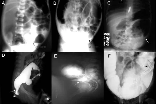
Long description
Plain abdominal radiographs and contrast enema images in Hirschsprung disease; arrows mark the transition zone between the dilated proximal bowel and the narrow distal segment.

A water-soluble contrast enema is the usual first imaging test in a suspected neonate. It can show reversal of the rectosigmoid ratio (the rectum narrower than the sigmoid), a "saw-tooth" spiculated rectal mucosa, and most reliably a transition zone between the narrow aganglionic distal bowel and the dilated ganglionated proximal bowel. In one surgical series, 75% of neonates with Hirschsprung disease had an abnormal rectosigmoid ratio, 85% had a transition zone and 50% had a saw-tooth rectal mucosa [3]. Radiographic signs can be absent early on, before the proximal bowel has had time to dilate, so a normal-looking enema does not rule the condition out. In older infants the enema can be both diagnostic and therapeutic by breaking down a functional obstruction.
Anorectal manometry is a useful adjunct, particularly outside the neonatal period. All patients with Hirschsprung disease have an absent rectoanal inhibitory reflex: the internal anal sphincter fails to relax on rectal balloon distension [3]. The test is more difficult in uncooperative young children and is not definitive on its own, so a positive result supports but does not replace histology.
Definitive diagnosis is made by rectal suction biopsy. The tissue is evaluated with haematoxylin and eosin and immunohistochemistry (calretinin and/or acetylcholinesterase staining). A suction biopsy is taken from above the dentate line, so it does not cause pain, and it must include the submucosa, because the Meissner plexus lies there [3]. When adequate submucosa is obtained, rectal suction biopsy or open biopsy is equally accurate, and the least invasive feasible method should be chosen [5]. Representative biopsies are taken from the posterior or lateral rectal wall at least 2 cm proximal to the dentate line, or 3 cm from the anal orifice, and must contain a representative amount of submucosa [5]. The reported sensitivity is 100% with 99.1% specificity [3]. Complications are rare (overall complication rate about 0.65%), but an insufficient specimen is obtained in roughly 10% of cases, in which case a full-thickness transanal biopsy under general anaesthesia is required [3].
Good preoperative care looks beyond the biopsy. Current European guidance recommends routinely looking for associated kidney and urinary tract malformations (CAKUT) and systematic assessment of nutritional status before surgery [5]. Malnutrition or a recent respiratory infection increases the risk of postoperative enterocolitis [4]. Many children arrive at surgery having already needed rectal irrigations or, in some series, a diverting stoma to decompress the bowel.
There is no Hirschsprung disease "score". The diagnosis is histologic: absence of ganglion cells from a representative rectal biopsy taken from the correct level. Supporting evidence (contrast enema transition zone, abnormal rectosigmoid ratio, absent rectoanal inhibitory reflex) increases confidence but cannot confirm or exclude the condition alone, particularly in the neonatal period.
Severity is usually expressed by the length of the aganglionic segment (rectosigmoid short-segment versus long-segment, total colonic or total intestinal disease) and by the presence of enterocolitis. For enterocolitis specifically, three grading systems exist. The American Pediatric Surgical Association Hirschsprung Disease Interest Group grade separates possible, definite and severe HAEC by history, examination and imaging, with management matched to grade; in that system, grade I HAEC is managed as an outpatient with oral metronidazole and hydration (with rectal irrigations considered), grade II with clear liquids or nothing by mouth plus oral or intravenous antibiotics with rectal irrigations, and grade III in hospital, often in intensive care, with bowel rest, intravenous antibiotics and rectal irrigations, plus surgical diversion if non-operative management fails or exploration if there is perforation [4]. Diagnostically, a HAEC score of 4 gives 83.7% sensitivity and 98.6% specificity for HAEC, compared with the older cut-off of 10 (41.9% sensitivity, perfect specificity) [4].
Suspected Hirschsprung disease in a neonate or infant
- Clinical suspicionNo meconium by 48 h, abdominal distension, bilious vomiting, or chronic constipation from birth
- Stabilise if sickFluids, nil by mouth, nasogastric decompression, rectal washout; treat suspected enterocolitis with antibiotics
- Contrast enemaLook for transition zone, abnormal rectosigmoid ratio, saw-tooth mucosa; may be normal early
- Anorectal manometryAbsent rectoanal inhibitory reflex supports the diagnosis (not definitive, especially in neonates)
- Rectal suction biopsyAt least 2 cm above the dentate line (or 3 cm from the anus); must include submucosa; H&E plus calretinin/acetylcholinesterase
- Question: Ganglion cells present in the specimen?
Yes
- Hirschsprung disease unlikelyConsider other causes of obstruction or constipation; review level and quality of biopsy
No
- Confirm with a second review or full-thickness biopsy if the specimen was inadequateAbout 10% of suction biopsies are non-diagnostic
- Define extent and plan pull-throughMap aganglionosis; screen for CAKUT and optimise nutrition
The first aim is to turn a functional obstruction into a controlled situation. In a sick neonate:
- Resuscitate: intravenous fluids and electrolytes, keep nil by mouth, decompress the stomach with a nasogastric tube.
- Relieve distension with gentle rectal washouts (irrigation). Perform the first irrigation cautiously; if there is severe distension or any concern about perforation, assess and stabilise first. Irrigations are done with warmed saline, 10 mL/kg aliquots via a soft wide-bore catheter with side holes, and in a child who has not yet had surgery the tube should be advanced past the transition zone if possible. Rectal digital examination can be both diagnostic and therapeutic, producing an explosive, foul-smelling stool and rapid decompression. [4]
- Start broad-spectrum antibiotics if enterocolitis is suspected.
- Investigate and then confirm histologically before major surgery.
The only definitive treatment is to resect the aganglionic segment and pull the normal ganglionated bowel down to the anus. Three classic operations are in common use (Swenson, Yancey-Soave and Duhamel), all with comparable outcomes and complication rates; the choice depends on surgeon experience and the length of the affected segment [5]. The procedure can be performed transanally for short-segment disease, often with a laparoscopic or open component to identify the level of aganglionosis and facilitate dissection. It may be done as a single-stage operation or after a diverting stoma.
For many babies the pathway is staged rather than single-stage: while awaiting definitive surgery, most children (about 77% in one European review) undergo rectal irrigations, about 22% undergo rectal dilatation or stimulation, and about a third require a diverting stoma [3]. Timing varies by centre: in one European review, pull-through was performed at diagnosis in about 33% of neonates and after 4 months of age or once the child exceeded 5 kg in about 67% [3]. Continuing modern efforts focus on minimally invasive, precision approaches to reduce surgical trauma while preserving long-term bowel function [6].
Intraoperatively the surgeon establishes the proximal (ganglionated) margin by biopsying at intervals and obtaining frozen sections before committing to the transection. Postoperatively, current European guidance has moved away from routine preventive anal dilatation; anal calibration is distinguished from therapeutic dilatation, and routine preventive dilatations are no longer recommended [5].
HAEC is the main cause of morbidity in Hirschsprung disease and the leading cause of mortality. It presents with fever, abdominal distension, diarrhoea (often explosive and foul), lethargy and, in severe cases, sepsis. Incidence is highly variable: 6% to 60% before pull-through surgery and 25% to 42% afterwards, with most prospective series reporting over 40% [4]. It occurs more commonly within the first 2 years of age [4]. Bowel perforation is seen in only 2% to 3% of patients [4].
Recognition is the hard part, because HAEC can mimic ordinary viral gastroenteritis or sepsis. Risk factors include Down syndrome (HAEC in around 50% versus 29% in patients without Down syndrome) [4], family history, male sex, delay in diagnosis, longer aganglionic segments (over 30 cm associated with postoperative HAEC; short-segment disease protective) [4], preoperative enterocolitis, malnutrition, hypoproteinaemia and respiratory infection [4].
Management follows the grade of severity: outpatient oral metronidazole with hydration and considered rectal irrigations for grade I; outpatient or inpatient care with clear liquids or fasting, rectal irrigations, oral or intravenous metronidazole plus consideration of ampicillin and gentamicin or piperacillin and tazobactam for grade II; and inpatient, often intensive care, with bowel rest, intravenous fluids, intravenous antibiotics and rectal irrigations for grade III, plus surgical diversion if non-operative management fails or laparotomy if there is perforation [4]. Rectal irrigation is the backbone of therapy at every grade and decompresses the bowel.
Many children achieve good continence but functional problems are common: constipation rates after surgery range from 4% to about 30% [3], increased stool frequency occurs in about 44% [3], and faecal soiling affects 35% to 50% with rectosigmoid disease (more with total colonic disease) [3]. Long-term follow-up therefore matters. Current European guidance emphasises individualised dietary advice, exclusion of mechanical obstruction (retained transition zone, stricture, anastomotic narrowing) when symptoms recur, management of internal sphincter achalasia (for example with botulinum toxin where appropriate), attention to sexual and fertility health, and structured transition to adult care [5]. At least 50% of adults operated on in childhood do not achieve normal bowel function scores, and up to 33% have obstructive symptoms and up to 69% some faecal soiling [2], so early counselling is honest and useful.
HAEC after surgery occurs in roughly 28% to 42% of patients [3], and up to 21% have four or more episodes [3]. It remains the leading cause of death, so any fever with abdominal distension in a child who has had a pull-through is treated as enterocolitis until proven otherwise, with antibiotics and irrigation.
- HAEC (above), the main cause of morbidity and the leading cause of death.
- Obstructive symptoms after pull-through from mechanical obstruction, retained aganglionosis or transition zone, hypoganglionosis, internal sphincter achalasia, proximal dysmotility or functional constipation. [2]
- Faecal soiling and incontinence, which affect quality of life and social functioning.
- Constipation and functional withholding, often needing repeat training and sometimes enemas.
- Anastomotic problems such as stricture or leak.
- Enterocolitis recurrence requiring repeated treatment.
Survival is excellent in modern practice, but bowel function is variable long-term. At least 50% of adults operated on in infancy do not reach healthy-population bowel function scores, up to a third have obstructive symptoms, and up to 69% have some soiling [2]. These figures are not a reason for pessimism; they are a reason for lifelong follow-up and for honest early counselling of families.
There is no primary prevention of Hirschsprung disease, although early recognition matters: the condition is considered in any newborn without meconium by 48 hours, and in any child with chronic constipation from birth that does not respond to laxatives. Genetic counselling is useful in familial cases; penetrance is variable and genetic testing is not yet guided by consensus recommendations [2].
- Down syndrome. Higher risk of Hirschsprung disease (more than 50-fold) [2] and a roughly 50% rate of HAEC versus about 29% in other patients [4], with more severe episodes. Expect more complex perioperative care and a lower threshold for investigation.
- Total colonic or total intestinal aganglionosis. Staging surgery, prolonged irrigation and nutrition support (often parenteral in the neonatal period), and worse long-term bowel function.
- Older children. Short-segment disease can present with chronic constipation and soiling. Manometry and biopsy remain useful, but suction biopsy can be non-diagnostic because the rectal mucosa is thicker; full-thickness biopsy may be needed.
- A water-soluble contrast enema and a careful clinical suspicion are often the only available pointers. Look for the same signs: no meconium by two days, distension, explosive stool on rectal examination.
- Rectal suction biopsy may not be available; a full-thickness biopsy or a carefully sited suction biopsy assessed by an experienced pathologist may be needed. Calretinin and acetylcholinesterase stains are not universally available, so plain haematoxylin and eosin with attention to absent ganglion cells in adequate submucosa is the fallback.
- Irrigations can be performed with a soft catheter and warmed saline; decompression and antibiotics are life-saving for suspected enterocolitis.
- Where pull-through surgery is not available locally, decompress, stabilise, and refer to a paediatric surgical centre early, because delayed diagnosis and perforation are the main causes of avoidable death.
- Hirschsprung disease is a congenital absence of ganglion cells in the distal colon, always involving the rectum, caused by failure of neural-crest cell migration.
- The two key embryologic pathways are RET-GDNF and EDNRB-endothelin-3; RET mutations account for about half of familial cases.
- Down syndrome carries more than a 50-fold increased risk. [2]
- Incidence is roughly 1 in 5000 live births; boys are affected about four times as often as girls; 80% to 90% become symptomatic as neonates. [3]
- Classic neonatal presentation is failure to pass meconium within 48 hours with abdominal distension, but 37% to 54% still pass meconium, so the diagnosis can be missed.
- Contrast enema may show a transition zone (85% of neonates) and an abnormal rectosigmoid ratio (75%); anorectal manometry shows an absent rectoanal inhibitory reflex.
- Rectal suction biopsy is the gold standard: 100% sensitivity and 99.1% specificity, provided the sample contains adequate submucosa taken at least 2 cm above the dentate line.
- Treatment is resection of the aganglionic segment and pull-through (Swenson, Yancey-Soave or Duhamel); many children need staged care with irrigations beforehand.
- Hirschsprung-associated enterocolitis (HAEC) is the main cause of morbidity and the leading cause of death; incidence ranges from 6% to 60% before and 25% to 42% after surgery. [4]
- Long-term bowel function is variable: at least half of adults operated on in childhood do not reach healthy-population bowel function scores; constipation, incontinence and obstruction are common but manageable.
- Routine preventive anal dilatation is no longer recommended; transition zones and residual motility disorders explain many postoperative symptoms.
Answer each one in your head first, then open the answer.
What is the embryologic cause of Hirschsprung disease and why is the distal bowel narrowed?
Show answerHide answer
During early gestation the neural-crest cells that should colonise the whole gut fail to complete their craniocaudal migration, leaving the distal bowel without ganglion cells (always including the rectum). The aganglionic segment lacks the nitrergic neurons that relax the gut and has excess cholinergic tone, so it is in spasm and narrowed rather than propulsive, creating a functional obstruction.
A 2-day-old term boy has not passed meconium, his abdomen is distended and he is vomiting bile. His mother is worried because the paediatrician said he passed a small stool after a rectal thermometer. Can Hirschsprung disease still be present?
Show answerHide answer
Yes. Although Hirschsprung disease classically presents with no meconium passed in the first 2 days, 37% to 54% of affected infants pass some meconium in the first 2 days. The rectal thermometer may have triggered an explosive decompression. Investigate with a contrast enema and, crucially, a rectal suction biopsy; a negative enema does not exclude the diagnosis. [2]
Which investigation is the gold standard for Hirschsprung disease, and what two technical points make a biopsy adequate?
Show answerHide answer
Rectal suction biopsy, evaluated with haematoxylin and eosin plus calretinin and/or acetylcholinesterase staining. It must be taken from above the dentate line (at least 2 cm proximal to it, or 3 cm from the anal orifice) and must include representative submucosa, where the Meissner plexus lies. Sensitivity 100% and specificity 99.1% when adequate tissue is obtained.
On a water-soluble contrast enema in a neonate with Hirschsprung disease, what three features may be seen, and which is the most reliable?
Show answerHide answer
Reversal of the rectosigmoid ratio (rectum narrower than sigmoid), a saw-tooth spiculated rectal mucosa, and a transition zone between narrowed distal and dilated proximal bowel. The transition zone is the most reliable, present in about 85% of neonates in one series, but signs can be absent before the proximal bowel has time to dilate.
Why is the rectoanal inhibitory reflex (RAIR) absent in Hirschsprung disease, and what must manometry not be allowed to do?
Show answerHide answer
RAIR is the relaxation of the internal anal sphincter on rectal distension, mediated by nitrergic neurons; these neurons are absent in the aganglionic rectum. Manometry supports the diagnosis, especially outside the neonatal period, but it is not definitive on its own and must not replace a rectal biopsy.
A 2-year-old with repaired Hirschsprung disease develops fever, abdominal distension and explosive diarrhoea. What is the likely diagnosis, and what are the first treatment steps?
Show answerHide answer
Hirschsprung-associated enterocolitis (HAEC), the main cause of morbidity and the leading cause of death. Start fluid resuscitation, bowel rest, and antibiotics (metronidazole, plus broader cover such as ampicillin and gentamicin or piperacillin and tazobactam if severe), decompress with rectal irrigation, and escalate to a surgical diversion if non-operative management fails or if there is perforation.
Which children have the highest risk of Hirschsprung-associated enterocolitis, and by roughly how much?
Show answerHide answer
Children with Down syndrome: HAEC occurs in around 50% of those with both Down syndrome and Hirschsprung disease, compared with about 29% in those without Down syndrome. Risk is also raised by longer aganglionic segments (over 30 cm), preoperative enterocolitis, malnutrition, hypoproteinaemia and respiratory infection; short-segment disease is protective.
Why can a child still have obstructive symptoms years after a technically successful pull-through operation?
Show answerHide answer
Because Hirschsprung disease is not just aganglionosis: retained transition zone, hypoganglionosis in residual bowel, excess nitrergic neurons, loss of the rectal reservoir, internal sphincter achalasia, stricture or withholding behaviour can all mimic or produce obstruction even after the aganglionic segment has been removed.
- Anand S, Kulkarni KK, Sodhani S. Hirschsprung Disease. StatPearls [Internet]. Treasure Island (FL): StatPearls Publishing; 2026. Accessed .
- Burns AJ, Goldstein AM. Causes and consequences: development and pathophysiology of Hirschsprung disease. World Journal of Pediatric Surgery 2024;7:e000903. doi:10.1136/wjps-2024-000903. Accessed .
- Mueller JL, Hotta R. Current and future state of the management of Hirschsprung disease. World Journal of Pediatric Surgery 2025;8:e000860. doi:10.1136/wjps-2024-000860. Accessed .
- Ziogas IA, Kuruvilla KP, Fu M, Gosain A. Hirschsprung-associated enterocolitis: a comprehensive review. World Journal of Pediatric Surgery 2024;7:e000878. doi:10.1136/wjps-2024-000878. Accessed .
- Rossi D, Irvine W, Granström AL, Mutanen A, Bakx R, Bjørnland K, et al. Updated European Reference Network for rare Inherited and Congenital Digestive and Gastrointestinal Anomalies guidelines for the management of rectosigmoid Hirschsprung's disease 2025. Journal of Pediatric Gastroenterology and Nutrition 2026;83:185-207. doi:10.1002/jpn3.70442. Accessed .
- Zheng K, Ma W, Li R, Sun D. Precision minimally invasive strategy for Hirschsprung's disease: from anatomical resection to whole-cycle functional management-a review. Frontiers in Pediatrics 2026;14:1916809. doi:10.3389/fped.2026.1916809. Accessed .
Figure credits
Educational content for medical students and doctors. It is not a substitute for clinical judgement or local guidelines.