Vesicoureteral reflux and urinary infection in children
How urine refluxes from bladder to kidney, how urinary infections present by age, how imaging finds reflux and scars, and how VUR is managed.
- Vesicoureteral reflux (VUR) is the backward flow of urine from the bladder up the ureter toward the kidney, most often when the child voids.
- Primary VUR is a congenital short intramural ureter; secondary VUR is driven by raised bladder pressure or obstruction, for example posterior urethral valves.
- VUR is graded 1 to 5 on a voiding cystourethrogram (VCUG); grades 3 to 5 are "dilating" and carry the real risk of upper urinary tract infection and renal scarring.
- Most childhood febrile urinary tract infections are single events caused by a virulent Escherichia coli strain and leave no scar; reflux matters most when it is high grade, recurrent, or combined with bladder dysfunction.
- Management follows risk: treat bowel and bladder dysfunction first, then use selective continuous antibiotic prophylaxis in infants and high-risk children, and reserve surgery for selected persistent high-grade reflux.
By the end of this page you should be able to:
- Distinguish primary from secondary VUR and explain why bladder and bowel dysfunction modify its course.
- Grade VUR from 1 to 5 from a VCUG description and state which grades are "dilating".
- Describe how a first or recurrent febrile UTI presents in infants, toddlers and older children.
- Choose the correct urine collection method and interpret dipstick results by age.
- Decide when renal ultrasound, VCUG, and dimercaptosuccinic acid (DMSA) scanning are needed.
- Explain the roles of continuous antibiotic prophylaxis and surgery, including their doses, benefits and controversies.
- Recognise posterior urethral valves and antenatal hydronephrosis as related sources of secondary reflux.
Reflux is the commonest congenital malformation of the urinary tract. It is considered here together with the childhood UTI that first brings it to light, because the two are inseparable in practice: serious reflux without infection rarely damages a kidney, and most kidney damage after a UTI occurs in children who have high-grade reflux or an underlying abnormality. Every abbreviation is expanded at first use and each investigation is explained by what it measures. [1]
DefinitionVesicoureteral reflux
VUR is the retrograde flow of urine from the bladder into the ureter and, in higher grades, into the renal pelvis and calyces, usually during voiding. It may be unilateral or bilateral and silent or symptomatic.
Primary VUR is a congenital deficiency of the ureterovesical valve. The intramural portion of the ureter is too short relative to its diameter, so the flap-valve mechanism fails when bladder pressure rises during micturition. Because this is a developmental problem of the ureterovesical junction, primary reflux tends to improve as the child grows and the intramural tunnel lengthens. A familial tendency is strong: siblings of an affected child have roughly a 25% to 30%, and offspring of an affected parent around 35%, chance of also having reflux, and concordance is high in twins. [3] [4] Among siblings the incidence is roughly 30%, although routine screening of an asymptomatic sibling with a normal renal ultrasound is not recommended. [1]
Secondary VUR is not due to a short tunnel but to raised intravesical pressure or obstruction downstream. The situations that produce it are posterior urethral valves, neurogenic bladder (for example spina bifida), bladder overactivity, dysfunctional voiding with constipation, an ectopic ureterocele, bladder exstrophy, and any significant bladder outlet obstruction. Here the ureterovesical junction may be structurally normal; reflux disappears only when the bladder pressure problem is controlled or the obstruction relieved. Distinguishing primary from secondary reflux matters, because treating "the reflux" without treating the bladder or the valves only delays the inevitable. [1]
Reflux nephropathy is the chronic kidney disease that can result from the combination of reflux and infection, or from severe congenital reflux alone. Acquired reflux nephropathy is more common in girls and is usually diagnosed after a febrile UTI; congenital reflux nephropathy is more common in boys, is often found antenatally or during hydronephrosis follow-up, and may occur with no prior infection. [6]
VUR is the commonest urological abnormality in neonates, found in roughly 1% of all newborns, rising to up to 15% in neonates with prenatally diagnosed hydronephrosis. A systematic review found that about 16% of neonates with ultrasound evidence of hydronephrosis eventually have VUR. When tested after a febrile UTI, 30% to 40% of children have some degree of reflux, compared with 17% of children without evidence of infection. Prevalence is 3 times higher in white than in Black patients, and twice as high in girls as in boys, except in antenatal hydronephrosis, where boys predominate. Sibling recurrence is about 30%. [1]
Urinary infection itself is common: by the age of 16 roughly 1 in 10 girls and 1 in 30 boys have had at least one UTI. [2] In childhood 85% to 90% of infections are due to Escherichia coli from the gastrointestinal tract. [2] VUR is frequently silent: many children have it, never have an infection, and it resolves unaided. [5]
Primary VUR has no modifiable risk factor; it is an embryological variant of the ureterovesical junction. Secondary VUR carries the risk factors listed above, and there are amplifying factors that increase the chance of infection and scarring in any child with reflux:
- Bladder and bowel dysfunction (BBD): detrusor overactivity, urge incontinence, withholding and chronic constipation. BBD is common in toilet-trained children presenting with UTI with or without primary VUR and increases the risk of febrile UTI and focal uptake defects on a radionuclide scan. [3]
- Uncircumcised male infants, in whom UTI risk is markedly higher. [1]
- Constipation and dysfunctional voiding, independent risk factors for recurrent infection. [2]
- A family history of renal disease or VUR, an antenatally detected renal abnormality, poor urine flow, an enlarged bladder, abdominal or spinal lesions, or high blood pressure, all of which suggest underlying pathology rather than a simple first infection. [2]
- Complete ureteral duplication, periureteral diverticula and other ureteral abnormalities, which reduce the chance of spontaneous resolution. [1]
A child with low-grade reflux, normal kidneys, normal bladder and bowel function, no UTI for 1 year and no symptoms may need no treatment at all. [1]
Three mechanisms link VUR to kidney damage.
1. The valve fails during voiding. The normal ureter is buried in the bladder wall so that rising pressure during voiding compresses it and creates a passive flap-valve. When the tunnel is short, that compression cannot occur and urine is driven retrograde up the ureter and, in higher grades, to the kidney. [1]
2. Reflux into kidney tissue. Not every refluxing kidney scars. Damage occurs when urine enters the renal parenchyma under pressure, with risk concentrated at the poles. Sterile high-pressure reflux can stunt renal growth, while infected reflux heals by cortical scarring with loss of nephrons. [1] [6]
3. High pressure and infection together. Sterile high-pressure reflux can stunt renal growth. Infected reflux causes acute pyelonephritis, which heals with a cortical scar and loss of nephrons. The risk of permanent damage rises with the grade of reflux, the number of infections, and the presence of high-pressure bladder dysfunction. [1] [6]
Reflux nephropathy occurs in 1% to 3% of children and is associated with 7% to 17% of children who develop end-stage kidney disease (ESKD) globally; it may account for up to 10% of ESKD in adults. End-stage renal failure in children attributed to reflux nephropathy accounts for roughly 5% of paediatric renal transplants. [1] [6]
UTI in children is notoriously quiet, and the younger the child the less specific the presentation. The following patterns should make a clinician test the urine. [2]
- Neonates and infants under 3 months: fever, vomiting, lethargy, irritability, poor feeding, jaundice, failure to thrive, or simply "not right". These children need urgent referral, urgent urine culture, and management as for a sick infant, because signs cannot be trusted and infection can be systemic. [2]
- Infants and toddlers aged 3 months to 3 years: fever is the hallmark, often without localising symptoms, sometimes with vomiting, abdominal pain, irritability or new bedwetting; a low threshold for testing urine is advised. [2]
- Older children: dysuria, frequency, urgency, loin or suprapubic pain, haematuria, enuresis in a previously dry child, or fever with systemic symptoms suggesting pyelonephritis. Fever over 38 degrees Celsius with loin pain or tenderness suggests acute upper UTI rather than simple cystitis. [2]
Signs and symptoms that change the likelihood of a UTI, summarised from NICE NG224, are shown in the following table. [2]
| Increases likelihood | Decreases likelihood |
|---|---|
| Painful urination; frequency; new bedwetting; fever; dark, cloudy or foul-smelling urine; haematuria; reduced fluid intake; abdominal, loin or suprapubic pain; capillary refill time longer than 3 seconds; a previous confirmed UTI | No dysuria; nappy rash; breathing difficulty; abnormal chest or ear findings; fever with a known alternative cause [2] |
The diagnosis of VUR combines three threads: prove the infection, image the kidneys and urinary tract, and grade any reflux.
Getting the urine right matters more than any other step, because a contaminated sample leads to both over-treatment and missed infection. NICE NG224 recommends: [2]
- Clean catch is the recommended method: the child passes urine into a container, ideally after cleaning the perineum; for a toddler a cleaned potty may be used. [2]
- If clean catch is not possible, other non-invasive methods such as commercial urine pads are acceptable. [2]
- Cotton wool balls, gauze, sanitary towels and adhesive urine bags are advised against, because contamination is common and leads to false-positive cultures. [2]
- Collect as soon as possible and ideally within 24 hours; storage and transport delays reduce reliability.
- Urine should be cultured within 4 hours, or refrigerated at 4 degrees Celsius (suitable for up to 48 hours); with boric acid preservative it remains suitable for up to 96 hours, with attention to the manufacturer's volume instructions. [2]
Urine testing is by dipstick for leukocyte esterase and nitrite, with culture (microscopy, culture and sensitivity) when indicated. [2]
- Under 3 months: suspected UTI requires urgent referral and urine culture; dipstick testing is not used at this age and culture is required. [2]
- Aged 3 months to 3 years with both leukocyte esterase and nitrite negative: do not give antibiotics and do not culture unless a higher-risk criterion applies. [2]
- Aged 3 months to 3 years with either test positive: culture the urine and start antibiotics. [2]
- Aged 3 years or older: both positive means treat as UTI; leukocyte esterase negative with nitrite positive means treat if the sample was fresh and culture it; leukocyte esterase positive with nitrite negative means culture without routine antibiotics unless clinical evidence of infection is strong; both negative means UTI is unlikely.
Sensitivity thresholds are practical, not absolute: nitrite predicts infection reasonably only on a fresh sample, leukocyte esterase can be falsely positive with non-urinary infection, and culture is the definitive test (a pure growth, usually around 100,000 colony-forming units per millilitre of voided urine, though local cut-offs vary). [2]
Renal ultrasonography evaluates renal anatomy, cortical thickness, hydronephrosis, ureteric dilatation, bladder wall thickness, diverticula, ureteroceles and post-void residual volume, and tracks renal growth over time. It is painless, inexpensive, radiation-free and gives immediate results. [1] It is recommended both initially and in follow-up, especially for high-grade reflux, hydronephrosis and renal growth monitoring. Its weakness is important: ultrasound lacks the sensitivity and specificity to detect high-grade VUR, and a normal ultrasound does not exclude reflux. [1]
Figure 1Vesicoureteral-reflux-004
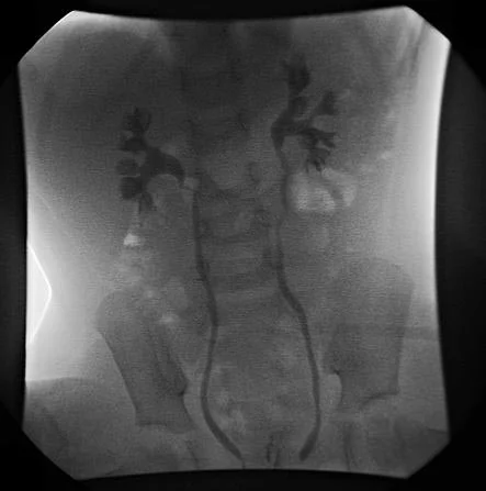
Long description
Voiding cystourethrogram showing bilateral grade III vesicoureteral reflux up the ureters.

The VCUG (also called MCUG) is the gold standard for diagnosing and grading VUR. A catheter fills the bladder with contrast; images are taken during filling, voiding and after voiding. VCUG gives detailed anatomy, detects bladder diverticula, identifies ureteral duplication and ureteroceles, shows bladder wall trabeculation, and allows international grading. [1]
Practical points from the AUA protocol and StatPearls guidance: more than one filling cycle (at least 2) should be used, because early or late reflux can be missed; diluted contrast at body temperature is instilled under gravity via a small urethral catheter without anesthesia (limited sedation for some children); spot images of the urethra during voiding, plus scout and post-void films, are required; and radiation should be kept as low as reasonably achievable. [1] A catheter can mask urethral pathology such as posterior urethral valves, so urethral images should also be taken after catheter removal. [1]
Guidelines from the AAP recommend evaluation for VUR in children aged 2 to 24 months after the second episode of febrile UTI, or earlier if renal ultrasonography is abnormal. [1] More generally, VCUG is recommended after a first febrile UTI if the ultrasound is abnormal, after a second UTI, or in the presence of other high-risk factors such as bladder and bowel dysfunction. [1]
Technetium-99m-labelled dimercaptosuccinic acid (DMSA) scintigraphy images renal cortical tissue and is the investigation of choice for renal scarring and differential renal function. It is recommended to assess kidney function, to quantify scarring after a documented infection, and to follow high-risk children. DMSA has higher sensitivity but lower specificity than ultrasound for detecting reflux. Neither DMSA nor ultrasound, alone or together, reliably replaces VCUG for diagnosing VUR. [1] Radionuclide cystography offers lower radiation and is useful for monitoring known reflux, especially grades III to V. [1]
VUR is graded 1 to 5 on the VCUG. [1]
| Grade | Appearance on VCUG |
|---|---|
| I | Reflux into the non-dilated ureter only |
| II | Reflux into ureter and renal pelvis, no dilatation |
| III | Mildly dilated ureter and pelvocalyceal system, with mild or minimal blunting of the calyces |
| IV | Ureter somewhat tortuous, mildly to moderately dilated, with blunted calyces but preserved papillary impression |
| V | Very tortuous, severely dilated ureter and markedly dilated collecting system, with loss of the fornices and no papillary impression |
The following algorithm summarises the NICE NG224 imaging and treatment pathway for a first febrile infection in a child older than 3 months. [2]
First febrile urinary tract infection in a child (NICE NG224, summary)
- Child with suspected UTIIllness, fever, pain, non-specific symptoms by age
- Collect urine as soon as possibleClean catch (or urine pad); avoid bags and cotton wool; culture within 4 hours or refrigerate
- Dipstick: leukocyte esterase and nitriteUnreliable and usually not needed under 3 months
- Question: Age and result
Under 3 months
- Refer urgently to paediatricsUrine culture; IV antibiotics as needed
3 months or older, dipstick positive
- Send urine for cultureStart oral antibiotics, usually trimethoprim or nitrofurantoin for uncomplicated lower infection
- Uncomplicated lower UTI: 3 days; pyelonephritis or systemic features: 7 to 10 daysIV co-amoxiclav, cefuroxime, ceftriaxone, gentamicin or amikacin if vomiting or severely unwell
3 months to 3 years, both tests negative
- No antibiotics for UTINo culture unless high risk or atypical; look for another cause
3 years or older, both negative
- UTI unlikelyDo not treat or culture; explore other causes
- Question: Response to treatment within 48 hours?
Yes, under 6 months, first episode
- Renal ultrasound within 6 weeksAbnormal scan: refer for possible MCUG and DMSA about 4 to 6 months later
Yes, 6 months or older, first episode
- No routine imaging or follow-upAdvice on fluids, toilet habits, constipation
Atypical or recurrent UTI
- Ultrasound by age (during acute episode or within 6 weeks)DMSA 4 to 6 months after infection; MCUG if ureteric dilatation, poor flow, non-E. coli infection or family history of VUR
- Atypical UTI (NICE NG224): seriously ill; poor urine flow; abdominal or bladder mass; raised creatinine; septicaemia; failure to respond to suitable antibiotics within 48 hours; infection with non-E. coli organisms. [2]
- Recurrent UTI: two or more febrile upper UTIs; or one febrile upper UTI plus one or more lower UTIs; or three or more lower UTIs. [2]
- VUR grades 3 to 5 are the "dilating" grades associated with the bulk of UTI and scarring risk, especially grades 4 and 5. Grade III is often classified at the lower end of the high-grade category. [1]
- Complicated reflux is more likely when there is concurrent obstruction, raised bladder pressure, bowel dysfunction, or severe grades IV and V. [1] [3]
Children under 3 months with suspected or confirmed UTI are referred urgently to secondary care. [2] For children over 3 months, antibiotics follow NICE antimicrobial guidance: an uncomplicated lower UTI is treated for 3 days, first-line oral trimethoprim or nitrofurantoin, with cefalexin, amoxicillin (where cultures show susceptibility) or nitrofurantoin as second choices; a complicated infection with pyelonephritis or systemic features is treated for 7 to 10 days, with oral cefalexin or co-amoxiclav, escalating to intravenous co-amoxiclav, cefuroxime, ceftriaxone, gentamicin or amikacin if the child is vomiting or severely unwell. Trimethoprim should not be used if there is risk of resistance (for example use in the previous 3 months); nitrofurantoin should not be used if estimated glomerular filtration rate is less than 30 millilitres per minute per 1.73 square metres, and only with caution up to 45. [2] Product labelling (BNF/MHRA) is stricter: nitrofurantoin is contraindicated if estimated glomerular filtration rate is less than 45 millilitres per minute per 1.73 square metres, with only a short 3 to 7 day course used with caution at 30 to 44 millilitres per minute per 1.73 square metres. [8] Children on prophylactic antibiotics should be treated with a different drug. [2] Asymptomatic bacteriuria is not treated. [2]
The EAU/ESPU approach agrees that initial management is medical for febrile UTI with high-grade VUR, with surgical care reserved for non-compliance with prophylaxis, breakthrough febrile UTIs despite prophylaxis, or symptomatic VUR that persists during long-term follow-up. [3]
This is the single most important modifier of VUR outcome, and it must be addressed before, or together with, any antibiotic. [1] [3]
- Bladder and bowel dysfunction: timed voiding every 90 to 120 minutes, double voiding, pelvic floor therapy and exercises, anticholinergics where indicated, and treatment of constipation with stool softeners. [1]
- Circumcision decision in uncircumcised infant boys with VUR, with shared decision-making about risks and benefits of UTI prevention, and careful foreskin care if not circumcised. [1]
- Hydration and avoidance of withholding, plus careful wiping in girls and addressing delayed bladder emptying. [2]
- Recognising and treating recurrent infection with the least possible antibiotic exposure.
The EAU/ESPU guidance is explicit that BBD is common in toilet-trained children presenting with UTI, with or without primary VUR, and increases the risk of febrile UTI and of focal uptake defects on a radionuclide scan; a practical approach to prophylaxis duration is to continue it until BBD has resolved. [3]
Prophylaxis is not indicated after a UTI when the urinary tract is normal, and is not routinely indicated after a first UTI. [2] [1] In high-grade VUR it has a clearer, evidence-supported role.
- The AUA Pediatric VUR Guidelines Panel recommends continuous prophylaxis for children aged 1 year or younger with VUR who have a history of febrile UTI, high-grade reflux (grades III to V), or concurrent bladder or bowel dysfunction. [1]
- Patients receiving most benefit are those with bladder or bowel dysfunction, uncircumcised boys, chronic constipation or diarrhoea, and grades IV and V reflux. [1]
- A randomised open-label trial in over 290 young infants (about 75% uncircumcised males, mean age 3 months) with high-grade VUR and no prior UTI showed that 2 years of prophylaxis significantly reduced the proportion with a first symptomatic UTI compared with placebo, but with more antibiotic-resistant organisms on culture; similar findings were reported in the larger RIVUR trial. [1]
- Strict adherence matters: children who comply are 2.5 times less likely to have a UTI and subsequent renal scarring than those with poor compliance. [1]
- Prophylaxis is generally considered optional rather than mandatory in non-high-risk children over 1 year, with individualised therapy after discussion with the family. [1]
- For children on prophylaxis, renal imaging (usually ultrasound) is recommended every 6 to 12 months, periodic VCUG or radionuclide cystography every 1 to 2 years, and regular blood pressure checks; imaging stops when reflux resolves on VCUG. [1]
The field genuinely disagrees here. The Emilia-Romagna Pediatric Urinary Tract Infections Study Group reviewed 25 years of literature and concluded that continuous prophylaxis is generally not recommended for most children with VUR, irrespective of grade or infection history, except where significant obstruction awaits definitive surgery, because the benefits in preventing UTI and scarring were relatively modest while the risk of promoting bacterial resistance was high. In the United States most practitioners still follow the AUA-style approach. Neither view should be presented as settled. [1]
Recommended prophylactic oral doses, which are lower than treatment doses and must be checked against the local formulary: [1]
- Amoxicillin 12.5 milligrams per kilogram daily (infants under 3 months). [1]
- Cephalexin 12.5 milligrams daily (neonates under 2 months, or as a second agent for breakthrough infection); from 3 months of age the NICE (BNFC) prophylactic dose is 12.5 milligrams per kilogram at night. [1] [2]
- Nitrofurantoin 1 milligram per kilogram daily (StatPearls: not under 1 month; not if glomerular filtration rate is less than 40 millilitres per minute; NICE and product labelling apply from 3 months and contraindicate it under 3 months). [1] [2] [8]
- Sulfamethoxazole 10 milligrams per kilogram plus trimethoprim 2 milligrams per kilogram daily (not under 1 month). [1]
- Trimethoprim 2 milligrams per kilogram daily (not under 2 months).
Prophylaxis can generally be discontinued when reflux has resolved or after successful surgery. [1]
Surgery is reserved for selected cases and is not first-line for most children, because many with even high-grade reflux resolve spontaneously during the first 5 years. [1] [4]
Indications (EAU/ESPU and modern reviews): [3] [4]
- High-grade VUR persisting despite correction of voiding disorders in toilet-trained children, when combined with recurrent febrile UTI. [4]
- Non-compliance with, or failure of, antibiotic prophylaxis, with breakthrough febrile infection.
- Progressive renal scarring or impaired renal growth on imaging.
- Symptomatic reflux persisting at long-term follow-up.
- In some infants, antibiotic prophylaxis or surgery is considered after recurrent UTI or progressive damage, but routine infant surgery is discouraged because of high spontaneous resolution and immature bladder function. [1]
Options:
- Endoscopic anti-reflux injection is minimally invasive but does not outperform prophylaxis in preventing new UTIs or scarring in most patients, and carries risks of recurrence, perforation, ureteral obstruction and anesthesia complications. [1]
- Open ureteral reimplantation by an intravesical or extravesical route has high cure rates in most series; the submucosal tunnel is extended to recreate the valve. [1]
- Laparoscopic and robotic-assisted approaches are increasingly used and show good resolution and complication rates comparable to open surgery, mainly via extravesical tunnels. [3]
A single breakthrough febrile UTI in a child on prophylaxis whose kidneys are healthy and unscarred is usually managed by changing the prophylactic agent rather than by surgery. [1]
Children who recover quickly from a first UTI and have a normal ultrasound do not need imaging follow-up after the age of 6 months. [2] Children with recurrent UTI need paediatric specialist follow-up even with normal imaging; those with abnormal imaging, impaired renal function, proteinuria or hypertension need specialist care. [2] For children with VUR on prophylaxis, renal ultrasound every 6 to 12 months, VCUG or radionuclide cystography every 1 to 2 years, and regular blood pressure checks is the standard. [1] When reflux resolves on VCUG, routine imaging can stop. [1]
- Renal scarring from acute pyelonephritis in high-grade reflux, usually polar.
- Chronic kidney disease and hypertension in the long term in a minority, more likely with bilateral scars, high pressure and recurrent infection. Scarring has been associated with severe hypertension, proteinuria, complications in pregnancy and progression to established renal failure, with risk greater when both kidneys are scarred. [2]
- Recurrent febrile UTI, particularly with bladder and bowel dysfunction.
- End-stage renal failure in a small minority, accounting for roughly 7% to 17% of children who develop ESKD globally. [6]
- Pregnancy complications later (hypertension, pre-eclampsia, bacteriuria) in women with prior reflux nephropathy. [2]
The natural history is favourable for most children. [1]
- About 75% (70% to 80%) of children with grades I and II resolve by age 5. [1]
- For grades III and IV, StatPearls reports 60% to 70% resolve over 5 years (best if reflux is unilateral and detected before age 2), whereas the 2026 EAU paediatric urology guideline reports 30% to 50% resolution of grades III to V within 4 to 5 years. [1] [9]
- In children over 5 years with bilateral reflux, resolution falls to 10% to 20% over 5 years. [1]
- Grade V rarely resolves without surgery. [1]
- Long-term follow-up shows permanent renal damage in about 40% of children presenting initially with moderate or severe hydronephrosis. [1]
Risk factors against spontaneous resolution include female sex, grades IV and V, ureteral duplication or diverticula, bladder and bowel dysfunction, and renal pelvis filling without bladder contraction. [1]
- A first febrile UTI should be treated promptly; prompt treatment after urine culture may reduce the risk of sepsis, acute kidney injury and, rarely, chronic kidney disease, and may reduce long-term risks such as hypertension in pregnancy. [2]
- Siblings of an affected child: baseline renal ultrasound is recommended, particularly under 3 years; VCUG is considered if abnormalities appear or infection occurs. Screening older, toilet-trained siblings without symptoms is optional. [1]
- Postnatal hydronephrosis, not sibling screening, drives the biggest screening burden (see the next section).
- General advice to families (NICE) includes not "holding on", timed toileting every 90 to 120 minutes, treating constipation, adequate fluids, and careful wiping. [2]
Posterior urethral valves (PUV) are obstructing membranes in the male urethra, the most common cause of congenital bladder outlet obstruction, with an incidence reported at roughly 1 in 5,000 to 8,000 live births. [7] They occur only in boys. The bladder outlet obstruction produces secondary VUR, a dilated and poorly compliant bladder, recurrent infection and renal impairment. [7] [1]
Diagnosis uses ultrasound, which may show a thick-walled bladder, bilateral (sometimes unilateral) hydronephrosis or hydroureter, scant amniotic fluid and the "keyhole" sign of a dilated posterior urethra. The MCUG is the most definitive study and shows tapering of the urethra. [7] Management starts with urinary catheterisation after birth to drain the bladder, followed by endoscopic valve ablation once the child is stable; if the urethra is too small a temporary vesicostomy may be performed. [7] Even after successful ablation up to one in five children may progress to renal failure, and long-term bladder monitoring is needed. [7] Fetal intervention is possible in specialist centres when amniotic fluid is dangerously low between 20 and 32 weeks' gestation. [7] The outcome of antenatal diagnosis before 24 weeks is particularly poor. [7]
Antenatal hydronephrosis itself is evaluated primarily by third-trimester ultrasound, using the anterior-posterior renal pelvic diameter (RPD). [1]
- Mild: RPD less than 10 millimetres. [1]
- Moderate: RPD 10 to 15 millimetres. [1]
- Severe: RPD greater than 15 millimetres. [1]
- Only RPD of 10 millimetres or more is generally considered clinically significant; postnatal ultrasound is usually delayed at least 48 hours (longer for mild unilateral cases) because early dehydration can underestimate dilatation, and follow-up ultrasound is arranged at 4 to 6 months. [1]
- Neonates with significant bilateral hydronephrosis, ureteral dilatation, bladder distension, or a markedly hydronephrotic solitary kidney (RPD 10 millimetres or more) need ultrasound within the first 48 hours and urological assessment. [1]
- Patients with bilateral hydronephrosis or unilateral hydronephrosis with ureteric dilatation or RPD greater than 15 millimetres should undergo VCUG. [1]
- Long-term follow-up shows permanent renal damage in about 40% of children initially presenting with moderate or severe hydronephrosis. [1]
These two headings often overlap in practice: VUR is one of the mechanisms by which antenatal hydronephrosis becomes postnatal renal damage, and children with antenatal hydronephrosis should be assessed for both reflux and obstruction.
- Infants have immature bladder function and a high rate of spontaneous resolution; conservative management with close monitoring dominates, with prophylaxis or surgery reserved for recurrent infection or damage. [1]
- Children with bladder and bowel dysfunction need treatment of the dysfunction alongside any reflux; without it, prophylaxis and surgery fail more often. [1] [3]
- Girls with VUR are more often affected by acquired reflux nephropathy after infection; long-term risks include hypertension and pregnancy complications. [6] [2]
- Boys with congenital (prenatal) reflux may have no prior infection but carry the risk of congenital nephropathy. [6]
- A basic renal ultrasound is enough to detect hydronephrosis, track renal growth and guide referral; it is available in most settings and needs no contrast. [1]
- VCUG and DMSA scanning may be unavailable; where they are, nephrology/urology referral to a centre that can perform them is appropriate, with interim prophylaxis for high-risk children.
- Clean-catch urine with dipstick testing, or early referral to culture where available, should be prioritised; delay in urine collection is a common avoidable cause of complications.
- Oral antibiotics (trimethoprim, nitrofurantoin, cefalexin) are used for uncomplicated lower infection; trimethoprim should be avoided if there is a risk of resistance (for example, if it has been used in the previous 3 months). Parenteral antibiotics are reserved for systemic illness. [2]
- Circumcision reduces UTI risk in infant boys where acceptable and safe.
- Treating constipation with cheap stool softeners and timed toileting costs little and improves most outcomes.
- VUR is backward flow from bladder to ureter and kidney, most often when the child voids.
- Primary VUR is a congenital short ureterovesical tunnel; secondary VUR is driven by pressure or obstruction downstream.
- Grades III to V are dilating reflux and carry the real risk of infection and scarring; grade V rarely resolves without surgery.
- VUR is common and usually silent; only 30% to 40% of children tested after a febrile UTI have reflux, and many low-grade cases never matter.
- Most childhood febrile UTIs are caused by E. coli and do not scar; risk concentrates in high-grade reflux with recurrent infection and bladder or bowel dysfunction.
- Urine must be collected cleanly (clean catch, not bags), ideally within 24 hours, and cultured when indicated. [2]
- Ultrasound screens and follows the kidneys and bladder; MCUG/VCUG grades reflux; DMSA defines scars and differential function.
- NICE imaging is risk-stratified by age, by response within 48 hours, and by atypical or recurrent infection.
- Continuous antibiotic prophylaxis helps in infants and high-risk children, but opinion is divided; strict adherence determines benefit.
- Treating bladder and bowel dysfunction is mandatory alongside any reflux programme.
- Surgery (endoscopic or open reimplantation) is effective but reserved for selected persistent, high-grade, or treatment-failing cases.
- Posterior urethral valves cause secondary VUR and need catheter drainage then valve ablation; antenatal hydronephrosis is graded by renal pelvic diameter.
- Long-term follow-up matters: scarring, hypertension, pregnancy complications and, rarely, chronic kidney disease.
Answer each one in your head first, then open the answer.
A 4-month-old boy has a temperature of 39 degrees Celsius and is irritable but feeding poorly. A urine dipstick is positive for leukocytes and nitrite. What should happen next, and when is imaging planned?
Show answerHide answer
Refer urgently to secondary care for IV antibiotics and urine culture; neonates and infants under 3 months need urgent referral, and the 3 to 5 month range needs culture before treatment is finalised. A renal ultrasound should be arranged within 6 weeks if it is the first UTI responding to treatment; MCUG and DMSA are reserved for abnormal ultrasound, second infection, atypical features or family history of VUR. Normal first-time responders aged 6 months or older need no imaging. [2]
What does each grade of VUR look like on MCUG, and which grades are called 'dilating'?
Show answerHide answer
Grade I: reflux into the non-dilated ureter only. II: ureter and renal pelvis without dilatation. III: mild ureter and pelvic dilatation with mild calyceal blunting. IV: tortuous, mildly to moderately dilated ureter with blunted calyces but preserved papilla. V: very tortuous, severely dilated ureter and collecting system with loss of fornices and no papilla. Grades III to V are dilating; grades IV and V carry the greatest scarring risk.
The parents of a child with grade IV VUR ask why you are treating constipation and a wetting problem before offering surgery. How do you explain?
Show answerHide answer
Secondary or amplifying VUR is often driven by raised bladder pressure and bowel dysfunction. Bladder and bowel dysfunction increases the risk of febrile UTI and scars, reduces spontaneous resolution and lowers surgical success. Treating it first (timed voiding, constipation management, anticholinergics where needed) is the step that modifies outcome; surgery is reserved for high-grade reflux that persists with recurrent febrile UTI despite this care.
A toilet-trained 7-year-old girl has a third lower UTI and normal renal ultrasound. She has no scarring. Does she need continuous antibiotic prophylaxis, surgery or just advice?
Show answerHide answer
Neither continuous prophylaxis nor surgery is automatic here. Third lower UTI is possible recurrence, so refer for paediatric assessment, DMSA 4 to 6 months after the most recent infection, and check bladder-emptying on ultrasound. Advise fluids, timed toileting, constipation treatment and careful wiping. Continuous prophylaxis (a single evening dose such as trimethoprim, nitrofurantoin, cefalexin or amoxicillin) is considered only if behavioural measures fail, after specialist advice; surgery (endoscopic or open reimplantation) is reserved for selected persistent high-grade reflux with recurrent febrile UTI or progressive damage. [2] [1]
Why does a short intramural ureter allow reflux only when the tunnel is short?
Show answerHide answer
The normal ureter runs through the bladder wall acting as a passive flap-valve. When that tunnel is short relative to diameter, compression cannot occur during voiding and urine is driven retrograde up the ureter, especially toward the kidney in the higher grades.
Posterior urethral valves, antenatal hydronephrosis, childhood fever and sepsis, urinary tract infection in adults, chronic kidney disease, hypertension, end-stage renal failure and renal transplantation (plain text; no links used, because only verified topics are linked).
- Leslie SW, Aeddula NR. Vesicoureteral reflux. StatPearls [Internet]. Treasure Island (FL): StatPearls Publishing; 2024 Apr 30.. Accessed .
- National Institute for Health and Care Excellence (NICE). Urinary tract infection in under 16s: diagnosis and management. NICE guideline [NG224]. London: NICE; 2022.. Accessed .
- Gnech M, 't Hoen L, Zachou A, Bogaert G, Castagnetti M, O'Kelly F, Quaedackers J, Rawashdeh YF, Silay MS, Kennedy U, Skott M, van Uitert A, Yuan Y, Radmayr C, Burgu B. Update and Summary of the European Association of Urology/European Society of Paediatric Urology Paediatric Guidelines on Vesicoureteral Reflux in Children. European Urology 2024;85(5):433-442. doi:10.1016/j.eururo.2023.12.005. Accessed .
- Goulin J, Demede D, Ranchin B, Mosca M, De-Mul A, Bidault Jourdainne V. What paediatricians need to know about modern urologic management of vesicoureteral reflux. Frontiers in Pediatrics 2025;13:1607019. doi:10.3389/fped.2025.1607019. Accessed .
- Tullus K, Shaikh N. Urinary tract infections in children. Lancet 2020;395(10237):1659-1668. doi:10.1016/S0140-6736(20)30676-0. Accessed .
- Aeddula NR, Baradhi KM. Reflux nephropathy. StatPearls [Internet]. Treasure Island (FL): StatPearls Publishing; 2023.. Accessed .
- Gharpure V. Posterior Urethral Valves in Neonate. Journal of Neonatal Surgery 2013;2:28.. Accessed .
- National Institute for Health and Care Excellence (NICE). BNF: nitrofurantoin. British National Formulary: nitrofurantoin.. Accessed .
- European Association of Urology. EAU Paediatric Urology Guidelines 2026. Paediatric Urology Guideline. Arnhem: EAU; 2026.. Accessed .
Figure credits
Educational content for medical students and doctors. It is not a substitute for clinical judgement or local guidelines.