Legg-Calve-Perthes disease
An idiopathic childhood avascular necrosis of the femoral head: presentation, imaging stages, differential diagnosis and containment-based management.
- Legg-Calve-Perthes disease (LCPD) is an idiopathic avascular necrosis of the capital femoral epiphysis in children, most often between 4 and 7 years of age (peak 5 to 6). [1]
- It declares itself with an afebrile, slowly worsening limp and hip, thigh or knee pain; the child does not look ill. [3]
- The key danger is not the pain (which usually settles within weeks) but progressive deformation of the soft femoral head while it is revascularising, a process that takes 2 to 4 years. [1]
- Plain radiographs evolve through the modified Waldenstrom stages; early disease may look normal, so MRI (perfusion MRI with gadolinium) is useful for early assessment. [1][2]
- Management is a race against time: protect the spherical femoral head in the younger, earlier stages (containment, limited weight-bearing), remodel severe hinge abduction later, and operate on late sequelae only when pain or function demand it. [1]
By the end of this page you should be able to:
- Explain why an idiopathic vascular insult to the femoral head produces a self-limiting but potentially deforming disease in children aged 2 to 14 years. [1]
- Recognise the typical presentation and distinguish it from transient synovitis and septic arthritis of the hip. [3]
- Read the modified Waldenstrom stages on plain radiographs and name the classifications used to judge prognosis and femoral head involvement. [1][2]
- Interpret plain radiographs, perfusion MRI and arthrography, stating what each adds. [2]
- Plan management by stage: observation and containment in early disease, surgical containment or pelvic osteotomy when extrusion is developing, remedial and then salvage surgery later. [1]
- State the principal prognostic factors, particularly age at onset, and give an honest appraisal of what the evidence supports. [1][2]
DefinitionLegg-Calve-Perthes disease (LCPD)
Idiopathic avascular necrosis of the capital femoral epiphysis in a child with an immature skeleton. The femoral head loses its blood supply, undergoes necrosis, fragmentation and gradual revascularisation and remodelling over 2 to 4 years. The aim of management is to preserve the spherical shape of the femoral head. [1]
"Perthes disease", "coxa plana" and "osteochondritis deformans juvenilis" are all names for the same condition. Pathologically it is not an osteochondritis but a vascular event: occlusion of the lateral epiphyseal vessels (or the medial circumflex artery feeding them) renders part or all of the epiphysis avascular, and the same child can revascularise spontaneously a few weeks later. [1]
Although the blood supply returns on its own, the bone has been weakened. In the early stages the femoral head deforms simply under body weight, and in the fragmentation stage the weakened trabeculae crumble. The self-limiting nature of the disease is its defining characteristic; the variable amount of deformation it leaves behind is its clinical significance. [1]
LCPD may affect children between 2 and 14 years of age, with the peak age of onset around 5 to 6 years. [1] It is between 1 and 11 per 100,000 children in the populations described, and males are affected about five times more often than females. [3][1] About 10% to 24% of patients have bilateral disease, usually not synchronous. [3]
- Age. The highest incidence is at 4 to 7 years, when the femoral epiphysis is supplied solely by the lateral epiphyseal branches of the medial circumflex artery. [1] Occurrence in adolescence is uncommon and carries a worse prognosis because revascularisation is slower and often incomplete. [1]
- Ethnicity. The disease predominantly affects white children from Northern Europe; equatorial regions and children of African ancestry have a very low incidence. [1]
- Geography and social deprivation. Incidence varies from 0.2 to 19.1 per 100,000 children aged 0 to 14 per year, equating to a lifetime risk of about 1 in 400 to 1 in 35,000. [1] Marked gradients exist within countries (Scotland reported more than twice the rate of London; northern and western Norwegian counties differ several-fold). [1] The most socioeconomically deprived households have been reported to have about four times as much disease as the most affluent, and secular incidence is falling as living standards improve. [1] In Merseyside the rate halved from 14.2 to 7.4 per 100,000 children per year between 1976 and 2009. [1]
- Heritability. Familial clustering is weak. Parents of affected children have a 0.5% to 0.8% chance of having had the disease and siblings a 2% to 4% chance; a Danish twin study found no LCPD in the co-twin of affected identical twins, implicating a shared environment rather than a genetic susceptibility. [1]
- Associations. Low birth weight shows a dose-response relationship with risk, and children with the disease have consistently been found to be shorter than their peers ("rostral sparing" growth disturbance), with congenital anomalies, behavioural hyperactivity and asthma more commonly recorded than expected. [1] Parental smoking has been associated in a dose-response manner. [1] The etiological factor itself remains elusive; thrombophilia studies have not supported a major coagulation defect. [1]
The cause of LCPD is idiopathic. The favoured mechanistic model is an interruption of the epiphyseal blood supply, not a primary thrombophilia. [1] No modifiable single risk factor is established, although the strong, street-by-street socioeconomic gradient and the associations with low birth weight, growth restriction, passive smoke exposure and congenital malformations point to an early environmental determinant, probably prenatal or in the first years of life. [1]
The only confirmed "risk factors" are therefore epidemiological associations, and they explain why the disease clusters in deprived communities but do not allow prediction in an individual child. [1]
At 4 to 7 years the capital femoral epiphysis receives its blood almost entirely from the lateral epiphyseal branches of the medial circumflex artery. Occlusion of either the artery or these end-vessels produces necrosis of part, or all, of the epiphysis. [1] Angiography, radioisotope studies and perfusion MRI confirm spontaneous revascularisation, usually recanalising the occluded vessels from the lateral side. If the epiphysis collapses before revascularisation completes, these channels may be obliterated and recovery is delayed by months to years; revascularisation can remain incomplete when onset is in adolescence. [1]
The full cascade matters for understanding both the presentation and the treatment rationale:
- Early, reversible deformation (modified Waldenstrom Stages Ia, Ib, IIa). Soon after infarction the mechanical properties of the epiphysis change. Weight-bearing produces an average 7.2% increase in femoral head width and a 12.4% decrease in height on standing, dimensions that return when the child lies down. [1] Epiphyseal cartilage hypertrophy and extrusion of the lateral part of the head initiate. [1]
- Irreversible deformation (Stages IIb, IIIa). Osteoclastic resorption of dead bone races ahead of osteoblastic new bone formation. The trabeculae crumble, the head collapses to a "mushroom" or is uniformly flattened, and extrusion beyond the acetabulum increases abruptly once it exceeds 20% of the head. [1] The femoral head is most vulnerable in Stage IIb. [1]
- Stabilisation (Stage IIIb onwards). The new woven bone remodels into lamellar bone aligned to resist load; no further passive deformation occurs. [1]
Severity also varies between clinical subtypes. A "discoid" epiphysis, completely sclerotic and flat before fragmentation, behaves like a whole-head infarct and has a uniformly poor outcome. [1] Three patterns of evolution are described in adolescents (late-onset, segmental collapse and destructive), all doing worse than classic childhood Perthes. [1]
The presentation is remarkably quiet: an afebrile child, not ill, develops a painless or mildly painful limp over weeks to months. [3] Pain is usually groin, lateral hip, thigh or medial knee (referred pain via the obturator nerve), and intensifies during and after activity. [3] There are no constitutional symptoms, which is the single most useful clinical pointer away from infection or malignancy. [3]
Functional clues include avoidance of running and jumping, difficulty putting on shoes, and a child who opts out of sport. In older children, hinge abduction pain appears later as the disease deforms.
- Gait: antalgic, often Trendelenburg (trunk shift over the affected hip) when the abductors are overpowered by leverage loss. [3]
- Hip range of motion: limitation of abduction and internal rotation is characteristic, and internal rotation loss is a sensitive marker of intra-articular hip pathology. [3]
- Pain at the end of the arc of motion rather than throughout (contrast septic arthritis). [3]
- Quadriceps and hip abductor weakness on resisted testing. [3]
- Leg-length inequality on the Galeazzi test, from early epiphyseal height loss; bilateral disease (10% to 24%) can mask it. [3]
- Early disease may be virtually painless with a subtle antalgic gait only.
- Hinge abduction. By Stage IIb or IIIa a "bump" on the lateral femoral head can no longer slide under the acetabular rim and impinges; abduction pain and restriction recur after an earlier lull. [1]
- Adolescent Perthes. Onset after about 11 years: slower revascularisation, poorer outcome. [1]
- Discoid epiphysis. Whole-head involvement, flat from the outset, poor regardless of treatment. [1]
The most important differential is the child with an acutely irritable hip. Transient synovitis (TS) and septic arthritis (SA) share the age range and the limp. LCPD is usually more indolent, afebrile and less locally tender, but cultures and aspiration must be used whenever the picture is unclear. [3]
| Feature | Legg-Calve-Perthes disease | Transient synovitis of the hip | Septic arthritis of the hip |
|---|---|---|---|
| Age | Usually 4 to 7 years (range 2 to 14) | Common in children | Any age; common in toddlers [1] |
| Onset | Weeks to months, insidious | Days, after a viral illness | Acute (days) |
| Systemic state | Afebrile, not ill | Low fever possible, well child | Febrile, toxic-appearing |
| Pain pattern | Groin, thigh, referred knee; worse with activity | Groin or thigh; pain at end of motion | Severe pain throughout full range |
| Hip position | Limited abduction and internal rotation | Flexed, abducted, externally rotated | Flexed, abducted, externally rotated |
| Motion | Limited at end of arc | Limited at end of arc | Pain throughout the arc |
| White cell count | Normal | Normal or mildly raised | More than 12,000 per microlitre very suggestive |
| ESR | Normal or mildly raised | Normal or mildly raised | More than 40 mm per hour suggestive [3] |
| CRP | Normal | Normal or mildly raised | More than 20 mg per litre increases likelihood [3] |
| Imaging | Sclerosis, fragmentation, flattening of proximal femoral epiphysis; may be normal early; bone scan positive early | Effusion only; normal bone | Effusion; MRI shows marrow and soft tissue changes |
| Management | Observation, containment, surgery by stage | Rest, NSAIDs, re-evaluate | Urgent joint aspiration and antibiotics |
The Kocher criteria are a clinical prediction tool that estimates the likelihood that a child with an acutely painful hip has septic arthritis rather than transient synovitis. [3] Each criterion present increases the probability; four out of four makes septic arthritis very likely, while a well child meeting none is more consistent with transient synovitis. [3]
| Criterion | Threshold in the source |
|---|---|
| Fever | Present (Kocher criteria component; no temperature cut-off in the source) [3] |
| Weight-bearing | Non-weight-bearing on the affected side |
| Erythrocyte sedimentation rate | More than 40 mm per hour |
| White blood cell count | More than 12,000 per microlitre |
| C-reactive protein (Caird addition) | More than 20 mg per litre increases likelihood |
A child with an acutely painful hip still needs broad consideration of trauma, osteomyelitis, Perthes disease, slipped capital femoral epiphysis, Lyme arthritis and juvenile idiopathic arthritis even when Kocher scores are low. [3]
Figure 1Roe-perthes
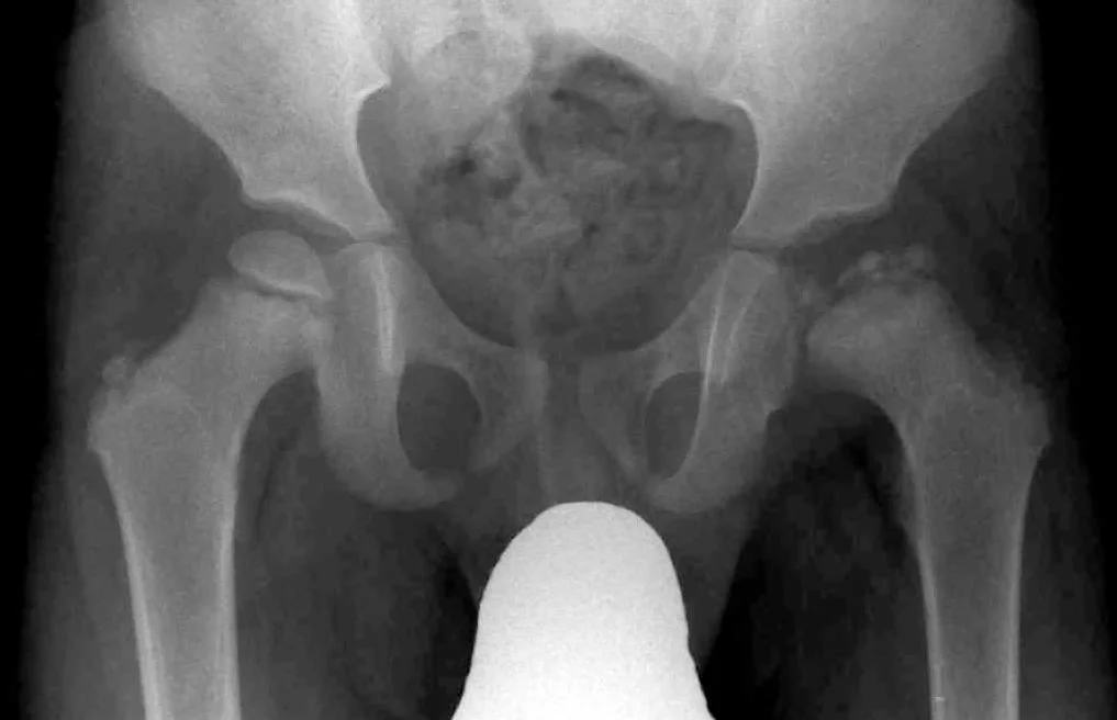
Long description
Hip radiograph of Legg-Calve-Perthes disease.

Figure 2LCPdisease2015
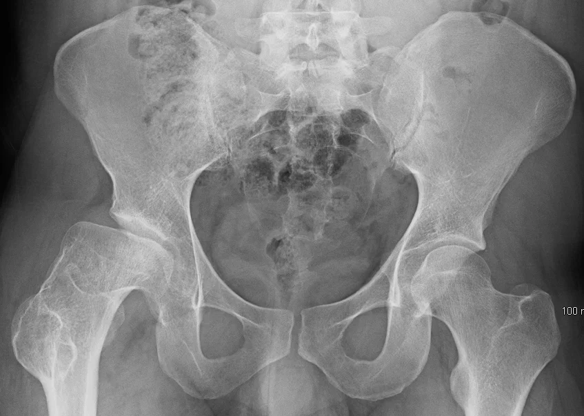
Long description
Radiograph of Legg-Calve-Perthes disease of the right hip.

Anteroposterior and frog-leg lateral films of the hips are the first investigations. In early disease they may be normal or show only a subtle sclerotic focus in the capital epiphysis. [3] With time they reveal the classic sequence: sclerosis, then fragmentation, then flattening and eventual re-ossification of the proximal femoral epiphysis. [3] If the radiograph is equivocal, repeat imaging after a few weeks often clarifies the picture. [3]
The modified Waldenstrom classification underpins both prognosis and treatment planning. [1]
| Stage | Main radiographic event | Susceptibility to deformation |
|---|---|---|
| Ia (early avascular necrosis) | Subchondral fracture, sclerosis, no collapse | Reversible (soft tissue-like) |
| Ib (late avascular necrosis) | More extensive sclerosis | Reversible |
| IIa (early fragmentation) | First vertical fissure through the epiphysis | Reversible |
| IIb (late fragmentation) | Brisk osteoclastic resorption, metaphyseal cysts, abrupt increase in extrusion | Irreversible; femoral head most vulnerable |
| IIIa (early reconstitution) | "Woolly" new bone at periphery, mushrooming | Still vulnerable |
| IIIb (late reconstitution) | Lamellar bone, trabeculae realigned | Not prone to further deformation |
| IV (healed) | Sclerotic femoral head of altered shape | End state |
The original (four-stage) Waldenstrom scheme was Stage I avascular necrosis, Stage II fragmentation, Stage III reconstitution, Stage IV healed. [1]
Three radiographic classifications are commonly used, and each measures something different (extent of epiphyseal infarction, lateral pillar height, and final shape).
Catterall classification is the oldest (1971), describing how much of the epiphysis is sclerotic and whether the metaphysis is involved. [2] It carries prognostic weight but has poor reproducibility. [2]
| Grade | Description | Prognosis |
|---|---|---|
| I | Very anterior involvement of epiphysis; no metaphyseal involvement | Excellent without treatment at any age |
| II | Anterior involvement less than 50%; possible metaphyseal involvement | Good under 4 years; about 50% good without treatment over 4 years [2] |
| III | Anterior involvement more than 50%; frequent metaphyseal involvement | Poor [2] |
| IV | Total epiphyseal involvement with metaphyseal involvement | Poor to bad |
Herring lateral pillar classification (1992) divides the femoral head into lateral (15 to 30% of width), central (50%) and medial (20 to 35%) pillars and grades involvement of the lateral one. [2] It was devised specifically because Catterall's scheme reproduced poorly, and it has the best intra- and inter-observer reliability of the three. [2] A B/C border stage exists when the lateral pillar keeps more than half its height but is thin (2 to 3 mm), less dense, or lower than the central pillar. [2]
| Group | Lateral pillar | Prognosis by Stulberg class |
|---|---|---|
| A | No involvement | Stulberg I and II in about 100% (excellent) |
| B | More than 50% of height maintained | Under 9 years: Stulberg I and II in 92% (fair); over 9 years: poor [2] |
| C | More than 50% of height lost | Stulberg II in 13%, III in 52%, IV or V in 35% (poor) [2] |
A useful caveat: the extent of collapse on which Herring's groups depend may not be visible until Stage IIb. [1]
Stulberg classification (1981) is applied at skeletal maturity and is strongly prognostic for the long-term fate of the hip. [2] Only Stulberg I and II (spherical or near-spherical heads) count as acceptable; Stulberg III gives mild to moderate arthritis and Stulberg IV and V progress to earlier degeneration. [1][2] The Mose method (spherical, crescent or non-spherical) is an alternative that similarly tracks long-term outcome. [2] A newer quantitative score, the Sphericity Deviation Score, regards a value of 10 or below as a spherical head. [1]
| Class | Shape | Prognosis |
|---|---|---|
| I | Spherical, congruent | Good |
| II | Spherical with less than 2 mm loss of height; coxa magna, short neck or oblique acetabulum | Good [2] |
| III | Aspherical but not flat; more than 2 mm loss of height | Mild to moderate arthritis [2] |
| IV | Flat head and flat acetabulum | Moderate arthritis (poor) |
| V | Flat head with normal neck and acetabulum | Severe early arthritis (bad) |
Perfusion MRI with gadolinium delineates the avascular (black, non-enhancing) portion of the epiphysis at presentation, before fragmentation, and a low residual perfusion index predicts greater later deformation and higher Herring group. [2] It also demonstrates extrusion, hinge abduction and the state of the articular cartilage, none of which plain films show. [2] Diffusion-weighted and ADC sequences have been correlated with Herring stage but not yet with final outcome. [2] Expect practical limitations: it is usually a research or specialist tool, not a first-line investigation, and no specific MRI prognostic sign is yet universally accepted. [2]
An arthrogram under anaesthesia assesses whether hinge abduction is reducible and whether the femoral head is congruent in abduction. The Laredo arthrographic classification has high prognostic value but is rarely used in routine practice. [2] Scintigraphy has no prognostic value. [2]
There is no blood test for LCPD. Tests are sent to exclude the mimics: full blood count, erythrocyte sedimentation rate, C-reactive protein, and joint aspiration when infection cannot be excluded (see Kocher criteria above). [3]
Management depends on three interacting judgements: the child's age (bone age more than chronological), how much of the epiphysis is involved and extruded, and which Waldenstrom stage the disease has reached. [1][2] Earlier intervention carries a much better chance of a spherical head: the odds of healing with an aspherical femoral head were reported to be 16.58 times greater when treatment began in Stage IIb or later. [1] Because containment attempted in Stage IIb no longer protects the femoral head from deformation, the therapeutic window is effectively Stages Ia to IIa. [1]
Early disease (Stages Ia, Ib, IIa): preventive intervention. The goal is to stop reversible deformation becoming permanent. Two approaches are combined:
- Containment. Keep the femoral head well seated in the acetabulum so that extruded portions are covered and the load is distributed. Non-operative containment (broomstick plaster, abduction casts or braces) attempts to reverse extrusion by hip abduction; surgical containment achieves the same aim with a proximal femoral varus osteotomy or a pelvic osteotomy (Salter innominate, or triple innominate). A shelf (labral support) or Chiari osteotomy covers the extruded head without reorienting the bones. [1]
- Weight-relief. Avoid weight-bearing until Stage IIIb (about 24 months from treatment start in children treated from onset), since loading during this window is what deforms the head irreversibly. [1] Prolonged non-weight-bearing combined with containment has reported 75% spherical heads at healing, versus 49% with only 6 months of protection. [1]
In a recent prospective study with surgical containment, prolonged abduction bracing was found to be ineffective and is not recommended; A-frame and Pogo stick braces have reported good results in smaller series, but robust comparative data are lacking. [1]
Surgical containment is generally favoured for the older child or whenever extrusion is present, provided the disease has not progressed beyond Stage IIa. [1] Pre-emptive containment is recommended in children older than 7 years at onset, even without visible extrusion, because extrusion invariably develops in them. [1] In younger children with limited involvement, observation with activity modification, physiotherapy for hip range of motion, and careful radiographic follow-up at regular intervals is a legitimate strategy. [1][2]
Trochanteric epiphysiodesis performed at the same time as containment may reduce greater-trochanteric overgrowth where capital physis arrest is a risk. [1]
Later presentation (Stages IIb, IIIa): remedial intervention. Containment no longer reliably prevents deformation, so the aims shift to relieving pain (often hinge abduction) and limiting further damage. Hinge abduction is distinguished as reducible (the head reduces into the acetabulum under anaesthesia) versus irreducible; a shelf acetabuloplasty suits the reducible form, while a proximal femoral valgus osteotomy repositions the head for the irreducible form. [1] The femoral head is seldom spherical by the end of the disease at this stage. [1]
Late and healed disease (Stage IIIb onwards): salvage. Whatever shape the head has at Stage IIIb is permanent. Surgery here addresses sequelae: femoral head reshaping via safe surgical dislocation, correction of intra- and extra-articular impingement, and management of the acetabular rim. [1] Long-term outcome data for hip-preservation surgery are not yet mature enough to promise delay of total hip replacement. [1]
Management algorithm for a child with LCPD
- Diagnose LCPD (hip pain or limp, characteristic radiograph or MRI)Establish Waldenstrom stage, Catterall and Herring grades, and age (prefer bone age)
- Question: Stage at presentation?
Early (Ia, Ib, IIa)
- Younger than 7 years with limited involvementObservation, activity modification, physiotherapy, review at regular intervals
- Older than 7 years, or extrusion presentPre-emptive or surgical containment
- Combine containment with weight-relief until Stage IIIb (about 24 months)Avoid bracing-only strategies; evidence for prolonged bracing is weak
IIb or IIIa
- Assess hinge abduction (arthrogram or exam under anaesthesia)
- Question: Reducible?
Yes
- Shelf acetabuloplasty (increase acetabular volume)
No
- Proximal femoral valgus osteotomy
IIIb or healed
- Salvage for symptoms or mechanical impingementFemoral head reshaping, osteochondroplasty, impingement correction
Femoral varus osteotomy is the most widely practised surgical containment and has the appeal of simplicity and a single incision; most studies report restoration of the neck-shaft angle without late genu valgum. [1] In many North American centres innominate (Salter) osteotomy, or triple pelvic osteotomy, is preferred, and combined femoral and pelvic osteotomies are used by some surgeons, without clear evidence of superiority over single procedures. [1] A second operation to remove metalwork is unavoidable with femoral osteotomy. [1] The surgeon also needs accessory procedures (trochanteric epiphysiodesis, hip contracture release) decided case by case. [1]
- Abduction braces appear ineffective in a prospective study, despite their theoretical grounding; the evidence base for most non-operative strategies is poor. [1]
- There is no Level I (randomised controlled) evidence for preventive containment. One recent prospective study found no benefit from surgical containment, conflicting with earlier North American and Norwegian studies. [1]
- No method of surgical containment is proven superior to the others. [1]
- Bisphosphonates and bone morphogenetic protein have shown promising results in animal models (suppressing osteoclastic resorption and enhancing new bone formation) and clinical trials are underway; they are not yet standard treatment. [1]
- Femoral head deformation: the core complication, classified by Stulberg at maturity; irreversible from Stage IIb. [1]
- Hinge abduction with recurrent pain and reduced range of motion, late in the disease. [1]
- Limb-length discrepancy from capital physeal arrest and coxa brevis ("greater trochanteric overgrowth"). [1]
- Coxa magna, short femoral neck, oblique acetabulum (Stulberg II features), usually with preserved function. [2]
- Early degenerative arthritis in Stulberg III to V hips, often leading to total hip replacement in early or mid-adulthood. [1][2]
- Osteochondritis dissecans of the femoral head after incomplete revascularisation. [1]
- Adduction contracture and trochanteric bursitis from altered biomechanics; botulinum toxin to adductors is sometimes used adjunctively. [1]
- Psychological burden of prolonged weight-bearing restriction in children started on non-operative programmes. [1]
Prognosis is dominated by four factors, in rough order of importance: age at onset, extent of epiphyseal involvement, lateral extrusion, and (in practice) compliance with treatment. [1][2]
- Age. The border between good and poor prognosis oscillates between 6 and 9 years. [2] Herring (2004) proposed 8 years chronologically or 6 years of bone age as the worse-prognosis threshold. [2] Children presenting before 6 years do much better on average, and one series found excellent outcomes with conservative treatment in them. [2] The age at onset distribution is log-normal, centred on 5 to 6 years. [1]
- Gender. Female sex is a less consistent risk factor, in part because of earlier bone maturation and shorter remodelling time at the same chronological age. [2] The female excess in severe disease may partly be an age-maturation effect rather than biology alone. [2]
- Body habitus. Overweight is a risk factor for poor outcome. [2]
- Clinical signs. Hip stiffness, especially adduction contracture or loss of abduction, worsens prognosis. [2]
- Extent of necrosis. Catterall grades III and IV and Herring group C predict poor outcomes, while group A predicts an almost universally spherical head. [2]
- Extrusion. More than 20% of the femoral head protruding beyond the acetabular margin is associated with poor outcome. [1]
- Discoid epiphysis. Even with early treatment, only about 25% end with a spherical head. [1]
- Adolescent onset. Prognosis is poor, especially the destructive pattern with negligible revascularisation. [1]
- Her versus environmental determinants. The clinical message distilled in the literature is that a stiff hip in an overweight girl over 6 is likely to finish with a poor outcome. [2]
Honest limits: beyond these broad factors, there is no reliable way to predict outcome in an individual hip early in the disease; "head-at-risk" Catterall signs are seldom visible in the stages where treatment decisions are actually made; and no randomised data settle how much of the reported benefit of containment is the treatment rather than the disease's natural tendency to do better in younger children. [1][2]
- Younger children (under 6). Favourable natural history; observation with monitoring is reasonable unless progression occurs. [2]
- Older children (school-aged and above). Extrusion develops almost inevitably; pre-emptive containment is recommended. [1]
- Girls. Earlier skeletal maturity compresses the remodelling window; treat bone age, not calendar age. [2]
- Bilateral disease. Subtle because the asymmetry used for diagnosis is absent; compare both hips carefully on every film. [3]
- Adolescents. Different patterns, worse biology; management is closer to adult femoral head collapse than to childhood Perthes. [1]
- Discoid and whole-head infarcts. Expect a poor result regardless; manage expectations and symptoms rather than expecting sphericity. [1]
- Plain anteroposterior and frog-leg lateral hip radiographs are available almost everywhere and are sufficient to diagnose established disease. [3]
- Early perthes may look normal; a child with persistent groin or knee pain and limited internal rotation with a normal radiograph needs repeat imaging after a few weeks or referral, not reassurance. [3]
- Close clinical follow-up substituted for MRI: pain continuing or recurring after a brief lull suggests hinge abduction or progression, both of which change management. [1]
- Non-weight-bearing and physiotherapy for abduction can be taught to families; bracing equipment and even plaster may be unavailable, in which case activity modification and monitored protected weight-bearing is the practical alternative. [1]
- Complex osteotomies, shelf procedures, and perfusion MRI require a paediatric orthopaedic service; the clinician's job in a low-resource setting is to recognise, stage as far as possible, refer early, and avoid harmful delays from repeated "watching" of what is already clearly beyond observation. [1][3]
- LCPD is idiopathic avascular necrosis of the capital femoral epiphysis in children, peak age 5 to 6 years, more common in boys, 10% to 24% bilateral. [1][3]
- The disease is self-limiting over 2 to 4 years; the threat to the child is deformation of the femoral head while it is soft, from Stage Ia to IIIa. [1]
- Presentation is a painless or mildly painful limp over weeks, referred knee or thigh pain, and limited hip abduction and internal rotation, without fever or systemic upset. [3]
- Modified Waldenstrom stages (Ia to IV) define the window for preventive containment (by Stage IIa at the latest). [1]
- Herring lateral pillar group A predicts a spherical head in almost all cases; group C predicts a poor outcome. [2]
- Only Stulberg I and II at skeletal maturity count as acceptable (spherical) results. [1][2]
- Containment (non-operative or surgical) plus weight-bearing protection until Stage IIIb (about 24 months) is the evidence-supported core of treatment, though evidence is largely non-randomised. [1]
- Age at onset is the strongest prognostic factor; extrusion above 20% and discoid epiphysis are the strongest radiographic red flags. [1][2]
- Septic arthritis must be excluded using the Kocher criteria before settling on a diagnosis of Perthes disease or transient synovitis. [3]
- Honestly, no treatment has been proven by randomised trial to change the outcome; management is a judgement based on age, stage and extrusion. [1]
Answer each one in your head first, then open the answer.
A 6-year-old boy has a painless limp of several weeks and medial knee pain. Hip radiographs show patchy sclerosis of the capital femoral epiphysis only. What is the most likely diagnosis, what is the immediate management priority, and what must you explain to the parents?
Show answerHide answer
Legg-Calve-Perthes disease. The immediate priority is to confirm the Waldenstrom stage (here likely Stage Ia/Ib) and to keep the femoral head contained and unloaded, with protected weight-bearing and hip range-of-motion physiotherapy, because deformation is reversible only while the disease is in its early stages. Parents should be told that the illness lasts 2 to 4 years, that pain is not the main problem, and that repeated radiographic checks are needed because the appearance will evolve. [1][3]
The same child is now older and radiographs show a Herring group C (more than 50% loss of lateral pillar height) with lateral extrusion well above 20%. How does this change your plan?
Show answerHide answer
The prognosis is now poor whatever is done. Surgical containment after Stage IIb rarely preserves sphericity. The aims shift to remedial measures: assess hinge abduction (arthrogram or exam under anaesthesia) and choose a shelf acetabuloplasty if reducible or a proximal femoral valgus osteotomy if irreducible, to relieve pain and preserve range of motion, followed by salvage surgery for symptoms or impingement later. Expectations should be reset accordingly. [1][2]
How do you distinguish early LCPD from transient synovitis on clinical grounds?
Show answerHide answer
Transient synovitis is usually post-viral, peaks within days, may have a low fever and causes pain at the end of the arc of motion; the child is relatively well. LCPD is afebrile and not ill, has an insidious onset over weeks, and produces loss of internal rotation and abduction with pain at the end of the arc. When infection cannot be excluded, use the Kocher criteria (fever, non-weight-bearing, ESR more than 40 mm per hour, WBC more than 12,000 per microlitre, plus CRP more than 20 mg per litre), and aspirate the hip if scores are high. [3]
Why is weight-bearing avoided for roughly 24 months rather than treated symptomatically until pain settles?
Show answerHide answer
In Stages Ia to IIIa the femoral epiphysis is mechanically soft: standing produces reversible widening and flattening of the head, and in Stage IIb weakened trabeculae crumble irreversibly. Avoiding load through Stage IIIb prevents the conversion of reversible deformation into permanent collapse; prolonged protection has reported 75% spherical heads versus 49% with only 6 months. [1]
A child is found to have complete whole-head sclerosis and flattening before fragmentation ('discoid' epiphysis). What outcome should you anticipate?
Show answerHide answer
Even with early treatment, only about 25% of such hips finish with a spherical femoral head, because the entire epiphysis is infarcted. Management then centres on symptom control, optimising range of motion, timing of any surgical intervention, and counselling the family about likely early arthritis. [1]
Which single factor most powerfully determines long-term outcome, and what caveat applies to all published treatment evidence?
Show answerHide answer
Age at onset (with bone age more informative than chronological age) is the strongest predictor. The caveat is that there is no randomised controlled trial validating preventive containment; prospective studies conflict, bracing strategies lack robust support, and outcomes may largely reflect which children were going to do well anyway. [1][2]
Coxa magna and hip remodelling after childhood avascular necrosis; slipped capital femoral epiphysis (a disease of adolescence that also presents with a limp); transient synovitis and septic arthritis of the hip; developmental dysplasia of the hip; osteomyelitis; bone scintigraphy and perfusion MRI in paediatric hip disease.
- Joseph B, Shah H, Perry DC. Epidemiology, natural evolution, pathogenesis, clinical spectrum, and management of Legg–Calvé–Perthes. Journal of Children's Orthopaedics 2023;17(5):385-403. Accessed .
- Rampal V, Clément J-L, Solla F. Legg-Calve-Perthes disease: classifications and prognostic factors. Clinical Cases in Mineral and Bone Metabolism 2017;14(1):74-82. Accessed .
- Adams Morancie N, Helton MR. Evaluating the Child With a Limp. American Family Physician 2023;107(5):474-485. Accessed .
Figure credits
Educational content for medical students and doctors. It is not a substitute for clinical judgement or local guidelines.