Slipped capital femoral epiphysis and the limping child
Adolescent hip slip: presentation, frog-leg X-ray signs, stable vs unstable, pinning, complications, and a differential of the limping child.
- Slipped capital femoral epiphysis (SCFE, also called slipped upper femoral epiphysis or SUFE) is the displacement of the capital femoral epiphysis on the femoral neck through the growth plate. It is the most common hip disorder of adolescence and a limp or knee pain in an overweight teenager should make you think of it. [2][6]
- The slip is usually gradual but occasionally presents suddenly. It is classified as stable when the child can still walk (with or without crutches) and unstable when they cannot, because that distinction predicts the risk of avascular necrosis of the femoral head. [5][6]
- Examination shows limited internal rotation of the hip and obligate external rotation when the hip is flexed. [2][6]
- Diagnosis is made on plain radiographs: bilateral anteroposterior and frog-leg lateral views for stable slips, with Klein's line on the anteroposterior view as a classic but insensitive sign. [2][6]
- The standard treatment of a stable slip is in situ fixation with a single screw, done promptly but without forcing reduction. Unstable slips have a much higher complication rate and are treated as an orthopaedic emergency. [5][6]
By the end of this page you should be able to:
- Recognise SCFE in an adolescent from the history and examination, including knee pain and the obligate external rotation sign. [2][6]
- Order and interpret bilateral hip radiographs, including the frog-leg lateral view, Klein's line and the Southwick and Wilson grading methods. [2][6]
- Distinguish stable from unstable slips and say what that means for prognosis and management. [5][6]
- Plan management, including in situ pinning, bilateral fixation in atypical or endocrine cases, and urgent referral for unstable slips. [4][5][6]
- Recall the complications (avascular necrosis, chondrolysis, contralateral slip) and their approximate rates. [5][6]
- Use an age-based differential table and a flowchart to work through a child with a limp. [1][2]
- List the endocrine and metabolic associations of atypical SCFE and the screening tests used. [4]
DefinitionSlipped capital femoral epiphysis (SCFE)
SCFE is the posterior and inferior displacement of the proximal (capital) femoral epiphysis relative to the femoral neck (metaphysis), through the open capital femoral growth plate (physis). The femoral neck moves up and forward; the epiphysis, which stays in the acetabulum, slips backward and downward. The condition is also called slipped upper femoral epiphysis (SUFE). [6]
The slip occurs through the zone of provisional calcification of the physis: the cartilaginous growth plate separates, leaving the epiphysis close to its normal position in the socket while the metaphysis rides up and rotates outward. [3] This single fact explains almost every clinical feature that follows. Early in the disease the radiograph can look nearly normal, with only subtle widening or irregularity of the physis, and medical attention may be sought for knee pain rather than hip pain. [3][6]
This is the classification that matters most for decisions. [5][6]
- Stable SCFE: the patient can walk, with or without crutches. About 90 percent of slips fall into this group. [6]
- Unstable (acute) SCFE: the patient cannot walk, even with crutches. [5][6]
The Loder classification was introduced in 1993 and changed practice because it separated two conditions that had been mixed together: stable slips, in which the physis is at least partially intact, and unstable slips, in which the physis has completely lost continuity. [5]
Two grading methods are used on the frog-leg lateral radiograph. [6]
Wilson method: relative displacement of the epiphysis on the metaphysis. [6]
| Wilson grade | Displacement |
|---|---|
| Mild | Less than one third of the metaphyseal width |
| Moderate | One third to one half |
| Severe | Greater than one half |
Southwick (slip angle) method: the lateral epiphyseal-shaft angle (LESA) on the frog-leg lateral view minus the same angle on the normal side. [6]
| Southwick grade | Slip angle |
|---|---|
| Mild | Less than 30 degrees |
| Moderate | 30 to 50 degrees |
| Severe | Greater than 50 degrees |
The simple morphological grouping (pre-slip, mild up to 30 degrees, moderate 30 to 60 degrees, severe greater than 60 degrees) conveys the same idea and is still quoted in some series. [3]
Clinical stability (ability to walk) does not reliably equal mechanical stability. In an intraoperative series, 13 of 24 hips whose patients could not weight-bear turned out to be mechanically stable, and 17 of 58 hips whose patients could weight-bear turned out to be mechanically unstable. [3] Every SCFE hip that still has an open physis must therefore be treated as potentially unstable, at least for the purpose of urgent referral and restricted weight-bearing. [3]
SCFE is the most common hip disorder of adolescence. [6]
- Prevalence around 10.8 per 100,000 children in one national sample, and a broader range of 1 to 10 per 100,000 in population series. [5][6]
- Age: most cases occur between 8 and 15 years, with average age at diagnosis of 13.5 years in boys and 12.0 years in girls. [6]
- Sex: more common in boys than in girls. [2][6]
- Ethnicity: more common in Black and Pacific Islander children, proposed to relate in part to body weight. [6]
- Laterality: bilateral (either simultaneous or, more often, sequential) in 18 to 50 percent of patients, the second hip usually slipping within about 18 months. [4][6]
- Season and vitamin D: in the northern United States there is a late-summer and autumn peak north of 40 degrees latitude, possibly related to impaired vitamin D synthesis from reduced sunlight. [6] Some authors report a high rate of vitamin D deficiency in affected children. [4]
- Trend: the condition appears to be increasing in incidence, which is consistent with the rising prevalence of childhood obesity. [3]
The basic problem is that a relatively weak growth plate is subjected to mechanical loads it can no longer resist. This usually happens during the pre-adolescent growth spurt, when the physis becomes more oblique and when hormonal changes alter its strength. [3]
- Obesity is the clearest risk factor. Approximately 63 percent of affected children have weight in the 90th centile or higher, and obesity has been causally linked to the slip in multiple population studies. [6]
- Growth surges during puberty coincide with the peak incidence. [6]
- There is, however, a subgroup of underweight or "skinny" SCFE patients with below-average body mass index (BMI) who have a relatively high rate of unstable slips. A normal build therefore does not exclude SCFE. [3][4]
About 3 to 10 percent of all SCFE cases (up to 41 percent in some series) are "atypical", meaning they are driven by an underlying endocrine, metabolic or other systemic disorder rather than by mechanical loading. [4] In one review the average age at diagnosis of an idiopathic slip was 12.8 years, versus 15.3 years in endocrine disease, 11.4 years in renal failure and 10.5 years in radiation-associated cases. [4] Atypical slips therefore tend to present either younger than 10 years or older than 16 years, and in shorter, lighter children. [4]
The recognised causes are:
- Hypothyroidism, the most common endocrinopathy in SCFE. Thyroid hormone receptors are present in physeal chondrocytes, and deficiency disorganises the hypertrophic zone. Children are often short, cold-intolerant, bradycardic, constipated and delayed in bone age. [4]
- Growth hormone deficiency or supplementation. The incidence of SCFE in children taking recombinant growth hormone is more than double that of the general population, with an average of 10 months from starting treatment to the slip; children with growth hormone deficiency itself are at higher risk than those treated for idiopathic short stature. [4]
- Hyperparathyroidism (primary or renal osteodystrophy). Parathyroid hormone disrupts endochondral ossification and the interdigitation between metaphysis and physis through osteoclastic resorption, so the physis loses its restraint against shear. [4] Renal osteodystrophy-associated SCFE is characteristically bilateral, severe and accompanied by bone deformity. [4]
- Hypogonadism (primary or secondary, including Turner syndrome and Kallmann syndrome) and panhypopituitarism (including craniopharyngioma, optic pathway glioma, and other tumours). [4]
- Multiple endocrine neoplasia type 2A/2B (medullary thyroid carcinoma and pheochromocytoma), in isolated case reports. [4]
- Radiation therapy: pelvic or total-body radiation. These slips usually appear a mean of about 6 years after radiotherapy and are more often mild, bilateral and found in younger, underweight children. [4]
- Valgus SCFE is an uncommon variant (around 4 percent of slips) in which the epiphysis moves lateroposteriorly. It presents about 2 years earlier than typical SCFE, is more common in girls, and around 22 percent of such cases are found to have an underlying endocrine disorder. [4]
Children falling outside the classic profile should have a structured endocrine and metabolic screen. The Loder age-height and age-weight tests give an objective trigger: [4]
- Presentation younger than 10 years or older than 16 years. [4]
- Height or weight below the 50th centile. [4]
- Either of these increases the likelihood of an atypical slip several-fold, and a normal height-and-weight profile in a 10 to 16 year old is strongly reassuring. [4]
- Bilateral slips and valgus slips also mandate screening. [4]
First-line screening tests are thyroid stimulating hormone (TSH, with free thyroxine if raised), parathyroid hormone (PTH), calcium and phosphate, and a comprehensive metabolic panel; vitamin D should be measured if PTH is abnormal. [4] Endocrinology referral is recommended even when the first screen is negative in a child with clinical features suggesting an underlying disorder. [4]
- Obesity reduction lowers the risk of a second slip: patients who lost weight after a unilateral SCFE had a much lower risk of a contralateral slip than those who gained or maintained weight. [3]
- Features that raise the risk of a contralateral slip include young age, obesity, and an increased alpha angle or posterior epiphyseal tilt on the unslipped side. [3]
- Prophylactic pinning of the unaffected hip is used in selected high-risk patients (young, endocrine/metabolic disorder, poor reliability of follow-up) but is not universal and remains controversial. [3][6]
Three things turn a weak growth plate into a slipped hip.
- Oblique and softened growth plate: around puberty the capital femoral physis tilts so that the shear force across it increases, and hormonal changes (sex hormones, thyroid hormones, growth hormone) alter its strength. [3][4]
- Mechanical load: body weight (often amplified by obesity) acts on this tilted plate. [3][6]
- Epiphyseal tubercle: a bony prominence at the metaphyseal-epiphyseal junction that normally stabilises the plate; its relative diminution in adolescence is part of the mechanism of failure. [3]
Once displacement starts, it is almost always an anterior and external rotational translation of the neck with posterior tilt of the epiphysis (the head stays in the socket and backs downward). [3] A rare valgus subtype runs the other way. [3][4]
From there the trouble unfolds in three steps.
- Immediate mechanical damage: the metaphysis is now prominent anteriorly and superolaterally, and this "cam" prominence jams against the acetabular rim and labrum from the very beginning of the slip. Cartilage and labral damage are present in virtually all hips with any SCFE, even mild ones, and appear to be related more to duration than to severity. [3]
- Physeal instability: as the displacement grows, the growth plate can lose all continuity, producing an unstable slip. Unstable slips carry a high rate of avascular necrosis of the femoral head because the retinacular blood vessels that supply the epiphysis are tethered and can be stretched or torn. [5][6]
- Remodelling: the neck can remodel slowly after stabilisation, but the intra-articular damage does not fully reverse and may present years later as femoroacetabular impingement and premature osteoarthritis. [3]
The presentation is often insidious and easily missed. Delays in diagnosis are common and matter, because worse deformity at presentation means worse long-term outcome. [3][6]
- Pain is usually vague and poorly localised to the hip, groin, thigh or knee. Knee or distal thigh pain is the presenting symptom in about 15 percent of cases. [2][6]
- Limp of days to months. Pain may be mild and intermittent at first. [2][3]
- Constitutional symptoms are usually absent. Fever, night pain at rest, or systemic upset should point away from SCFE toward infection or neoplasm. [2]
- Fatigue, obesity, short stature or signs of an endocrinopathy can accompany atypical cases. [4]
- A clear history of trauma is unusual; SCFE is more often a fatigue-type failure of the physis than an acute injury. [6]
- Gentle or chronic slips may have little or no pain at all, so a "painless limp" in an adolescent still deserves an examination and radiograph. [3]
- Limited internal rotation of the affected hip is the most frequent abnormal physical finding (seen in 64 of 66 examined patients in one early series). [3]
- Obligatory external rotation: with the hip passively flexed to 90 degrees the leg rotates outward, because the neck has slipped outward and the capsule is still attached to the epiphysis. This is a highly characteristic sign. [2][6]
- Loss of abduction and flexion of the hip. [2]
- A Trendelenburg gait or weak hip abductors. [2]
- Leg-length discrepancy on the Galeazzi test from superior migration of the femoral neck. [2]
- Inability to bear weight in an unstable slip. [5][6]
- Examine both hips and compare range of motion; it is easy to miss a bilateral slip. [6]
- Look for the stigmata of an endocrinopathy or metabolic disease if the profile is atypical. [4]
A limp in a child has many causes. Age is the most useful first filter. [1][2]
| Age group | Typical causes |
|---|---|
| Any age | Fracture and soft-tissue injury; septic arthritis; osteomyelitis; cellulitis; juvenile idiopathic arthritis; foreign body; developmental dysplasia of the hip; neoplasm; neuromuscular disease; non-accidental injury |
| 1 to 3 years | Transient synovitis of the hip (rare under 3); septic hip; developmental dysplasia of the hip; toddler's fracture (spiral fracture of the tibia); immunisation reaction; cerebral palsy |
| 3 to 10 years | Transient synovitis; Legg-Calve-Perthes disease; leukemia; osteomyelitis; Köhler bone disease; Baker cyst; tarsal coalition; dermatomyositis |
| 11 to 19 years | Slipped capital femoral epiphysis; Osgood-Schlatter disease; osteochondritis dissecans; chondromalacia patellae; overuse and stress injury; sprain or strain; tendinopathy; tumour (benign or malignant) |
The conditions most likely to be confused with SCFE at presentation are transient synovitis, septic arthritis, Legg-Calve-Perthes disease, osteomyelitis, tumour and trauma. The table below summarises the separating features. [1][2][6]
| Condition | Typical age | Onset | Systemic upset | Key findings |
|---|---|---|---|---|
| Septic arthritis | Any; common in toddlers | Acute, 3 to 5 days | Febrile, toxic; joint held flexed, abducted and externally rotated; pain throughout range of motion | Incidence 4 to 5 per 100,000 children per year; ESR usually more than 40 mm per hour, WBC more than 12,000 per microlitre, CRP more than 20 mg per litre; urgent aspiration |
| Transient synovitis | Under 10 (rare under 3) | Days, often after viral illness | Well, afebrile; groin or thigh pain; pain at the end of the range of motion | Incidence about 0.2 percent per year in children; Kocher criteria to separate from sepsis |
| Perthes disease (avascular necrosis of femoral head) | 4 to 9 years | Insidious over weeks to months | Afebrile and well; groin, thigh or referred knee pain; restricted internal rotation and abduction; Galeazzi sign | Incidence 1 to 11 per 100,000 children; sclerosis and fragmentation of proximal femoral epiphysis on radiograph |
| SCFE | 10 to 15 years | Insidious, sometimes acute on chronic | Afebrile unless infection; obesity or endocrinopathy; limited internal rotation; obligate external rotation on flexion; knee pain in about 15 percent | Incidence about 4.8 to 10.8 per 100,000 children; widening of physis, Klein's line abnormality, decreased epiphyseal height |
| Osteomyelitis | Any age | Subacute | Fever possible; localised bony tenderness, warmth, swelling, redness | Incidence about 9 per 100,000 children; point tenderness, raised inflammatory markers; MRI if suspected |
| Leukaemia | Any childhood age | Variable | Night pain, anorexia, weight loss; abdominal mass, hepatosplenomegaly, lymphadenopathy, bony tenderness | Most common childhood cancer (about 4,000 new cases per year in the US); look for low-normal platelets or low WBC even without blasts |
| Malignant bone tumour (e.g. osteosarcoma) | Child to young adult | Weeks | Night pain, severe localised pain | Incidence 4 to 7 per million per year; palpable bony mass usually around knee |
| Non-accidental injury | Any age, especially non-ambulant child | Variable | Inconsistent history; lower limb fracture in a child not yet walking | Careful history, examination and skeletal survey (see child abuse and neglect) |
| Toddler fracture (spiral tibia) | 1 to 3 years | After a twist or fall | Point tenderness over tibial shaft; normal early radiograph possible | Repeated radiography in 2 to 3 weeks if suspected |
Any of the following should prompt urgent same-day assessment or referral, rather than watchful waiting: [1]
- Fever, toxic appearance, or refusal to bear weight.
- Night pain, weight loss, anorexia, fatigue or night sweats.
- Severe pain, anxiety, agitation or neurovascular compromise after injury.
- Suspicion of non-accidental injury or an inconsistent history.
- A child older than 9 years with painful or restricted hip movement, particularly loss of internal rotation, to exclude SCFE.
- Redness, swelling or stiffness of a joint (infection or inflammatory arthritis).
- A limp persisting beyond a few days in a young child, or an adolescent with risk factors for SCFE (obesity, endocrinopathy).
Approach to a child with a limp
- Child presents with a limpTake history (trauma, fever, recent illness, night pain, weight change); note age; inspect shoes and gait
- Is there trauma or concern for abuse?Osteomyelitis/septic arthritis and non-accidental injury must be considered in any age
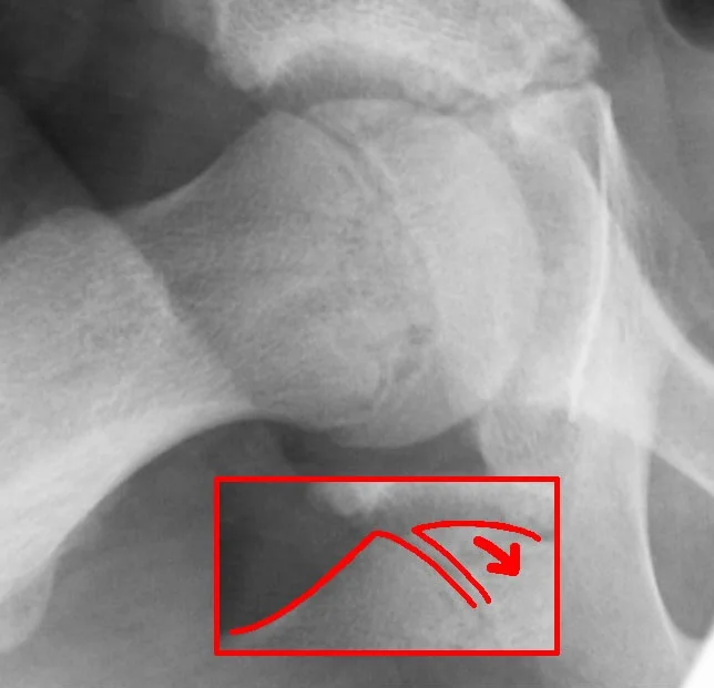
Figure 1Slipped capital femoral epiphysis
Long description
Radiograph of a slipped capital femoral epiphysis.

Plain radiographs are the diagnostic standard. [3][6]
- Bilateral anteroposterior and frog-leg (Lauenstein) lateral views are required for a stable slip. [2][6]
- In an unstable slip the hip will not allow abduction, so anteroposterior and cross-table lateral views of the involved side are compared with the uninvolved side. [6]
- Inform the radiologist that SCFE is suspected; otherwise a subtle early slip may be reported as normal. [6]
Radiographic signs of SCFE (none is individually diagnostic; the frog-leg lateral is usually the first to show the slip): [2][6]
- Widening or irregularity of the physis compared with the other side.
- Decreased height of the capital epiphysis.
- Loss of the intersection of the lateral epiphysis by Klein's line, a line drawn along the superior border of the femoral neck: in a normal hip the epiphysis projects above it; in SCFE it sits on or below it. Klein's line is classical but insensitive, especially for mild slips, so a normal line does not exclude the diagnosis.
- The Steel sign, a double density on the anteroposterior view from superimposition of the posteriorly slipped epiphysis on the metaphysis.
- Prominence of the lesser trochanter from external rotation of the femur.
- Bilateral views are mandatory because slips can be bilateral, and a second slip may be silent. [2][6]
- Ultrasound may show a small effusion or a subtle metaphyseal step-off, and can demonstrate the slip before radiographic change in a "pre-slip". [3]
- MRI with appropriate sequences shows early physeal changes, perfusion and (in established disease) cartilage status. It is useful when the radiograph is normal but suspicion is high. [3][2]
- The patient must be made non-weight-bearing immediately (crutches or a wheelchair) and referred the same day to an orthopaedic surgeon experienced with SCFE. [6]
- Slips that are unstable, or any patient who cannot bear weight, require urgent or semi-urgent fixation, not a routine outpatient appointment. [5][6]
- Do not manipulate or force a reduction, especially in stable slips, because aggressive reduction is a recognised cause of avascular necrosis. [5]
- Realignment osteotomy has a higher complication rate and is reserved for selected severe slips. [5]
In situ fixation with a single screw across the physis is the standard treatment worldwide. [5][6] It is a simple technique, prevents further slip, and has low recurrence and complication rates. [6] A systematic review of all published stable slip treatments found a pooled avascular necrosis rate of about 1.4 percent for single-screw pinning in situ, versus 11 percent for physeal osteotomy and 3 percent for Ganz surgical dislocation. [5] Ganz surgical dislocation gave the highest patient satisfaction (87 percent excellent outcomes) but was used for more severe slips and carries roughly double the avascular necrosis rate of pinning in situ. [5]
Most children with mild to moderate stable slips treated by in situ fixation have good to excellent long-term outcomes. [6] Screws designed to allow continued femoral neck growth may be advantageous over standard screws, and subsequent removal of the metalwork is discussed with the treating surgeon. [5]
Unstable SCFE is a much more severe injury. Osteonecrosis occurs in an estimated 20 to 50 percent of patients with the unstable form, and up to 60 percent have been reported in some series. [5][6] Fixation is usually achieved in situ. There is genuine controversy about the timing of surgery, whether gentle reduction should be attempted, and whether traction is useful. [6] A large systematic review concluded that open reduction and internal fixation (Parsch technique) gave the best results for unstable slips if performed on an urgent basis (within 24 hours). [5] Bilateral and prophylactic measures in high-risk (endocrine or metabolic) cases follow the atypical-case guidance above. [4]
Prophylactic pinning is controversial but is advocated by many paediatric orthopaedic surgeons for patients at high risk of a contralateral slip: children younger than 10 years (or with significantly delayed skeletal maturity), endocrine or metabolic disease, radiation-associated slips, and patients with poor follow-up. [4][6] With standard idiopathic SCFE most centres do not fix the contralateral hip prophylactically. [6]
- Avascular necrosis (AVN) of the femoral head: essentially a complication of unstable slips and severe displacement. Reported in 20 to 50 percent of unstable slips (up to 60 percent in some series), and much less in stable slips (about 1.4 percent with single-screw pinning). AVN leads to early degenerative arthritis of the hip. [5][6]
- Chondrolysis: acute loss of articular (hyaline) cartilage of the hip, presenting with joint stiffness and pain. Incidence has fallen from 5 to 7 percent to about 1 to 2 percent with modern techniques. It can occur after surgery, with hip spica cast treatment, or in untreated severe slips. [6]
- Contralateral slip: a new slip in the opposite hip, occurring in 18 to 50 percent of patients, usually within 18 months. Risk is higher with obesity, young age at first slip and a suspicious contralateral radiographic appearance. [4][6]
- Femoroacetabular impingement and long-term osteoarthritis: the metaphyseal prominence damages the labrum and anterior acetabular cartilage from the time of the slip. OA rates in long-term follow-up of stable slips vary by treatment, from 0 percent after Ganz dislocation to 3 percent after pinning in situ, but clinical impingement-type symptoms are common. [3][5]
- Slip progression or loss of fixation: more likely when fewer than three threads engage the epiphysis, or in younger children with soft bone. [4]
Outcome is governed mainly by four things. [3][5][6]
- Stability of the slip: stable slips do far better than unstable ones.
- Severity and duration of the slip: cartilage damage begins early and accumulates; a mild slip diagnosed promptly has an excellent outlook after fixation.
- Age and remodelling potential: younger children remodel more; older adolescents with severe slips are more likely to have permanent deformity and impingement.
- Body habitus: obesity raises both the risk of the initial slip and the risk of a contralateral slip. [3][6]
Mild to moderate stable slips treated by in situ fixation usually have good to excellent long-term function. [6] Even so-called mild slips can produce symptomatic femoroacetabular impingement in adult life, which is why referral for assessment of residual deformity is offered after the growth plate has closed. [3]
- A clinical suspicion plus a single plain radiograph is enough to diagnose most established slips. Bilateral anteroposterior and frog-leg lateral views are the investigation of choice; if they are not available, even a single lateral view of the painful hip is far better than none. [2][6]
- Klein's line is a bedside-ish radiographic aid: draw it yourself with a ruler along the superior neck and check that the epiphysis sits above it. It may be negative in mild slips, so a normal line does not exclude the diagnosis. [6]
- Referral and immobilisation are the treatments. A confirmed slip should be protected from weight-bearing (crutches, a wheelchair, bed rest, or a hip spica if no traction is available) and sent urgently for orthopaedic care; slipping further before fixation is what causes avascular necrosis. [5][6]
- Pinning in situ is the worldwide standard because it needs only a single screw and fluoroscopy or simple imaging guidance; complex osteotomies are for specialist centres. [5][6]
- Early slips are radiographically subtle (widened physis only). An adolescent with a limp, hip or knee pain, or limited internal rotation and a normal radiograph needs close review, restricted load and repeat imaging, not reassurance. [3][6]
- Endocrine disease can be missed but the basic screen (TSH, PTH with calcium/phosphate) is cheap; hypothyroid and malnourished children in particular can present this way, and children with renal failure are at high risk. [4]
- SCFE is the most common adolescent hip disorder; think of it in any limping, overweight teenager, and in any adolescent with knee pain. [2][6]
- It classifies as stable when the child can walk and unstable when they cannot, and this distinction predicts the risk of avascular necrosis. [5][6]
- Limited internal rotation and obligate external rotation on hip flexion are the cardinal signs; they can be the only finding. [2][6]
- Diagnosis needs bilateral anteroposterior and frog-leg lateral radiographs; check Klein's line but do not rely on it alone, and grade the slip (Wilson or Southwick). [2][6]
- Most slips happen between 10 and 15 years and occur more often in boys and in Black and Pacific Islander children, related in part to body weight. [6]
- Around 63 percent of affected children have weight in the 90th centile or higher. [6]
- Knee (or distal thigh) pain is the presenting complaint in about 15 percent of cases. [6]
- Bilateral slips occur in 18 to 50 percent of patients, usually within 18 months; protect the contralateral hip. [6]
- Unstable slips carry a 20 to 50 percent (up to 60 percent in some series) rate of avascular necrosis; stable slips about 1.4 percent with single-screw pinning. [5][6]
- In situ single-screw fixation is the standard for stable slips; isolated reduction is avoided. Ganz dislocation gives higher patient satisfaction for severe slips but at roughly double the avascular necrosis rate. [5][6]
- Chondrolysis now occurs in 1 to 2 percent after surgery (formerly 5 to 7 percent); it can also occur with a hip spica or untreated severe slips. [6]
- Endocrine or metabolic disease (hypothyroidism, growth hormone, hyperparathyroidism, hypogonadism, panhypopituitarism, renal osteodystrophy, radiation) accounts for 3 to 10 percent of SCFE (up to 41 percent in some series); screen children younger than 10 years, older than 16 years, underweight for age, with bilateral or valgus slips. [4]
- Avascular necrosis is the complication to fear most in unstable disease; chondrolysis and contralateral slip are the ones to guard against in stable disease. [5][6]
Answer each one in your head first, then open the answer.
A 13-year-old boy has had a limp for six weeks and now complains of right knee pain. He is overweight. On examination he has a Trendelenburg gait and loss of internal rotation of the right hip, with the leg drifting into external rotation when you flex the hip. What is the most likely diagnosis and what is the first investigation?
Show answerHide answer
Slipped capital femoral epiphysis. Knee pain is the presenting complaint in about 15 percent of cases, and the combination of an overweight adolescent, loss of internal rotation and obligate external rotation on flexion is characteristic. The first investigation is bilateral anteroposterior and frog-leg lateral radiographs of both hips; inform the radiologist that SCFE is suspected, as early slips may show only physeal widening. [2][6]
On a frog-leg lateral radiograph you measure the epiphyseal-shaft angle difference between the affected and normal sides. How do you grade the slip and what does the grade tell you about treatment?
Show answerHide answer
Using the Southwick method, less than 30 degrees is mild, 30 to 50 degrees moderate and greater than 50 degrees severe; the Wilson method instead grades by displacement (less than one third, one third to one half, or more than one half of the metaphyseal width). The grade guides management: pinning in situ with one screw is standard for mild and most moderate stable slips; severe stable slips may be discussed for Ganz surgical dislocation, trading a higher risk of avascular necrosis (about 3 percent) for better patient satisfaction when surgical expertise is available. [5][6]
A child with SCFE can barely put their foot to the floor and refuses to bear weight even with crutches. Why does this change management so dramatically?
Show answerHide answer
This is an unstable slip. The growth plate has lost continuity, and the retinacular vessels supplying the femoral head are under tension, so avascular necrosis of the femoral head occurs in an estimated 20 to 50 percent of patients (up to 60 percent in some series), compared with about 1.4 percent after single-screw pinning of a stable slip. The child needs emergency or semi-urgent orthopaedic care, weight-bearing protection, and fixation without forced reduction. [5][6]
A 9-year-old underweight girl presents with bilateral SCFE and no history of trauma. What investigations should precede or accompany surgery?
Show answerHide answer
Screen for atypical (endocrine or metabolic) SCFE: TSH with free thyroxine if raised, parathyroid hormone with calcium and phosphate, a comprehensive metabolic panel, and vitamin D if PTH is abnormal. Bilateral fixation is recommended when an underlying endocrinopathy or renal disease is present, and endocrine or nephrology involvement is required for medical management. Growth hormone and gonadal axes need assessment if the screen is negative but clinical suspicion persists. [4]
A teenager's anteroposterior hip radiograph shows a normal Klein's line. Can SCFE be excluded?
Show answerHide answer
No. Klein's line is classical but insensitive, particularly in mild slips, because a mild slip can sit on or just above the line. The frog-leg lateral view is usually the first to show the slip, so a normal anteroposterior view (including a normal Klein's line) never excludes SCFE. Obtain bilateral films and repeat or image further (ultrasound or MRI) if clinical suspicion remains. [2][6]
Using the flowchart approach, what distinguishes septic arthritis from transient synovitis in a limping child under 10 years?
Show answerHide answer
Septic arthritis is an emergency that destroys the joint within 24 to 48 hours; the child is febrile and toxic, with severe pain throughout the full range of hip movement and the joint held flexed, abducted and externally rotated. Transient synovitis is benign and self-limiting (3 to 10 days), often follows a viral illness, affects a well, afebrile child, and causes pain only at the end of the arc of movement. The Kocher criteria (fever, non-weight-bearing, ESR more than 40 mm per hour, WBC more than 12,000 per microlitre, plus CRP more than 20 mg per litre) quantify the risk; aspiration is required when sepsis cannot be confidently excluded. [1][2]
Related reading: transient synovitis of the hip; septic arthritis of the hip and Kocher criteria; Legg-Calve-Perthes disease; developmental dysplasia of the hip; juvenile idiopathic arthritis; child abuse and neglect; childhood bone tumours and leukaemia (as causes of a limp); osteomyelitis; vitamin D deficiency; hypothyroidism; growth hormone deficiency; hyperparathyroidism and renal osteodystrophy; femoroacetabular impingement. Plain text cross-references only.
- Raja H, Khan SA, Waheed A. The limping child — when to worry and when to refer: a GP’s guide. British Journal of General Practice 2020;70(698):467. doi:10.3399/bjgp20X712565. Accessed .
- Adams Morancie N, Helton MR. Evaluating the Child With a Limp. American Family Physician 2023;107(5):474-485. Accessed .
- Millis MB. SCFE: clinical aspects, diagnosis, and classification. Journal of Children's Orthopaedics 2017;11:93-98. Accessed .
- Lindgren AM, Lieber AM, Shah SA, Thacker MM. Management of Atypical Slipped Capital Femoral Epiphysis. Journal of the Pediatric Orthopaedic Society of North America 2023 (published 2024);5(3):727. Accessed .
- Naseem H, Chatterji S, Tsang K, Hakimi M, Chytas A, Alshryda S. Treatment of stable slipped capital femoral epiphysis: systematic review and exploratory patient level analysis. Journal of Orthopaedics and Traumatology 2017;18(4):379-394. Accessed .
- Peck D. Slipped Capital Femoral Epiphysis: Diagnosis and Management. American Family Physician 2010;82(3):258-262. Accessed .
Figure credits
Educational content for medical students and doctors. It is not a substitute for clinical judgement or local guidelines.