Drug interactions and autonomic receptors overview
How drugs interact at receptors and autonomic targets, explained simply.
Figure 1MIT 20.201 Mechanisms of Drug Actions, Fall 2005 - Types of Chemical Bonds in Ligand-Receptor Interactions
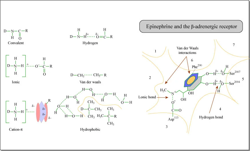
Long description
Types of Chemical Bonds in Ligand-Receptor Interactions. Format Diagram Credit MIT OCW. MIT OpenCourseWare Course of Origin 20.201 Mechanisms of Drug Actions, Fall 2005 MIT Course Instructor Dedon, Peter Tannenbaum, Stev

Drug interactions occur when one drug alters the effect of another, and understanding them requires a clear grasp of how drugs act at receptors. At its core, pharmacology rests on the idea that most drugs produce their effects by binding to specific molecular targets—usually receptors, enzymes, or ion channels. When two or more drugs are given together, they can influence each other's actions in predictable ways, and these patterns are classified into several distinct types.
The autonomic nervous system (ANS) is a particularly important arena for drug interactions because it controls involuntary functions such as heart rate, blood pressure, digestion, and glandular secretion. The ANS has two main divisions: the sympathetic ("fight or flight") and parasympathetic ("rest and digest") branches. Each division uses specific neurotransmitters—noradrenaline for most sympathetic postganglionic fibres and acetylcholine for parasympathetic fibres—that act on distinct families of receptors.
Autonomic receptors fall into two broad structural classes. The first are ligand-gated ion channels, which are fast-acting receptors that directly open an ion pore when the neurotransmitter binds. The second are G protein-coupled receptors (GPCRs), which are slower and work through intracellular signalling cascades. The P2Y receptors illustrate this well: they are GPCRs with seven transmembrane domains, an extracellular N terminus, and an intracellular C terminus. P2Y1, P2Y2, P2Y4, and P2Y6 receptors couple positively to phospholipase C via Gq/11 proteins, while P2Y12–14 receptors couple negatively to adenylyl cyclase via Gi proteins; P2Y11 receptors are dually coupled to both Gq/11 and Gs. [2]
DefinitionReceptor types
Autonomic receptors are either ionotropic (ligand-gated ion channels, fast) or metabotropic (GPCRs, slower, using second messengers). Both are targets for drug interactions.
The clinical relevance of these distinctions is profound. A drug that blocks a receptor (an antagonist) can reverse the effect of an agonist, while two drugs acting through different receptors on the same cell may produce effects that are greater than the sum of their parts. Understanding these principles allows prescribers to anticipate and avoid harmful combinations, or to exploit beneficial ones.
Inter-individual drug response (IVDR) is a complex, multifactorial phenomenon where patients exhibit varying sensitivities to pharmacological interventions. Understanding these variations is essential for precision medicine, as it allows clinicians to optimise drug therapy and minimise the risk of adverse drug reactions (ADRs). [6]
DefinitionIVDR
Inter-individual drug response (IVDR) refers to the variability in how different patients respond to the same pharmacological treatment, influenced by a combination of genetic, environmental, and physiological factors.
The factors contributing to IVDR are diverse and often overlapping:
- Genetics: Polymorphisms in genes encoding drug-metabolizing enzymes, transporters, and pharmacological targets directly alter drug metabolism and sensitivity. The gut microbiota can develop resistance to drugs over time, making them less effective in treating infections. [6]
- Disease state: Underlying conditions can alter organ function, drug receptor expression, or metabolic pathways.
- Drug-drug interactions: The concomitant use of multiple medications can alter the absorption, distribution, metabolism, and excretion (ADME) of drugs.
- Environmental factors: Exposure to toxins, pollutants, diet, smoking, and exercise can modulate how drugs are processed.
- Pharmacomicrobiomics: This field describes the interactions between gut microbiota and drug response. Microbiota can influence PK and pharmacodynamics (PD) through several mechanisms: [6]
- Metabolic alteration: Microbiota can produce enzymes, such as beta-lactamases, that degrade drugs like beta-lactam antibiotics, rendering them ineffective.
- Target modification: Microbiota may modify drug targets in the host, reducing susceptibility to pharmacological effects.
- Resistance induction: Over time, microbiota can develop resistance or acquire resistance genes, impacting the efficacy of antimicrobial treatments.
| Factor | Primary Mechanism |
|---|---|
| Genetics | Variations in enzymes, transporters, and targets |
| Age/Gender | Differences in body composition, hormones, and organ function |
| Microbiota | Alteration of drug metabolism and target modification |
| ADME | Drug-drug interactions affecting absorption and clearance |
Research into pharmacomicrobiomics often integrates systems-level data, including pharmacometabolomics and pharmacometabonomics, to study the metabolites generated by host-microbiota interactions. For instance, in prostate cancer treatment, the drug abiraterone (ABI) has been shown to remodel gut microbiota, depleting certain species while promoting the proliferation of anti-inflammatory bacteria like A. muciniphila, which subsequently elevates vitamin K2 production.
Clinical pearlMicrobiota and efficacy
The gut microbiota can directly influence drug efficacy by producing enzymes that metabolise drugs, such as the production of beta-lactamases that break down beta-lactam antibiotics. [6]
- Synergy is defined as a combination effect greater than Loewe additivity – the drug-with-itself expectation. [1]
- The Combination Index at 50% effect is CI₅₀ = (Cₓ/IC₅₀ₓ) + (Cᵧ/IC₅₀ᵧ); CI < 1 = synergy, CI = 1 = additivity, CI > 1 = antagonism.
- If a single agent cannot reach the chosen effect level, set its effective concentration to the top tested dose; all CI values become upper limits. [1]
- The CI is best suited for fixed dose-ratio combination curves.
- Synergy scores like "S" have no natural scale and are only useful for relative comparisons within a screen.
- Group data are reported as mean ± s.e.m., with two-tailed t-tests used routinely.
- In pancreatic cancer, perineural invasion occurs in approximately 80–100% of patients and correlates with poor survival.
- The tumour–nerve interaction involves CAFs, TAMs, and PSCs, forming a vicious circle that drives malignant behaviour.
Answer each one in your head first, then open the answer.
What is the Loewe additivity baseline and why is it important for defining synergy?
Show answerHide answer
It is the expected response if a drug were combined with itself at equivalent doses; synergy is defined as a combination effect greater than this additive expectation.How do you calculate the Combination Index at 50% effect and what does a value of 0.8 mean?
Show answerHide answer
CI₅₀ = (Cₓ/IC₅₀ₓ) + (Cᵧ/IC₅₀ᵧ), where Cₓ and Cᵧ are the concentrations in the 50%-effective mixture and IC₅₀ values are from single-agent curves. A value of 0.8 indicates synergy (CI < 1).What happens to the CI if one drug alone cannot reach 50% effect at any tested dose?
Show answerHide answer
The effective concentration is set to the top tested dose, and all CI values become upper limits rather than exact values.Why is the CI not reliable when the dose ratio of the two drugs varies across the experiment?
Show answerHide answer
The CI is best suited for analysing fixed dose-ratio combination curves; varying ratios make the CI difficult to interpret.What is the clinical significance of perineural invasion in pancreatic cancer?
Show answerHide answer
It occurs in approximately 80–100% of pancreatic cancer patients and is correlated with poor survival and decreased quality of life.
Additive, permissive, synergistic, potentiation, antagonism, tachyphylaxis, autonomic receptor types
Additive interaction occurs when two drugs with the same effect produce a combined response the same as adding their separate effects together. For example, combining two antihypertensive drugs that lower blood pressure through different mechanisms often produces an additive reduction. This is the simplest type of interaction and is generally predictable if the dose–response curves of both drugs are known.
Permissive interaction describes a situation where one drug is required for another to exert its full effect, even though the first drug alone may have little observable action. A classic example is the permissive role of glucocorticoids in allowing catecholamines to produce their full bronchodilator and vasopressor effects. Without the permissive hormone, the second drug's response is blunted.
Synergistic interaction (sometimes called supra-additive) occurs when the combined effect of two drugs is more than their separate effects added together. This is more than simple addition—the drugs amplify each other. For instance, combining a sedative with an opioid analgesic can produce respiratory depression far greater than either drug alone would predict. Synergy can be dangerous but is also exploited therapeutically, as when combining antibiotics that attack different steps of a bacterial pathway.
Potentiation is a specific form of synergy where one drug has little or no effect of its own but enhances the effect of another drug. The classic example is probenecid, which has minimal direct pharmacological action but potentiates penicillin by blocking its renal tubular secretion, thereby raising penicillin concentrations. Potentiation can also occur at the receptor level, where one drug increases the affinity or efficacy of another at its receptor.
Antagonism is the opposite of synergy: one drug opposes or blocks the effect of another. There are several subtypes. Competitive antagonism occurs when an antagonist competes with an agonist for the same binding site; increasing the agonist concentration can overcome it. Non-competitive antagonism occurs when the antagonist binds elsewhere (an allosteric site) or irreversibly, so that increasing agonist concentration cannot fully overcome the block. Functional (physiological) antagonism occurs when two drugs produce opposite effects through different receptors or pathways—for example, a vasoconstrictor opposing a vasodilator. The concept extends beyond pharmacology: functional antagonism of receptor systems has been observed following deletion of a metabolic enzyme, including reduced activity of nicotinic receptors in acetylcholinesterase−/− mice and impairments in 5-HT1A receptors in monoamine oxidase A−/− mice. [5]
Exam trapCompetitive vs non-competitive
Competitive antagonism is surmountable by raising agonist concentration; non-competitive antagonism is not. This distinction matters clinically when choosing reversal strategies.
Tachyphylaxis is the rapid development of tolerance—a diminishing response to a drug after repeated or continuous administration over a short period. It occurs when receptors are desensitised or internalised quickly. Chronic inactivation of an enzyme that degrades a lipid transmitter can lead to receptor downregulation and desensitisation in the nervous system, demonstrating that both classical neurotransmitters and lipid messengers are capable of causing tolerance following chronic inactivation of their cognate degradative enzymes. Tachyphylaxis is common with drugs acting on GPCRs, such as β-agonists used in asthma, and limits their long-term efficacy. [5]
| Interaction type | Definition | Example |
|---|---|---|
| Additive | Combined effect = sum of individual effects | Two antihypertensives lowering BP |
| Permissive | One drug required for another's full effect | Glucocorticoids permitting catecholamine action |
| Synergistic | Combined effect > sum of individual effects | Sedative + opioid causing profound respiratory depression |
| Potentiation | One drug enhances another's effect with little own effect | Probenecid potentiating penicillin |
| Antagonism | One drug opposes another's effect | β-blocker opposing β-agonist |
| Tachyphylaxis | Rapid tolerance after repeated dosing | β-agonist desensitisation in asthma |
Autonomic receptor types are best understood by their neurotransmitter and signalling pathway. The sympathetic system acts mainly through adrenergic receptors (α1, α2, β1, β2, β3), all GPCRs. The parasympathetic system acts through muscarinic receptors (M1–M5), also GPCRs, and nicotinic receptors, which are ligand-gated ion channels. Within the GPCR family, the P2Y receptors illustrate the diversity of signalling: P2Y1, P2Y2, P2Y4, and P2Y6 couple to phospholipase C via Gq/11, while P2Y12–14 couple to adenylyl cyclase inhibition via Gi. This means that drugs acting at different subtypes can have opposing effects on the same second messenger pathway, setting the stage for functional antagonism. [2]
The clinical relevance of receptor subtypes is illustrated by the P2Y receptors in pain pathways. P2Y1 and P2Y2 receptors are typically expressed in small, nociceptive cells, whereas the P2Y4 subtype seems to be present in large neurons involved in the transmission of tactile allodynia. This differential distribution means that a drug targeting P2Y4 might relieve allodynia without affecting acute pain signalling—a form of receptor-selective therapy. [2]
Approach to analysing a drug interaction
- Identify the receptors involvedDetermine which receptor types each drug acts on (e.g., adrenergic, muscarinic, P2Y).
- Question: Do both drugs act at the same receptor?
Yes
- Assess competitive vs non-competitive antagonismCan increasing agonist overcome the block?
No
- Assess downstream signalling convergenceDo they share second messengers (e.g., cAMP, Ca2+)?
- Question: Do they converge on the same pathway?
Yes
- Consider synergy or functional antagonismSame direction = synergy; opposite = functional antagonism.
No
- Consider additive or permissive effectsIndependent pathways may still summate.
A practical approach to any drug interaction is to ask three questions. First, which receptors are involved, and are they the same or different? Second, do the drugs converge on the same intracellular signalling pathway? Third, is the interaction desirable (therapeutic synergy) or harmful (toxic potentiation)? Answering these questions allows the prescriber to predict whether a combination will be additive, synergistic, antagonistic, or permissive.
Clinical pearlClinical pearl
When adding a second drug, always ask: does it act at the same receptor (competition), the same pathway (synergy or functional antagonism), or an independent pathway (additive)? This framework prevents most prescribing errors.
G-protein coupled receptors (GPCRs) are central to autonomic signalling, where ligand binding triggers intracellular cascades. A key mechanism involves the activation of phospholipase C (PLC), which hydrolyses phosphatidylinositol 4,5-bisphosphate (PIP2) to generate inositol 1,4,5-trisphosphate (IP3) and diacylglycerol (DAG). IP3 binds to receptors on the endoplasmic reticulum, causing the release of stored calcium ions into the cytoplasm. This rise in intracellular calcium ([Ca2+]i) acts as a critical second messenger, amplifying the signal for cellular responses such as neurotransmitter release or enzyme activation. The involvement of these pathways is confirmed by pharmacological studies; for instance, UTP-induced calcium responses in dorsal horn astrocytes were shown to be sensitive to thapsigargin and pertussis toxin, with antagonism by PPADS and suramin, demonstrating the functional expression of P2Y2 receptors linked via G protein coupling to the release of [Ca2+]i via the phospholipase C (PLC)/IP3 pathway. [2]
DefinitionPLC/IP3 Pathway
The G-protein coupled activation of phospholipase C leads to the production of IP3, which triggers calcium release from intracellular stores, serving as a primary second messenger system.
The specificity of these interactions can be modulated by selective antagonists. For example, 2-MeSAMP selectively blocks the P2Y12 receptor-type, while ARC69931MX acts as a selective antagonist at P2Y12 and P2Y13 receptors. Understanding these selectivities is vital for distinguishing between different GPCR subtypes in complex tissues. In sensory neurons, ATP increases [Ca2+]i in 30% of dorsal horn neurons via P2Y receptor activation, a finding derived from the lack of effect by the selective P [2]
| Agent | Selectivity |
|---|---|
| 2-MeSAMP | Selectively blocks P2Y12 |
| ARC69931MX | Selective antagonist at P2Y12 and P2Y13 |
| Suramin | Blocks P2Y2 but not P2Y4 |
The therapeutic implications of GPCR interactions are evident in multi-target drug strategies. The anti-inflammatory synergy between prednisolone and nortriptyline illustrates how coordinated activity on primary targets can enhance therapeutic selectivity. Prednisolone activates glucocorticoid receptors, while nortriptyline inhibits autocrine pathways via norepinephrine transporters and beta-adrenergic (ADRB2) receptors. Because ADRB2 receptors are more highly expressed in lymphocytes than in the liver and pituitary, this synergy increases the therapeutic window over major glucocorticoid toxicities. This example underscores how understanding the distribution and coupling of autonomic receptors allows for the optimization of drug combinations to minimize side effects while maximizing efficacy. [1]
GPCR Signalling Cascade
- Ligand binds GPCRe.g., ATP or UTP
- G-protein activationAlpha subunit dissociates
- PLC activationHydrolyses PIP2
- IP3 and DAG productionSecond messengers generated
- Ca2+ releaseIP3 opens ER channels
In the context of pain modulation, P2Y2 receptors are crucial for nociception through TRPV1 channels. ATP-induced thermal hyperalgesia is abolished in mice lacking TRPV1, whereas it is preserved in P2Y1 receptor-deficient mice, indicating the specific role of P2Y2. Furthermore, suramin, which blocks P2Y2 but not P2Y4, abolished the effect of UTP on TRPV1 potentiation. These findings demonstrate that G-protein coupled signalling directly influences ion channel function, linking autonomic receptor activation to sensory processing. The complexity of these pathways is further highlighted by the role of autonomic nerves in pancreatic cancer, where their interaction with tumor cells plays an irreplaceable role in disease development. While the exact functions of autonomic neurotransmitters in the tumor microenvironment require further study, the foundational G-protein mechanisms remain consistent across diverse physiological and pathological contexts. [2] [4]
Pain transmission begins at peripheral nociceptors—specialised sensory nerve endings that detect noxious stimuli. In the skin, P2Y2 receptors on the terminals of capsaicin-sensitive, P2X3-expressing cutaneous sensory neurons effectively stimulate nociceptive transmission. These receptors respond to ATP released from damaged cells, triggering depolarisation and action potential generation. [2]
At the level of the dorsal root ganglion (DRG), ATP can excite neurons by inhibiting the M-type potassium current. The M-current is a low-threshold, non-inactivating, voltage-sensitive K+ current that is weak or undetectable at resting membrane potential and increases with membrane depolarisation, inducing spike frequency adaptation and acting as a dynamic brake on neuronal excitability. When P2Y receptors inhibit this current, the brake is released, promoting depolarisation and action potential firing. This effect involves G protein-mediated reactions, as intracellularly applied GDP-β-S, an inhibitor of G protein-mediated reactions, prevented the effect of ATP. Since agonists of M-currents have been shown to be antinociceptive, P2Y receptors inhibiting these currents may stimulate pain transmission. [2]
Within the spinal cord dorsal horn, the central terminals of DRG neurons release glutamate and substance P to transmit signals to second-order neurons. This release is tightly regulated by voltage-activated calcium channels (VACCs). P2Y1 receptors at the central terminal inhibit N-type calcium channels, which play a major role in neurotransmitter release, thereby decreasing sensory transmitter (glutamate) release and reducing spinal pain transmission. The inhibition is mediated through the βγ subunit of the G protein, since a strongly depolarising pre-pulse known to dissociate Gβγ from the VACC prevented the inhibitory effect of ATP. Interestingly, in vivo studies show that intrathecal administration of the P2Y2/4 agonist UTP and the P2Y1 agonist ADP-β-S inhibits pain transmission, suggesting P2Y receptors inhibit glutamate release via VACC inhibition. [2]
Clinical pearlP2Y receptors: a double-edged sword
P2Y receptors can both promote pain transmission (by inhibiting M-currents in DRG neurons) and inhibit it (by reducing glutamate release at central terminals). The net effect depends on receptor subtype and location.
Glial cells in the spinal cord also shape pain transmission. These cells become activated in response to substances released by primary afferent terminals (ATP, SP, CGRP, and glutamate) and by spinal cord neurons (nitric oxide, prostaglandins). ATP-induced activation of glial cells is mediated by both P2X and P2Y receptors. Once activated, glial cells release neurotransmitters and neuromodulators including ATP, which can further stimulate neighbouring glial cells and neurons, altering pain transmission—both by enhancing SP and glutamate release from central terminals and by increasing excitability of spinal cord neurons. [2]
NoteGlial cells vary by condition
The role of glial cells in pain transmission is context-dependent. For example, Schwann cells promote pain through NOX1-dependent hydrogen peroxide release in sciatic nerve injury models, yet IL-6 secretion by Schwann cells in pancreatic cancer is linked to decreased abdominal mechanical sensitivity. [4]
In pathological states such as pancreatic ductal adenocarcinoma, pain transmission is amplified. Abdominal pain occurs in approximately 80% of PDAC patients. Neurogenic inflammation begins early—intrapancreatic neurogenic inflammation is present as early as PanIN2 stage. DRG axons release substance P and CGRP by up-regulating TRPV1, mediating neurogenic inflammation and stimulating nociceptive nerve fibres; the action potential then travels along the spinal cord, activates secondary neurons, and transmits pain signals to the brain. Central sensitisation may contribute to intractable pain that resists even aggressive interventions. [4]
Pain transmission pathway
- Noxious stimulusTissue damage releases ATP
- Peripheral nociceptor activationP2Y2 receptors on capsaicin-sensitive, P2X3-expressing terminals
- DRG neuron excitationP2Y receptors inhibit M-current → depolarisation
- Question: Central terminal release?
P2Y1 activation
- N-type Ca²⁺ chan
Micturition is the process by which the bladder stores and periodically voids urine, and it depends on a finely balanced interplay between the autonomic nervous system, somatic motor neurons, and local sensory signalling. The bladder wall (detrusor muscle) receives parasympathetic innervation via the pelvic nerves, where acetylcholine acts on muscarinic M3 receptors to trigger contraction during voiding. In contrast, sympathetic innervation via the hypogastric nerves releases noradrenaline, which activates β-adrenergic receptors to relax the detrusor and promote urine storage, while α-adrenergic receptors at the bladder neck and urethra increase outlet resistance. Somatic control of the external urethral sphincter is mediated by the pudendal nerve through nicotinic acetylcholine receptors at the neuromuscular junction.
Sensory feedback is critical to coordinating this cycle. Stretch receptors in the bladder wall signal fullness, and purinergic signalling contributes to this sensation. ATP and ADP act on P2Y receptors, which are expressed across sensory neurons: immunostaining studies show that P2Y1 receptor antibodies stained over 80% of the DRG, NG and TG neurons[1]. These receptors are functionally relevant, as ATP, ADP and UTP were shown to inhibit voltage-activated calcium channels (VACCs) in cultured small-diameter (20–35 µM) DRG neurons from the rat, indicating the functional presence of P2Y1, P2Y2 and/or P2Y4 receptors on these cells[1]. This inhibition of calcium channels reduces neurotransmitter release, modulating afferent input. [2]
DefinitionP2Y receptors
P2Y receptors are G protein-coupled receptors activated by extracellular nucleotides such as ATP, ADP and UTP; they modulate neuronal excitability and calcium signalling in sensory pathways.
The functional consequences of P2Y activation are notable. UTP evoked a slow-onset depolarization and sustained firing in dissociated DRG neurons from the rat that persisted for tens of seconds after removal of UTP from the medium[1]. In a subsequent study, UTP activated sustained action potential firing in 50% of C fibers using the skin nerve preparation from the adult mouse[1]. Interestingly, UDP had no effect, pharmacologically excluding the involvement of P2Y6 receptors[1]. The majority (70%–80%) of UTP-sensitive C- and AM fibers respond strongly to capsaicin, whereas UTP-insensitive fibers were largely capsaicin-insensitive[1]. Most of the UTP-sensitive C- and AM fibers (80%–100%) also responded to the P2X agonist α,β-meATP, indicating that P2Y and P2X receptors are widely co-expressed on these fibers[1]. [2]
Clinical pearlP2Y and P2X co-expression
Because P2Y and P2X receptors are co-expressed on most sensory fibers, drugs targeting one receptor type may have overlapping or synergistic effects on bladder sensation.
Therapeutic implications arise from modulating these pathways. The ADP analog ADP-β-S, applied intrathecally to rats, prolonged the nociceptive threshold in the tail-flick test, indicating analgesia[1]. Mechanistically, ATP was found to inhibit the N-type VACCs and the subsequent increase in [Ca2+]i in rat DRG neurons, with no other Ca2+ channel types altered[1]. This effect of ATP was antagonized by the P2Y1 selective antagonists MRS 2179 and PPADS and was abolished by intra [2]
Exam trapSelectivity in vivo
A drug may be molecularly selective in vitro, but in vivo it may not be therapeutically selective, either because of undesirable activity at effective doses or because the biological system responds to compensate. [1]
This principle matters clinically: achieving therapeutic selectivity has been a major obstacle in drug discovery, and overcoming compensatory mechanisms often requires high drug doses that can induce unwanted effects in other tissues. For micturition, this means that targeting a single receptor may be insufficient; combination strategies may offer better control, though synergistic side-effects remain a concern. [1]
| Receptor | Location | Action | Effect on micturition |
|---|---|---|---|
| Muscarinic M3 | Detrusor | Contraction | Promotes voiding |
| β-adrenergic | Detrusor | Relaxation | Promotes storage |
| α-adrenergic | Bladder neck/urethra | Contraction | Increases outlet resistance |
| Nicotinic | External sphincter | Contraction | Maintains continence |
| P2Y1 | Sensory neurons | Inhibits VACCs | Modulates afferent signalling |
Micturition reflex pathway
- Bladder fillingStretch receptors activated
- Question: Afferent signal sufficient?
No
- Storage continuesSympathetic tone dominant
Yes
- Parasympathetic activationACh on M3 receptors
- Sphincter relaxationSomatic inhibition
- VoidingDetrusor contraction
Drug interactions and autonomic receptor dysfunction converge in several clinical diseases where the balance between sympathetic and parasympathetic tone is disturbed, and where polypharmacy creates predictable adverse effects.
Pancreatic cancer and autonomic innervation
Pancreatic ductal adenocarcinoma (PDAC) is notable for perineural invasion (PNI), which correlates with poor prognosis. The tumour microenvironment contains both sensory and autonomic nerves. Interestingly, parasympathetic nerves exert opposite effects depending on tumour type: unlike in prostate cancer, parasympathetic signalling inhibits Kras-mutant pancreatic cancer. Enhanced cholinergic signals in PDAC directly suppress MAPK/EGFR and PI3K/AKT pathways via type 1 muscarinic receptors (CHRM1), and also inhibit tumour stem cell transformation. This aligns with clinical observations suggesting an antitumour effect of the vagus nerve. [4]
For medical students, this illustrates a key principle: autonomic receptor activation is tissue- and context-specific. A drug that blocks muscarinic receptors (e.g., atropine, or anticholinergics used for bladder overactivity) could theoretically interfere with this protective cholinergic signalling in PDAC patients—an interaction worth considering in palliative care.
Cancer-associated pain and analgesic drug interactions
Pain in PDAC significantly impairs quality of life. The widely recommended analgesia approaches include systematic analgesic medications and celiac plexus/ganglia neurolysis. Neurolysis is reserved for when analgesic therapy is ineffective or produces unacceptable adverse effects. This is a classic example of a non-pharmacological intervention used to circumvent drug-related toxicity—an important concept when considering drug interactions, as opioid-related side effects often limit dose escalation. [4]
Endocannabinoid system and analgesic tolerance
The endocannabinoid system offers a clinically relevant model of receptor adaptation. Acute blockade of monoacylglycerol lipase (MAGL), which elevates 2-arachidonoylglycerol (2-AG), produces analgesia; however, sustained MAGL inactivation leads to loss of this effect—a form of tolerance. In contrast, sustained pharmacological disruption of fatty acid amide hydrolase (FAAH), which elevates anandamide, produces persistent analgesia without tolerance or changes in CB1 receptor expression or function. [5]
Clinical pearlReceptor adaptation differs by ligand
Brain CB1 receptors adapt differently to sustained elevations of the two principal endocannabinoids: tolerance develops to 2-AG elevation (MAGL inhibition) but not to anandamide elevation (FAAH inhibition). [5]
This has therapeutic implications: FAAH inhibitors may offer sustained analgesia with low dependence risk, whereas MAGL inhibitors risk tolerance and withdrawal. For clinical practice, this explains why some cannabinoid-based analgesics lose efficacy over time while others remain stable—a drug interaction at the receptor level that affects dosing strategies. [5]
Combination therapy selectivity and toxicity
Combination therapies are increasingly favoured because greater selectivity is anticipated. However, synergistic therapies are often mirrored by synergistic toxicity when side-effect mechanisms overlap with efficacy pathways. Large-scale studies show a statistical bias towards greater selectivity for synergies, meaning combinations are more specific to particular cellular phenotypes than single drugs. This specificity narrows further as combination order increases, limited by biological complexity. [1]
Exam trapSynergy cuts both ways
Do not assume that combining drugs with synergistic efficacy automatically improves the therapeutic index—if the toxicity mechanism is closely related to the efficacy mechanism, synergistic toxicity is likely.
For autonomic pharmacology, this matters when combining drugs that affect blood pressure, heart rate, or bronchial tone. For example, combining a beta-blocker with a non-dihydropyridine calcium channel blocker for hypertension can produce synergistic bradycardia and heart block—a predictable interaction where the toxicity mechanism (negative chronotropy) mirrors the efficacy mechanism.
Investigating drug interactions at autonomic receptors requires a combination of functional assays, receptor localisation studies, and careful pharmacological dissection. The choice of investigative tool depends on whether you are asking where a receptor is expressed, what it does when activated, or how drugs modify its signalling.
Cell viability and ATP-based assays
A cornerstone of drug interaction studies is the measurement of cell viability, particularly when assessing whether a drug combination is selectively toxic to pathogens or cancer cells rather than to host tissues. The ATPlite assay quantifies cellular ATP levels: cells are incubated with ATPlite reagent for 10 minutes before data collection with a luminometer. Higher ATP levels correlate with greater cellular viability. This approach allows researchers to compare host cell viability in the presence of virus and drug treatment against uninfected cells, providing a quantitative measure of drug safety. [1]
This technique has revealed therapeutically-aligned selective combinations—pairs of drugs whose synergy against a target does not come at the cost of increased host toxicity. For example, the antibacterial synergy between ribavirin (a metabolism inhibitor) and disulfiram (used for alcohol avoidance) has almost no effect on human cell viability. Similarly, the antiviral synergy of cepharanthine and benzamil shows no detectable synergy against host cell viability, and the anti-inflammatory synergy between prednisolone and nortriptyline against TNF-α secretion shows no corresponding increase in toxic effects as measured by PBMC metabolic viability. In each case, the synergy increases the likely safe treatment window—a key goal when combining drugs that act on autonomic or other receptor systems. [1]
Clinical pearlWhy viability matters
A drug combination that enhances efficacy but also enhances host cell death has a narrowed therapeutic window. ATP-based viability assays let you distinguish true synergy from additive toxicity.
Receptor localisation and transport studies
Immunohistochemistry (IHC) can demonstrate where autonomic and sensory receptors are physically located. For instance, P2Y1 receptor immunoreactivity is evenly distributed throughout the cytoplasm of cells and randomly distributed throughout ganglia. A powerful technique is nerve ligation: ligating the vagus nerve leads to accumulation of staining adjacent to the ligature—central to the proximal ligature and peripheral to the distal ligature—indicating anterograde and retrograde transport respectively. This implies that P2Y1 receptors are transported to and expressed functionally on sensory nerve terminals. [2]
Exam trapLocalisation ≠ function
Demonstrating receptor protein in a tissue does not prove it is functional there. Functional studies—such as calcium imaging or electrophysiology—are required to confirm that the receptor responds to agonists.
Electrophysiological and calcium imaging approaches
In spinal cord slices, recordings are performed in a submersion chamber superperfused with artificial cerebrospinal fluid saturated with 95% O₂/5% CO₂, containing 125 mM NaCl, 2.5 mM KCl, 1.25 mM NaH₂PO₄, 26 mM NaHCO₃ and 12 mM glucose at pH 7.4[1]. Where necessary, 50 μM picrotoxin is added to block GABA_A receptors, and a cut between CA3 and CA1 suppresses epileptiform activity[1]. These conditions allow clean measurement of synaptic responses. [3]
Calcium imaging in cultured microglia can identify receptor subtypes: application of ATP and 2-MeSATP in Ca²⁺-free solution induced an increase in [Ca²⁺]ᵢ, whereas the P2X₁,₃,₄-selective β,γ-MeATP had no effect. This suggests a P2Y receptor mediates the response, since P2X receptors typically require extracellular calcium. Similarly, in acutely isolated neurons that lost their dendrites, there was no P2Y response, and PPADS blocked the effect of 2-MeSATP—pharmacological fingerprinting that distinguishes receptor subtypes. [2]
Pharmacological dissection of receptor subtypes
The P2Y receptor family illustrates how agonists and antagonists discriminate subtypes. P2Y₁, P2Y₁₁, P2Y₁₂ and P2Y₁₃ are sensitive to ADP and ATP; P2Y₄ and P2Y₆ respond to UDP and UTP; P2Y₂ responds to both ATP and UTP; and P2Y₁₄ responds to uridine sugars such as UDP-glucose. In sensory neurons, ATP and UTP—but not 2-MeSATP or α,β-meATP—augmented capsaicin-evoked release of substance P and CGRP. This effect was antagonised by suramin but not PPADS, and inhibited by the PKC inhibitor bisindolylmaleimide, indicating ATP acts at P2Y receptors to sensitise neurons in a PKC-dependent manner. [2]
| Receptor | Preferred agonists |
|---|---|
| P2Y₁, P2Y₁₁, P2Y₁₂, P2Y₁₃ | ADP, ATP |
| P2Y₄, P2Y₆ | UDP, UTP |
| P2Y₂ | ATP and UTP |
| P2Y₁₄ | UDP-glucose, UDP-galactose |
Behavioural correlates
Intrathecal administration of UTP and UDP produced antinociceptive effects on acute mechanical and thermal nociception in normal rats via P2Y₂, P2Y₄ and P2Y₆ receptors. Crucially, UTP and UDP caused no motor deficit in the inclined plane test, confirming the effect was antinociceptive rather than due to motor impairment. This illustrates how behavioural assays complement molecular studies to establish functional relevance. [2]
Investigating a drug interaction at a receptor
- Choose assay systemCell line, primary culture, or slice preparation
- Question: Question: localisation or function?
Localisation
- ImmunohistochemistryAssess distribution; ligation studies for axonal transport
Function
- Viability or calcium assayATPlite for toxicity; Ca²⁺ imaging for receptor activation
- Pharmacological fingerprintAgonist selectivity + antagonist blockade (e.g. suramin vs PPADS)
- Confirm in vivoBehavioural assay; rule out motor confounds
Drug interactions in the autonomic nervous system arise when two or more agents act on the same receptors, enzymes, or signalling pathways, producing effects that differ from what each would do alone. Understanding these interactions is clinically essential because patients frequently take multiple medications that target autonomic receptors, and the resulting synergy or antagonism can be exploited therapeutically or cause harm.
DefinitionDrug interaction
A drug interaction occurs when the effect of one drug is altered by the presence of another, through pharmacokinetic (absorption, distribution, metabolism, excretion) or pharmacodynamic (receptor-level) mechanisms.
When drugs act on multiple targets, the combined effect can be greater than expected from each drug alone. Analyses of drug combinations from thirteen phenotypic screens relevant to six disease areas show that synergies operate in more narrow biological contexts than single drugs. This multi-target cooperativity underlies much of the therapeutic selectivity seen in combination therapy. For example, a drug that blocks both muscarinic and nicotinic receptors may produce broader autonomic blockade than a selective agent, but also greater risk of adverse effects. [1]
The experimental basis for this comes from thirteen sets of combination data comprising 94,110 dose matrices, drawn from screens in six different disease areas. These screens varied in design—number of drugs, aspect ratio of drug lists, sampling density, and choice of assays—as well as in the agents themselves. Some screens compared disease-related phenotypes to models of healthy cell viability, testing for therapeutic selectivity, while others investigated mechanistic selectivity, where differential response profiles across phenotypes highlight particular mechanisms. [1]
Clinical pearlClinical pearl
Combination therapy targeting different autonomic receptor subtypes can achieve therapeutic selectivity—killing or modifying the diseased pathway while sparing healthy tissue—but only when the synergy is context-specific, not when drugs simply add their effects everywhere.
A useful illustration comes from the endocannabinoid system, which modulates autonomic and pain pathways. Chronic blockade of monoacylglycerol lipase (MAGL) with JZL184 (40 mg/kg, i.p.) elevates 2-arachidonoylglycerol (2-AG), which tonically activates CB1 receptors. Over six days, mice treated daily with JZL184 developed marked tolerance to the antinociceptive effects of WIN55,212-2. However, when rimonabant (3 mg/kg, i.p.), a CB1 receptor antagonist, was given concurrently with JZL184, the antinociceptive responses to WIN55,212-2 were close in magnitude to those in control animals. Rimonabant treatment (10 mg/kg, i.p.) also ameliorated brain CB1 receptor adaptations in chronically JZL184-treated mice, as judged by CP55,940-stimulated [35S]-GTPγS binding. [5]
Exam trapCommon trap
Do not assume that all tolerance is receptor desensitisation. Chronic MAGL blockade produced a larger degree of cross-tolerance to CB1 agonists than genetic disruption of MAGL, suggesting that metabolic changes—such as reductions in arachidonic acid—may also contribute. [5]
The pattern of cross-tolerance is region-specific. Chronic MAGL disruption caused strong cross-tolerance to the antinociceptive and hypothermic effects of THC and WIN55,212-2, but minimal cross-tolerance to cannabinoid-induced catalepsy. This is consistent with the lack of CB1 receptor desensitisation in the caudate putamen and globus pallidus, regions associated with catalepsy. Conversely, tolerance to antinociception was attributed to significant desensitisation of CB1 receptors in the periaqueductal grey (PAG), a brain area strongly implicated in cannabinoid-induced antinociception. [5]
| Treatment | CB1 receptor function | Behavioural consequence |
|---|---|---|
| Chronic JZL184 (40 mg/kg) | Desensitisation in hippocampus, cingulate cortex, PAG | Tolerance to antinociception and hypothermia |
| Chronic JZL184 + rimonabant (3 mg/kg) | Adaptations prevented | Antinociceptive responses near control |
| Chronic PF-3845 (10 mg/kg) | No significant change in IPSC depression | No cross-tolerance to CB1 agonists |
Not all enzyme inhibitors produce the same receptor adaptations. Chronic dosing with JZL184 caused a modest elevation in anandamide (~three-fold) two hours after final treatment, likely reflecting partial blockade of FAAH. This was much lower than the 15-fold rise in brain anandamide observed in mice treated for one or six days with the selective FAAH inhibitor PF-3845 (10 mg/kg, i.p., once per day). PF-3845 did not alter brain 2-AG levels after acute or chronic treatment. Repeated in vivo administration of PF-3845 did not significantly alter CP55,940-induced depression of inhibitory postsynaptic currents in hippocampus or cingulate cortex, whereas chronic JZL184 did. The CB1 receptor antagonist AM251 (2 µM) completely blocked CP55,940-induced depression of IPSCs in both brain regions. [5]
NotePractical note
When prescribing drugs that affect autonomic or cannabinoid pathways, consider whether the patient is already taking an agent that acts on the same receptor. Concurrent use of a receptor antagonist can prevent tolerance but may also block the desired therapeutic effect.
The provided evidence does not contain data regarding specific clinical dosing, demographic thresholds, or global health disparities for drug interactions involving autonomic receptors. Consequently, a standard clinical review of special populations (such as the elderly, pediatric, or renal impairment groups) cannot be constructed from these specific passages. Instead, this section focuses on the mechanistic nuances observed in experimental models and cellular contexts that inform our understanding of receptor biology and potential interaction sites.
In experimental oncology, mechanistically selective synergies offer insights into biology relevant to specific phenotypes. For instance, a strong synergy between LY 294002 and camptothecin was observed in lung-derived H460 cells compared to colon-derived Colo-205 cells. This reveals an interaction between phosphoinositide 3 kinase and DNA topoisomerases that occurs only in some cancer contexts. Such context-dependent interactions highlight that drug responses are not uniform across all tissue types or cell lines, a critical consideration when extrapolating preclinical data to diverse patient populations.
In the study of sensory ganglia, satellite glial cells (SGCs) are closely apposed to neurons and are affected in animal models for pain. Specific P2Y receptor agonists, such as UTP, 2-MeSADP, and ADP-β-S, caused an increase in intracellular Ca2+ concentration in SGCs, indicating the functional presence of P2Y1, P2Y2, or P2Y4 receptors on these cells. This functional presence suggests that glial cells are not merely passive support structures but active participants in the autonomic and sensory pathways, potentially serving as targets for modulating pain states.
The endocannabinoid system further illustrates complex regulatory mechanisms. 2-AG acts as a full CB1 receptor agonist, whereas anandamide acts as a partial agonist. Bulk brain levels of 2-AG are much higher than anandamide, which may allow 2-AG to achieve greater occupancy of CB1 receptors in vivo. Furthermore, the enzymes MAGL and FAAH are found in different neuronal populations and subcellular compartments, specifically pre- and postsynaptic locations, respectively. These anatomical distinctions differentially affect endocannabinoid signaling pathways, suggesting that therapeutic interventions targeting these systems must account for precise cellular localization to avoid unintended effects on synaptic plasticity. [5]
NoteContext-dependent synergy
Synergistic drug interactions are not universal; they depend on the specific cellular context, such as lung versus colon cancer cell lines, revealing unique biological interactions.
The regulation of synaptic plasticity by the endocannabinoid system demonstrates opposing effects based on the duration of enzyme inhibition. Acute inhibition of MAGL, but not FAAH, potentiates depolarization-induced suppression of inhibition (DSI) in neurons of the hippocampus and cingulate cortex. However, repeated administration of the MAGL inhibitor JZL184 led to profound DSI deficits in these neuronal populations. These impairments in short-term synaptic plasticity are consistent with observed alterations in CB1 receptor function, as deletion or antagonism of this receptor has been shown to abolish DSI. This supports a model where prolonged elevations of endogenous 2-AG cause functional antagonism of CB1 receptors. [5]
In the context of chronic pain, activated microglia and astrocytes in the spinal cord can create and maintain chronic pain states. P2Y receptor agonists were found to inhibit cytokine release from activated spinal cord microglia, with a rank order of inhibitory potency: 2-MeSATP = ATP > ADP-β-S = ADP ≫ UTP > UDP. By inhibiting microglia overstimulation through the reduction of cytokine release, P2Y receptors could interrupt the development and continuation of chronic pain. This mechanism highlights a potential therapeutic avenue for managing pain in populations where chronic inflammatory states are prevalent. [2]
Regarding receptor distribution, double immunofluorescence histochemistry showed that 30% of P2Y1 receptor-immunoreactive cells also showed NF200 immunoreactivity in dorsal root ganglia, nodal ganglia, and trigeminal ganglia. NF200 stains large-diameter neurons with myelinated axons. The P2Y1 receptor was also co-localized with the neuropeptide calcitonin gene-related peptide (CGRP) in more than 60% of cells in these ganglia. Additionally, P2Y1 receptors were highly co-expressed in IB4-containing neurons, with 80%, 60%, and 20% of IB4 cells being immunoreactive for P2Y1 in dorsal root, nodal, and trigeminal ganglia, respectively. These specific co-expression patterns indicate that P2Y1 receptors are not uniformly distributed but are concentrated in specific neuronal subpopulations, which may influence the efficacy of targeted therapies in different anatomical regions. [2]
| Ganglion Type | P2Y1/NF200 Co-expression | P2Y1/CGRP Co-expression | P2Y1/IB4 Co-expression |
|---|---|---|---|
| Dorsal Root Ganglia | 30% | >60% | 80% |
| Nodal Ganglia | 30% | >60% | 60% |
| Trigeminal Ganglia | 30% | >60% | 20% |
Endocannabinoid Modulation of DSI
- Acute MAGL InhibitionPotentiates DSI in hippocampus and cingulate cortex
- Question: Chronic Administration?
Yes
- Profound DSI DeficitsFunctional antagonism of CB1 receptors
No
- Maintained PlasticityNo profound deficits observed
Drug interactions are not merely a list of pairs to memorise; they are predictable consequences of pharmacokinetic and pharmacodynamic principles. The single most useful mental model for the exam is the distinction between additivity, synergy and antagonism, and the quantitative tools used to tell them apart. When two drugs act on the same pathway, the examiner wants to know whether the combination does something more than simply giving more of one drug.
The formal definition of synergy rests on the concept of Loewe additivity – the response you would expect if a drug were simply combined with itself. If a mixture of two drugs produces a greater effect than the equivalent dose of either drug alone (i.e., more than the additive expectation), that is synergy. If it produces less, that is antagonism. The key exam point is that synergy is not just "two drugs together work well"; it is a mathematically defined excess over additivity.
The Combination Index (CI) is the workhorse calculation. For a 50% effect level, the CI₅₀ is calculated as:
CI₅₀ = (Cₓ/IC₅₀ₓ) + (Cᵧ/IC₅₀ᵧ)
where Cₓ is the concentration of drug X in the 50%-effective mixture, and IC₅₀ₓ is the concentration of drug X needed for 50% effect when given alone. The same logic applies to drug Y. A CI of 1 means the combination is exactly additive (no benefit over single-agent dose escalation). A CI below 1 indicates synergy; a CI above 1 indicates antagonism. In the exam, you may be given two drugs, their individual IC₅₀ values, and the concentrations in a mixture that achieves 50% effect – you must plug the numbers into this formula and interpret the result.
A crucial practical caveat: if a single agent never reaches the chosen effect level (e.g., it cannot achieve 50% inhibition even at the highest tested dose), then we set its effective concentration to the top tested dose, and all CI values become upper limits rather than exact values. This means you cannot conclude synergy with certainty – only that the true CI is at or below the calculated number. This is a favourite exam trap. [1]
The CI is best suited for analysing fixed dose-ratio combination curves – that is, experiments where the two drugs are combined in a constant proportion across a range of total doses. If the ratio varies from point to point, the CI becomes difficult to interpret. The isobologram is the graphical counterpart: it plots the doses of drug X and drug Y that together produce a fixed effect (e.g., 50% inhibition). The line connecting the single-agent IC₅₀ points on the axes is the line of additivity. Points below the line indicate synergy; points above indicate antagonism. [1]
One further subtlety: synergy scores such as "S" do not have a natural scale, so they are only useful for relative comparisons within a screen, not as absolute measures of strong synergy. In other words, an S value of 2 in one experiment is not directly comparable to an S value of 2 in another; it only tells you that this combination is more synergistic than another combination tested in the same batch. Do not quote an absolute "synergy threshold" for S.
When interpreting experimental data in exam vignettes, remember that group data are routinely reported as mean ± s.e.m. and statistical differences are estimated using the t-test (paired or independent), with two-tailed tests used routinely. Sample pairing is used where appropriate, e.g., when monitoring real-time changes against baseline or comparing cells in paired recordings. This matters because a drug interaction study that claims synergy must show the difference is statistically significant, not just numerically lower than 1. [3]
Clinical pearlThe CI shortcut
CI < 1 = synergy; CI = 1 = additivity; CI > 1 = antagonism. If a single drug cannot reach the effect level, its effective concentration is set to the top tested dose and the CI becomes an upper limit.
The most common mistake in drug interaction questions is confusing potency with efficacy. A combination may be more potent (lower doses needed for the same effect) without being more efficacious (higher maximum effect). The CI₅₀ specifically addresses potency at the 50% effect level. If the question asks about maximum effect, the CI is not the right tool.
A second pitfall is ignoring the Loewe additivity baseline. Students often declare "synergy" simply because the combination works better than either drug alone. That is not sufficient – the combination must work better than the drug-with-itself additive expectation. For example, if drug A alone at dose 10 gives 40% effect, and drug B alone at dose 10 gives 40% effect, an additive expectation would be roughly 80% (if effects are independent and non-overlapping). If the combination of A 10 + B 10 gives 85%, that is only slightly above additivity; if it gives 95%, that is synergy. The exam will test this distinction.
A third pitfall concerns the upper limit issue. When a single agent does not reach the chosen effect level, the CI becomes an upper limit. Students who forget this will quote a CI of 0.7 and claim synergy, when in fact the true CI could be anywhere from 0.7 down to 0 – or the combination might not even be synergistic. Always check whether both single agents achieved the effect level before interpreting the CI.
A fourth pitfall is misapplying the CI to non-fixed dose ratios. The CI is best suited for fixed dose-ratio combination curves. If the experiment used varying ratios, the CI values are not reliable. Similarly, the isobologram requires a fixed effect level; drawing conclusions across different effect levels is invalid.
A fifth pitfall is treating synergy scores as absolute. The "S" score has no natural scale and is only useful for relative comparisons within a screen. Do not compare S values across different screens or claim a combination is "strongly synergistic" based on an S value alone. [1]
A sixth pitfall, more clinical, is forgetting that drug interactions can be pharmacokinetic (affecting absorption, distribution, metabolism, excretion) rather than pharmacodynamic (affecting the receptor or pathway). The CI and isobologram address pharmacodynamic interactions at the effect site. A drug that inhibits another's metabolism may shift the dose-response curve without true synergy – the combination may appear synergistic when it is merely increasing the effective concentration of one drug.
Exam trapThe 'works better' fallacy
"Better than either drug alone" is not synergy. Synergy means better than the Loewe additive expectation – the drug-with-itself level. Always compare against additivity, not against monotherapy.
In clinical practice, the concept of synergy guides combination therapy in oncology, antimicrobial treatment, and anaesthesia. The isobologram and CI are used in preclinical drug development to decide whether two agents merit further study in combination. For example, in pancreatic cancer, where perineural invasion is identified in approximately 80–100% of patients and is correlated with poor survival and decreased quality of life, combination regimens targeting both tumour cells and the neural microenvironment are being explored. The tumour–nerve interaction involves not just cancer cells and neurons but also cancer-associated fibroblasts (CAFs), tumour-associated macrophages (TAMs), and pancreatic stellate cells (PSCs), which interact to form a vicious circle leading to malignant behaviour. When designing combinations that target multiple components, the CI helps determine whether the drugs act synergistically or merely additively. [4]
In the tumour microenvironment, Schwann cells can be activated by hypoxia, tumour cells, and T-cell-secreted factors such as IL-6, undergoing phenotypic and functional reprogramming similar to reversible dedifferentiation, with up-regulation of neurotrophins. A drug that blocks Schwann cell activation might be combined with a conventional cytotoxic agent. Before clinical use, the combination would be tested in fixed dose ratios and analysed with the CI to confirm synergy.
Neuroplasticity in pancreatic cancer – nerve hypertrophy and density changes in and adjacent to the pancreatic parenchyma – is a target for intervention. Interestingly, nerve density in the tumour centre is significantly lower than normal pancreas, contrary to traditional belief. This has implications for drug delivery: a combination that normalises the neural microenvironment might improve the penetration of cytotoxic drugs. Again, the CI would be used to assess whether the combination is synergistic.
Beyond oncology, the enriched environment paradigm shows that moderately positive stimulation can increase the infiltration and vitality of natural killer cells through the mediation of sympathetic nerves, presenting a tumour-inhibitory phenotype. This illustrates the autonomic nervous system's role in immune modulation – relevant when considering drugs that affect sympathetic tone in combination with immunotherapies. A beta-blocker combined with an immune checkpoint inhibitor might be assessed for synergy using the same CI framework.
In practical prescribing, the CI concept translates to a simple question: does adding drug B to drug A allow a dose reduction of both while maintaining effect? If yes, the combination is at least additive, possibly synergistic. If the CI is below 1, the combination is more potent than dose escalation of either drug alone. This is clinically valuable when dose-limiting toxicity prevents simply increasing the dose of a single agent.
Assessing a drug combination for synergy
- Obtain dose-response curves for drug X alone, drug Y alone, and fixed-ratio combinationsUse the same effect endpoint (e.g., 50% inhibition).
- Question: Does each single agent reach the chosen effect level (e.g., 50%)?
Yes
- Calculate CI₅₀ = (Cₓ/IC₅₀ₓ) + (Cᵧ/IC₅₀ᵧ)Use concentrations from the 50%-effective mixture.
- Question: Is CI < 1?
Yes
- SynergyCombination more potent than additivity.
No (CI ≥ 1)
- Additivity or antagonismCI = 1 is additive; CI > 1 is antagonistic.
No
- Set effective concentration to top tested doseAll CI values become upper limits.
- Interpret cautiouslyCannot confirm synergy; true CI may be lower.
In anaesthesia, the synergy between sedatives and opioids is well known; the CI framework allows anaesthetists to combine lower doses of each, reducing respiratory depression. In antimicrobial therapy, synergistic combinations (e.g., a beta-lactam with an aminoglycoside) are chosen when the CI is below 1, allowing lower doses and reduced toxicity. In all these settings, the principle is the same: the combination must beat the drug-with-itself additive level.
- Lehár J, Krueger AS, Avery W, et al. Synergistic drug combinations tend to improve therapeutically relevant selectivity. Nature biotechnology 2009;27(7):659-666. doi:10.1038/nbt.1549. Accessed .
- Gerevich Z, Illes P. P2Y receptors and pain transmission. Purinergic signalling 2004;1(1):3-10. doi:10.1007/s11302-004-4740-9. Accessed .
- Henneberger C, Papouin T, Oliet SH, Rusakov DA. Long-term potentiation depends on release of D-serine from astrocytes. Nature 2010;463(7278):232-236. doi:10.1038/nature08673. Accessed .
- Wang J, Chen Y, Li X, Zou X. Perineural Invasion and Associated Pain Transmission in Pancreatic Cancer. Cancers 2021;13(18):4594. doi:10.3390/cancers13184594. Accessed .
- Schlosburg JE, Blankman JL, Long JZ, et al. Chronic monoacylglycerol lipase blockade causes functional antagonism of the endocannabinoid system. Nature neuroscience 2010;13(9):1113-1119. doi:10.1038/nn.2616. Accessed .
- Zhao Q, Chen Y, Huang W, et al. Drug-microbiota interactions: an emerging priority for precision medicine. Signal transduction and targeted therapy 2023;8(1):386. doi:10.1038/s41392-023-01619-w. Accessed .
Figure credits
Educational content for medical students and doctors. It is not a substitute for clinical judgement or local guidelines.