Drug metabolism and elimination kinetics
How the liver chemically alters drugs and how the body clears them, using phase I and II reactions and cytochrome P450
Figure 1Hepatic drug metabolism
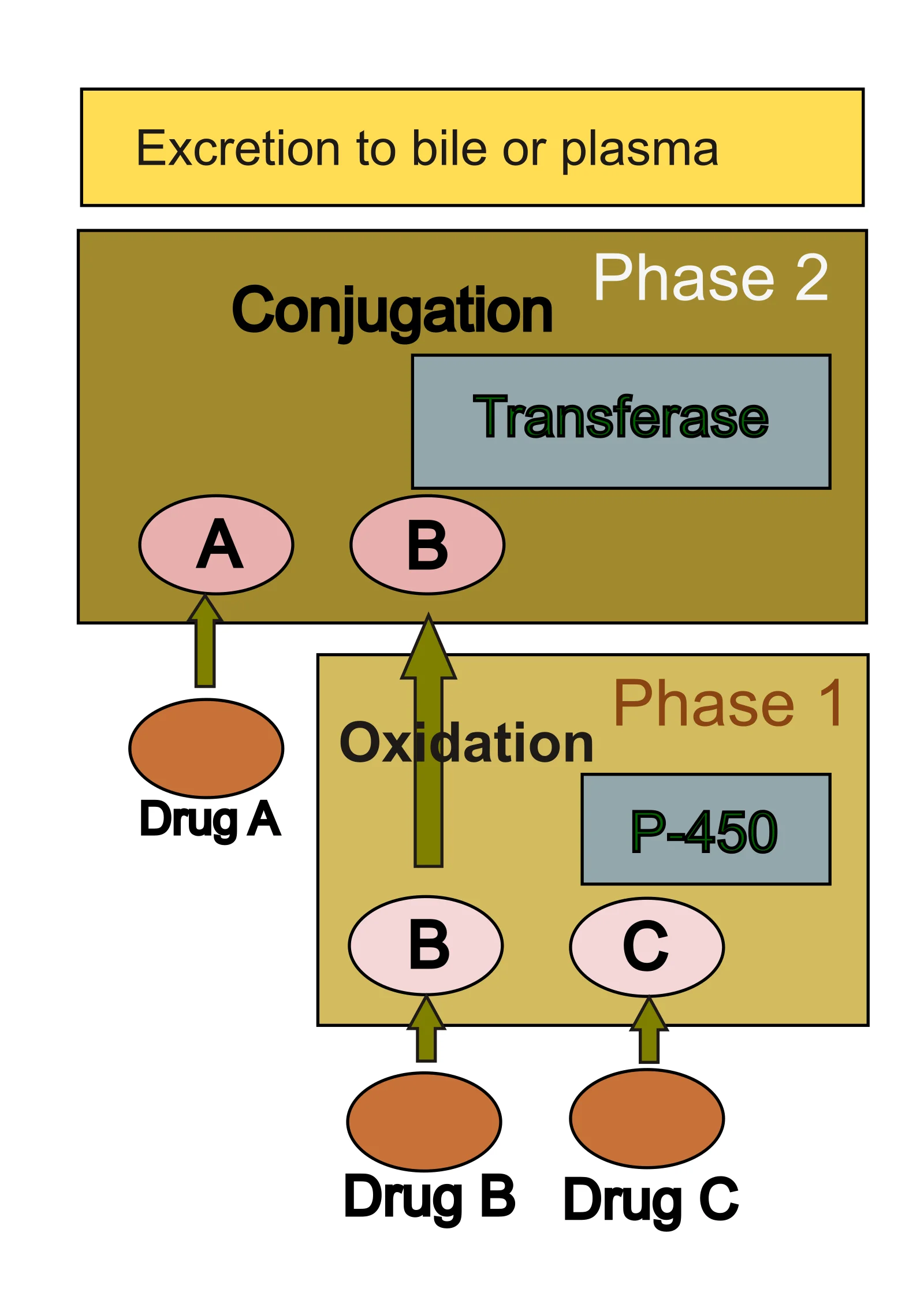
Long description
Drug metabolism in liver. transferases are : glutathione, sulfate, acetate, glucoronic acid. P-450 is cytochrome P-450 enzymes. 3 different pathways are depicted for Drugs A,B and C.

DefinitionWhy lipophilicity matters
Lipophilic drugs linger in the body because they partition into fat rather than dissolving readily in water; this prolonged retention is what creates the risk of toxicity, and it is why the body invests so heavily in converting them to water-soluble forms before excreting them[1].
Cytochrome P450 (CYP) enzymes are the workhorses of this process. They are membrane-bound hemoproteins[1], mainly expressed in the liver, although kidney, placenta, adrenal gland, gastrointestinal tract and skin also contribute[1]. The human genome encodes at least 57 CYPs, organised into 18 families and 43 subfamilies[1].
The scale of the CYP contribution is striking. Almost 80% of oxidative metabolism and approximately 50% of the overall elimination of common clinical drugs can be attributed to one or more CYPs from families 1 to 3[1]. CYPs are involved in more than 90% of the reported enzymatic reactions of drug metabolism[1], and they account for roughly 75% of drugs processed by enzymes[1]. Among the 57 putatively functional human CYPs, the CYP1, 2 and 3 isoforms are mainly responsible for the metabolism of about 80% of clinical drugs[1]. [4]
Clinical pearlInterindividual variation
Genetic polymorphisms and epigenetic changes in CYP genes, together with environmental factors, may be responsible for interindividual variation in drug response[1].
Metabolism is usually divided into phase I and phase II reactions[1], and the sections that follow set out what each accomplishes and how they are distinguished in practice.
Drug metabolism is the process of altering drug molecules chemically after they enter the body[5]. This chemical transformation is essential because the lipophilic nature of many drugs allows them to remain in the body for extended periods, which increases the risk of toxicity[5]. By converting lipophilic centers into hydrophilic centers, metabolism increases water solubility, facilitating the excretion of the drug through urine or bile[5]. This process is generally divided into phase I and phase II reactions[5].
The cytochrome P450 (CYP) superfamily of membrane-bound hemoproteins is central to these processes[5]. These enzymes are the most diverse catalysts known in biochemistry and are responsible for nearly 80% of oxidative metabolism and approximately 50% of the overall elimination of common clinical drugs[5]. Beyond their role in elimination, CYPs influence drug action, safety, bioavailability, and drug resistance[5]. These metabolic activities occur not only in primary metabolic organs like the liver but also at local sites of action[5].
Interindividual variation in drug response is a significant clinical challenge, often resulting from the interplay of genetic, epigenetic, and environmental factors[5]. Genetic polymorphisms in CYP genes can lead to substantial differences in therapeutic efficacy between ethnic groups and individuals[5]. Furthermore, environmental factors such as gender, age, nutriture, and disease states can modulate CYP activity[5].
DefinitionDDGI
DDGI is the combined effect of a genetic variant and a perpetrator drug on multiple drug metabolic pathways, which should be considered when prescribing a perpetrator drug to a patient stabilized with a victim drug[5].
Special populations require careful consideration due to altered physiological states. For example, body wasting and cachexia modify organ function and body composition, which significantly impacts pharmacokinetics.
Clinical pearlClinical impact of cachexia
Hepatic metabolism and renal drug elimination are significantly reduced in cachexia, which could increase risk of dose-related adverse events. [6]
In clinical practice, the interaction between drugs and genetics is increasingly relevant. When a perpetrator drug induces or inhibits one metabolic pathway while a genetic variant alters another, the resulting variation in plasma drug concentrations is termed a drug-drug-gene interaction (DDGI)[5]. Despite its clinical importance, research into the prevalence and evaluation models for DDGI remains limited[5].
DefinitionThe two phases
Phase I reactions introduce or unmask a reactive group on the drug molecule, most often by oxidation, whereas phase II reactions attach an endogenous polar group to that new or pre-existing handle; together they render the molecule sufficiently water soluble for excretion[1].
Phase I metabolism is dominated by CYPs, the most diverse catalysts known in biochemistry[1]. Oxidation is the canonical phase I reaction and CYP families 1 to 3 carry out almost 80% of oxidative metabolism of common clinical drugs[1]. The practical consequence is the conversion of lipophilic centres to hydrophilic centres, which increases water solubility and so permits elimination in urine or bile[1].
Exam trapMetabolism does not always terminate activity
The general statement that metabolism decreases therapeutic effects[1] is not a safe rule to apply drug by drug. CYP-mediated metabolism can also generate active or toxic species, and the same enzyme system governs drug action, safety, bioavailability and drug resistance through metabolism in metabolic organs and local sites of action[1].
Phase II metabolism is the second stage of this sequence and the passage material describes it as the paired partner of phase I within the same overall biotransformation process, which divides into phase I and phase II reactions[1]. Where phase I has introduced a polar group, phase II conjugates an endogenous substrate to it; where the parent drug already carries a suitable group, phase II can act directly. The result in both cases is a highly water-soluble conjugate that the kidney or the biliary system can excrete, completing the conversion from a fat-soluble xenobiotic into a form the body can clear.
| Feature | Phase I | Phase II |
|---|---|---|
| Principal chemistry | Oxidation, reduction, hydrolysis | Conjugation with an endogenous group |
| Dominant enzymes | Cytochrome P450 (CYP) families 1 to 3 | Conjugating transferases |
| Position in the sequence | First, introduces or unmask a reactive handle | Second, attaches a polar group to that handle |
| Shared purpose | Render lipophilic drugs water soluble so they can be excreted in urine or bile | Render lipophilic drugs water soluble so they can be excreted in urine or bile |
NoteInteractions and elimination
CYPs can be inhibited or induced by concomitant drugs and circulating metabolites, and this influences treatment outcomes through drug-drug interaction (DDI), drug-gene interaction (DGI) and drug-drug-gene interaction (DDGI)[1]. CYP induction of active parent drugs can increase their metabolism and elimination, diminishing therapeutic effect, whereas CYP inhibition can cause drug accumulation, reduced metabolism, possible clinical toxicity or enhanced pharmacological effects[1].
DefinitionFirst-order elimination
A constant fraction of drug is removed per unit time, so the elimination rate is proportional to the plasma concentration: dC/dt = -k x C. The half-life is therefore constant at any dose.
DefinitionZero-order elimination
A constant amount of drug is removed per unit time, whatever the concentration: dC/dt = -k0. Metabolic capacity is saturated, so increasing the dose does not increase the elimination rate.
Most drugs at ordinary therapeutic doses follow first-order kinetics. The same proportion of drug disappears in each equal time interval, so a semilogarithmic plot of concentration against time is a straight line. Because a fraction is cleared, doubling the dose doubles the concentration and the half-life stays the same.
Zero-order kinetics appear when an elimination pathway is saturated. A fixed amount is cleared per hour, so the half-life lengthens as concentrations rise and the time to eliminate the drug becomes unpredictable. Small dose increases can then produce disproportionately large rises in plasma concentration, which is why saturable elimination risks accumulation.
| Feature | First-order | Zero-order |
|---|---|---|
| Amount removed per hour | Variable, proportional to concentration | Fixed |
| Half-life | Constant | Increases as concentration rises |
| Cause | Ordinary capacity | Saturated enzymes or transporters |
Metabolism removes many drugs, so saturable metabolism matters in practice. The CYP1, CYP2 and CYP3 families handle around 80% of clinical drugs, with CYP3A4 and CYP2D6 contributing to over 50% of CYP-related drug metabolism. CYP3A4 and CYP3A5 metabolise over 30% of drugs used today and are the most abundant CYP in the human body. CYP2D6 handles nearly 25% of clinical drugs. [1]
Clearance (CL) and volume of distribution (Vd) describe drug disposition: CL is the volume of plasma cleared per unit time, and Vd describes how widely a drug distributes beyond plasma. These parameters were measured in a rat cachexia model: nine male Wistar-Han rats received a single oral dose of midazolam and propranolol, while 10 rats received a single intravenous dose of iohexol; dried blood samples were obtained within 2 h of dosing and CL and Vd were derived by non-linear mixed effects modelling. Iohexol is excreted only by glomerular filtration. [2]
Clinical pearlGenetic variation changes elimination
CYP2D6 polymorphisms affect the metabolism of about 50% of the drugs it handles. Loss-of-function variants reduce elimination and raise plasma concentrations; gain-of-function variants enhance elimination and lower them. [1]
Exam trapCachexia alters elimination
Hepatic metabolism and renal elimination rose transiently at the cancer stage, then deteriorated rapidly once cachexia evolved. More cautious dosing is needed in cancer patients with wasting and cachexia. [2]
DefinitionWhat urine pH means
Urine pH is the negative logarithm of hydrogen ion activity in urine. It sets the ionisation state of weak electrolytes in tubular fluid, controlling how much drug is reabsorbed or excreted, and is therefore a modifiable determinant of clearance.
Urine pH is not fixed; it shifts with diet, systemic acid–base balance and renal tubular function. Because most drugs are weak electrolytes, these shifts matter. An estimated 95% of drugs have an ionisable group, yet only 62.9% of drugs were ionisable at pH 2–12. The great majority are weak bases or weak acids: 75% of drugs are weak bases and 20% are weak acids; the remainder are non-ionics, ampholytes and alcohols. [4]
The governing relationship is the Henderson–Hasselbalch equation. For a weak acid, pH = pKa + log([ionised]/[unionised]); for a weak base, pH = pKa + log([unionised]/[ionised]). The unionised form is lipid soluble and crosses the tubular epithelium, so it is reabsorbed. The ionised form is water soluble and stays in the tubular fluid, so it is excreted. Because the relationship is logarithmic, small changes in urine pH produce large shifts in the ionised-to-unionised ratio. Alkalinisation therefore drives a weak acid into its ionised, excretable form, while acidification drives a weak base into its ionised, excretable form. This is ion trapping.
NoteIllustrative example
A weak acid with pKa 4.5 is about 99.9% ionised at urine pH 7.5 (ratio 1000 to 1), but only about 9% ionised at urine pH 3.5. These figures are illustrative, not patient data.
| Drug type | Acidic urine | Alkaline urine |
|---|---|---|
| Weak acid | More unionised, more reabsorption | More ionised, more excretion |
| Weak base | More ionised, more excretion | More unionised, more reabsorption |
Urine pH and drug excretion
- Weak electrolyte reaches the tubuleIts ionisation depends on urine pH and its pKa.
- Question: Weak acid or weak base?
Weak acid
- Alkalinise the urineThe ionised form predominates and is excreted.
Weak base
- Acidify the urineThe ionised form predominates and is excreted.
Clinical pearlDisease changes excretion
In cachexia, renal function may become impaired and slow the excretion of drugs and their metabolites. Cachexia also alters absorption, distribution and drug-metabolising enzyme function.
Clinical pearlMetabolism varies between patients
Interindividual variation in drug metabolism arises from genetic and epigenetic variants and environmental factors.
Exam trapWhat pH manipulation cannot do
Altering urine pH only traps weak electrolytes. Drugs already fully ionised at physiological pH, or cleared mainly by metabolism, show little change in excretion, and the effect differs between patients.
Most drugs are weak acids or weak bases, so they do not dissociate completely in water. Each molecule instead sits in an equilibrium between an ionised (charged) form and a unionised (uncharged) form, and the position of that equilibrium is set by the drug's pKa and the pH of its surroundings. Because only the unionised form crosses lipid membranes readily, this equilibrium governs drug absorption, distribution and clearance. [4]
DefinitionpKa
The pKa of a drug is the pH at which its ionised and unionised forms are present in equal amounts. For a weak base, the protonated cation BH+ dissociates as BH+ = B + H+, with Ka = [B][H+] / [BH+].
Weak acids include the carboxylic acids, which may carry one, two or three dissociable protons (HnA), and the phenols, whose acidity depends on the substituent of the aromatic ring. Phenolic pKa values range from 4 to 11, so a phenol may be largely unionised in the stomach yet substantially ionised in plasma. Paracetamol, N-(4-hydroxyphenyl)acetamide, is a phenolic drug whose pKa can be determined alongside 4-nitrophenol. [4]
Weak bases are commonly nitrogen heterocycles or aromatic amines. Aniline, 2- and 4-aminopyridine and methylpyridine derivatives behave as protonated cations in equimolar HCl. Amino acids behave as zwitterions (AB±) and can be titrated together with organic acids and bases. The same titration approach works in the presence of biological buffers such as Mes. [4]
| Class | Examples | Behaviour |
|---|---|---|
| Carboxylic acids (HnA) | mono-, di- and tri-protic acids | ionisation tuned by pH |
| Phenols (O–H acids) | paracetamol, 4-nitrophenol | pKa 4–11 depending on substituent |
| Aromatic amines | aniline, 2- and 4-aminopyridine | protonated BH+ in acid |
| N–H heterocycles | imidazole, benzotriazole, benzimidazole | behave as N–H acids [4] |
| Amino acids (AB±) | amino acids, organic acids and bases in one mixture | zwitterionic; pKa from a single titration curve [4] |
Ion trapping follows directly from the equilibrium: a weak acid placed in an alkaline compartment ionises and is held there, while a weak base placed in an acidic compartment is protonated and retained. This pH-dependent partitioning of weak multiprotic acids can be tuned simply by varying the pH. The same principle underlies pH-dependent renal excretion of acidic and basic drugs. [3]
Potentiometric titration, with numerical processing of the titration curve, allows pKa values of weak acids and bases to be measured directly in mixtures without separating the individual compounds. [4]
DefinitionDefinition
Ion trapping is the pH-driven, passive accumulation of a drug in the fluid compartment where the pH favours its ionised form. Only the unionised fraction crosses a lipid membrane, so once ionised the drug is effectively trapped on that side.
Most drugs are weak acids or weak bases and exist in equilibrium between an unionised and an ionised form. The position of that equilibrium is set by the pH of the surrounding fluid and by the drug's pKa. From first principles, for a weak acid: pH = pKa + log([ionised]/[unionised]); a weak base follows the mirror-image relationship. Each unit change in pH shifts the ionised-to-unionised ratio by an order of magnitude.
Clinical pearlDirection of trapping
A weak acid is trapped where the fluid is more alkaline than its pKa; a weak base is trapped where the fluid is more acidic. Acids leave the acid, bases leave the base.
The kidney filters plasma and then adjusts tubular fluid pH, making urine pH the main physiological lever. Alkalinising the urine converts a weak acid such as salicylate into its ionised salt, which is poorly reabsorbed and is therefore excreted. Acidifying urine has the mirror-image effect on weak bases but is far less safe in practice. The same principle operates across other interfaces, including the placenta and lysosomes, where weak bases accumulate.
Exam trapWhat ion trapping is not
Ion trapping is passive and pH-dependent. It is not active transport, it does not change the total amount of drug in the body, and it only redistributes drug between compartments. It fails when specific transporters dominate.
| Property | Acidic fluid | Alkaline fluid |
|---|---|---|
| Weak acid | More unionised; crosses membrane; reabsorbed | More ionised; trapped; excreted |
| Weak base | More ionised; trapped; excreted | More unionised; crosses membrane; reabsorbed |
Illustrative worked example. Salicylate has a pKa near 3.5. At urine pH 5 the ionised-to-unionised ratio is about 32; at pH 8 it is about 32,000, a thousand-fold rise in the ionised fraction. Raising urine pH therefore markedly increases salicylate excretion.
NoteClinical framing
Ion trapping governs the renal arm of elimination, while metabolism governs the hepatic arm; together they determine how quickly a drug and its metabolites disappear from plasma.
Ion trapping: a teaching outline
- Identify the drugWeak acid or weak base? Note the pKa.
- Question: Does this fluid favour the ionised form?
Yes
- Ionised drug is trappedCannot cross the lipid membrane; stays in that fluid.
- Excretion risesE.g. salicylate in alkaline urine.
No
- Unionised drug crossesPassive diffusion; reabsorption or distribution.
DefinitionpKa and the acidity constant
The acidity constant, Ka, describes how readily a weak acid releases a proton; pKa is its negative logarithm, pKa = -log10 Ka. A low pKa means a strong acid that is mostly ionized at physiological pH, whereas a high pKa means a weak acid that stays largely un-ionized.
For succinic acid, two acidity constants and two pKa values exist, because the molecule is diprotic. The first deprotonation occurs at the lower pKa; the second, at the higher. Passage P145 gives pKA,1 = 4.2 and pKA,2 = 5.6 for succinic acid. [3]
| Quantity | Value |
|---|---|
| pKA,1 | 4.2 |
| pKA,2 | 5.6 |
Ionization follows the Henderson-Hasselbalch relationship, pH = pKa + log10([A-]/[HA]) for a monoprotic acid. At pH equal to pKa, exactly half the molecules are ionized. As pH rises above pKa, the ionized form predominates; as pH falls below pKa, the un-ionized form predominates. For succinic acid, both ionizable groups have pKA,1 = 4.2 and pKA,2 = 5.6. At physiological pH, both groups lie above their pKa values, so the molecule is largely doubly ionized. [3]
Illustrative example (no reference needed): for a weak acid with pKa 4.2 at pH 7.4, the ionized-to-unionized ratio is 10^(7.4 - 4.2) = 10^3.2, roughly 1600 to 1, because each pH unit multiplies the ratio by ten.
The effective pKa is not always fixed. Choi et al. monitored the ionization states of a fluorescent pH indicator and found that its more ionized form (z = −3) is more abundant in the PEC phase than in the supernatant at one specific pH value. They interpreted this as a shift in the effective pKA value caused by favorable electrostatic interactions with the PEC phase. [3]
Predicting ionization from pH and pKa
- StartKnow the pH and the pKa of the ionizable group.
- Question: Is the pH above the pKa?
Yes
- Group is mostly ionized.
No
- Group is mostly un-ionized.
Charge matters because lipid membranes are far more permeable to un-ionized drug. A weakly acidic drug therefore absorbs best in the acidic stomach. In the kidney, ionized drug is trapped in tubular fluid and cleared more rapidly. For a basic drug, the pKa refers to its conjugate acid, and the same pH-pKa comparison predicts the fraction ionized.
Elimination kinetics describe the mathematical relationship between the concentration of a drug and the rate at which it is removed from the body. While many drugs follow first-order kinetics, where the rate of elimination is proportional to the concentration, others exhibit Michaelis-Menten (M-M) kinetics, where the rate is limited by enzyme saturation[5].
Ethanol serves as a primary model for understanding how metabolic pathways shift based on substrate concentration. The liver utilizes two key isozymes: ADH 1, which has a low Km, and ADH 3, which has a very high Km[5].
- Low concentrations: ADH 1 is the dominant enzyme, typically following M-M kinetics as it approaches saturation[5].
- High concentrations: As ADH 1 reaches capacity, ADH 3 contributes increasingly to elimination, often following first-order kinetics[5].
In certain models, the relationship between the normalized area under the curve (AUC) and the ethanol dose is linearly proportional[5].
Illustrative calculation: If a patient receives a dose of 2 g/kg and the normalized AUC is 10 units, then at a dose of 4 g/kg (assuming linear proportionality), the normalized AUC would be 20 units. However, this linearity breaks down when the dose exceeds 3 g/kg, as the decrease in liver ADH activity leads to a non-linear increase in AUC[5].
Exam trapNon-linear toxicity
When ethanol doses are high, a decrease in liver ADH activity can cause a non-linear increase in AUC, which sustains metabolism through ADH and elevates toxicity[5].
Several studies have suggested that the elimination of blood ethanol involves first-order kinetics[5]. In clinical settings, understanding these shifts is vital, as the transition from M-M to first-order kinetics can lead to unexpected drug accumulation if the metabolic capacity is overwhelmed[5].
Metabolic Pathway Shift
- StartEthanol enters the liver
- Question: Is concentration low?
Yes
- Dominant metabolism by ADH 1Follows M-M kinetics
No
- Shift to ADH 3Follows first-order kinetics
- Most drugs are eliminated by first-order kinetics: a constant fraction, not a constant amount, is removed per unit time, so the half-life stays constant.
- Clearance = rate of elimination / plasma concentration; Vd = total body drug content divided by its plasma level; half-life follows from the ratio between them.
- CYP induction is relatively common among the CYPs that oxidise xenobiotics and is mostly transcriptional and epigenetic. [1]
- The aryl hydrocarbon receptor system regulates CYP1A1, CYP1A2, CYP1B1 and CYP2S1, and three orphan nuclear receptors have also been identified.
- Irreversible inhibition is caused by reactive metabolite intermediates, binds covalently, and is restored only by new enzyme synthesis.
- There are three types of irreversible inhibitors: group-specific reagents, substrate analogues and suicide inhibitors.
- Reversible inhibition can be reversed; irreversible inhibition cannot. [1]
- Induction and inhibition change clearance, so the same dose can produce a different concentration and a different effect.
Answer each one in your head first, then open the answer.
Why does the half-life of a first-order drug stay constant when the dose is doubled?
Show answerHide answer
A constant fraction, not a constant amount, is eliminated per unit time, so the time for the concentration to fall by half is independent of the starting concentration.A patient's drug level falls two weeks after a new medicine is started. Is inhibition or induction more likely?
Show answerHide answer
Induction, because CYP induction is mostly transcriptional and epigenetic and needs new enzyme protein, so the effect builds over days.What is the key difference between reversible and irreversible CYP inhibition?
Show answerHide answer
Reversible inhibition can be undone, whereas irreversible inhibition involves a strong covalent bond and enzyme activity returns only when new enzyme is synthesised.Name the three types of irreversible inhibitors.
Show answerHide answer
Group-specific reagents, substrate analogues and suicide inhibitors.Which CYP isoforms does the aryl hydrocarbon receptor system regulate?
Show answerHide answer
CYP1A1, CYP1A2, CYP1B1 and CYP2S1.
Enzymes, organs and body composition reshape drug exposure, so one fixed dose can heal one patient, harm another. Four themes follow: inherited enzyme variation, immune-mediated liver injury, cardiovascular CYP expression and wasting illness.
DefinitionPharmacogenetic polymorphism
The large variation in genetics in the population, which makes the clinical issues regarding CYP2D6 considerable. [1]
CYP2D6 prefers basic molecules, whereas CYP2C9 prefers compounds with slightly acidic properties. The Clinical Pharmacogenetics Implementation Consortium (CPIC) has published guidelines translating genetic test results into actionable prescription decisions. [1]
Some toxicity is immune-mediated: tienilic acid is a substrate and a mechanism-based inactivator of CYP2C9, and some patients treated with it develop liver injury, while some present with liver–kidney microsomal antibodies. [1]
All CYP2 isoforms are predominantly expressed in the human liver except CYP2J2, reported to be primarily a cardiovascular CYP. CYP2J2 is associated with hypoxia, cardiotoxicity and coronary artery disease. [1]
Chronic pharmacotherapy is a mainstay of chronic disease management, usually with a fixed therapeutic pattern throughout the disease; the single exception is chemotherapy, where guideline-driven dosing is adjusted to body weight, parameters of body composition, and organ function. The target daily dose may not be equally safe and efficient once a patient develops body wasting with or without cachexia. [2]
Clinical pearlThe organs of elimination waste with the body
Renal function and hepatic clearance are reduced in a rat model of cancer cachexia, modifying major metabolic and elimination routes; clinically, higher concentrations and toxicity risk could follow. [2]
Patients with lower muscle mass are at higher risk for chemotherapy toxicity, from changed pharmacokinetics or higher susceptibility of sarcopenic patients. In one study, changes in pharmacokinetic parameters between D0 and D10 were not related directly to changes in body composition; a part of the administered drug could have distributed into the tumour on D10, which had to be removed before body composition measurement, so this could have masked the effect. [2]
Exam trapDo not blame muscle loss for everything
Absence of a body-composition effect on pharmacokinetics does not prove wasting is harmless.
NoteWhy human data are messy
Regulated conditions are impossible in a clinical study with patients; cancer stage and clinical management could have influenced results. [2]
From enzyme to patient
- Patient on chronic therapyFixed therapeutic pattern throughout the disease.
- Question: Body wasting or cachexia?
Yes
- Reassess exposureReduced renal function and hepatic clearance raise toxicity risk; fixed doses may no longer be equally safe and efficient.
No
- Continue, screen for pharmacogenetic riskPharmacogenetic variation in CYP2D6; CPIC guidelines guide prescribing.
Drug metabolism and elimination kinetics are investigated along complementary lines: the enzymes that chemically transform a drug, and the parameters that describe how its concentration changes with time. Both are measured, not assumed, and both shape how a drug is used in practice.
DefinitionThe two core kinetic parameters
Volume of distribution (Vd) and clearance (CL) are the primary parameters estimated in a one-compartment model. Vd describes the apparent space into which a drug disperses, while clearance describes the body's capacity to remove it.
Cytochrome P450 (CYP) enzymes drive drug metabolism, and their presence can be demonstrated directly in tissue. CYPs have been detected in tumours, as well as in cancer cells and cell lines. Most antitumour agents that exert antitumour efficacy in cancer cells have been observed to be metabolised by the CYP1, CYP2 and CYP3 families. The mapping is specific: flavonoids by CYP1b1, tamoxifen by CYP2D6, docetaxel and cyclophosphamide by CYP3A4/5, thalidomide by CYP2C9 and CYP2C19, and paclitaxel by CYP2C8. Of note, the expression of CYPs in tumour cells was usually aberrant, compared with adjacent normal tissues. This matters clinically: the expression of CYPs in tumour cells may play an important role in antitumour therapy. [1]
A one-compartment model estimates volume of distribution and clearance (Vd and CL, respectively). A two-compartment model is more detailed: it estimates the volumes of the central and peripheral compartments and the distribution and elimination clearances (V1, V2, Q and CL, respectively). With oral dosing, a first-order absorption rate constant was additionally estimated. [2]
Clearance is a functional parameter: it does not say how much drug is removed from a particular organ, only the volume of fluid cleared of drug per unit time. Volume of distribution, by contrast, is an apparent volume that can exceed the volume of the body for drugs that bind tissue extensively.
Exam trapOral dosing alone cannot give absolute bioavailability
As the absolute bioavailability (F) cannot be estimated with oral dosing alone, the structural parameters were reported as Vd/F and CL/F or V1/F, V2/F, CL/F and Q/F. [2]
Pharmacokinetic data are noisy, so variability is built into the model rather than ignored. Random effects incorporating inter-animal and inter-occasion (D0 vs. D5 vs. D10) variability were included as exponential terms assuming log-normal parameter distributions. Residual variability was described by additive and proportional error models. [2]
| Symbol | Meaning |
|---|---|
| Vd | Volume of distribution |
| CL | Clearance |
| V1, V2 | Central and peripheral compartment volumes |
| Q | Distribution clearance |
| F | Absolute bioavailability |
DefinitionThe pathway in brief
Phase I reactions introduce reactive or polar groups such as -OH, -COOH, -NH2 and -SH into drugs that cannot be excreted; phase II conjugation by transferases such as UDP-glucuronosyltransferases, sulfotransferases and glutathione S-transferases then makes them excretable.
Many drugs do not merely travel through metabolism; they reprogramme it. Phenobarbital induces CYP2B genes through the constitutive androstane receptor (CAR), the pregnane X receptor (PXR) activates CYP3A genes in response to varied chemicals including synthetic and natural steroids, and the peroxisome proliferator-activated receptor (PPAR) mediates induction of the CYP4A fatty acid hydroxylases. CAR and PXR are the major nuclear receptors related to CYP induction. External factors such as diet, prior exposure to other drugs, and tobacco and alcohol consumption can also influence CYP expression and activity. CYP2D6 in the liver usually increases quickly to adult levels after birth and keeps constant with age, whereas disease states can reduce the metabolic capacity of drugs. [1]
The clearest therapeutic exploitation of this pathway is in oncology. CYP1B1 is recommended as a novel oncological therapeutic target, and CYP1B1 inhibitors have been proposed to overcome treatment resistance in a number of tumour cell lines. Other CYPs have emerged as potential targets and indicators, such as CYP2J2 for breast cancer and CYP2W1 for colon cancer. Targeting CYPs in preclinical and clinical trials for chemoprevention and chemotherapy has become an effective way to improve antitumour treatment outcomes. Low CYP expression in tumour cells may reduce activation of antitumour agents, while overexpression may rapidly devitalise tumour agent substrates, which may be associated with treatment resistance, tumour relapse and poor prognosis. [1]
Drugs also serve as probes of the pathway. In a rat study, midazolam, propranolol and iohexol were given by gavage: 200 μL of a solution containing 647 mg/mL iohexol and 2.5 mg/mL propranolol, equal to 1 mg midazolam and 0.5 mg propranolol per rat. On day 5, all three drugs showed a non-significant trend towards increased clearance (CL) and volume of distribution (Vd). By day 10, midazolam and propranolol CL and midazolam Vd had significantly decreased from baseline by 80.5%, 79.8% and 72.0% respectively (P less than 0.05 for all), while iohexol CL fell by 29.8%, related to body weight loss (Pearson's r = 0.837, P = 0.019). [2]
| Measure | Change from baseline |
|---|---|
| Midazolam clearance | decreased by 80.5% |
| Propranolol clearance | decreased by 79.8% |
| Midazolam volume of distribution | decreased by 72.0% |
| Iohexol clearance | decreased by 29.8% |
The activity of drug-metabolising enzymes differs from person to person and from population to population. The expression and activity of cytochrome P450 enzymes (CYPs) can vary considerably among individuals and ethnicities, and inherited variation in CYP genes is a major explanation for these individual differences. [1]
DefinitionPharmacogenomics
Pharmacogenomics is the study of how inherited differences in drug-handling genes alter enzyme activity, and therefore drug response, between individuals and populations.
The human CYP allele nomenclature committee home page collects more than 350 functionally polymorphic CYPs. The highest amounts of allelic variants are described for CYP2D6 (63 alleles), CYP2B6 (28 alleles), and CYP2A6 (22 alleles). Each named allele is a distinct genetic variant that can change how much active enzyme a patient produces. [1]
| Enzyme | Reported alleles |
|---|---|
| CYP2D6 | 63 |
| CYP2B6 | 28 |
| CYP2A6 | 22 |
CYP polymorphisms involve multiple allelic variants, the frequencies of which vary among different populations. A variant that is common in one ethnic group may be rare in another, so the proportion of poor, intermediate, extensive and ultra-rapid metabolisers differs around the world. [1]
Clinical pearlGlobal perspective
Dosing evidence generated in one population cannot be assumed to transfer unchanged to another; allele frequencies, not just body weight or age, shape expected enzyme activity.
Special populations, in this context, are groups whose expected metabolism differs from the reference population used in licensing studies. Ancestry is one such axis: a patient whose forebears come from a population with a high frequency of a loss-of-function allele may clear a CYP2D6 substrate slowly even at a standard dose.
Exam trapDo not treat a standard dose as universal
If a drug is mainly cleared by a highly polymorphic enzyme, the same milligram dose can produce therapeutic failure in an ultra-rapid metaboliser and toxicity in a poor metaboliser.
Other special populations also deserve attention. Neonates have immature hepatic enzymes, so many drugs are cleared more slowly in early life. Pregnancy alters body composition, plasma protein binding and hepatic blood flow, and the placenta itself expresses drug-metabolising enzymes. Older adults may lose hepatic mass and renal function.
Thinking about metabolism across populations
- Identify the drugWhich CYP enzyme mainly clears it?
- Consider the patientAncestry, ethnicity and any pharmacogenetic results.
- Question: Does the patient's population carry a high frequency of a relevant allele?
Yes
- Expect shifted activityHigher or lower clearance than the reference population.
No
- Use standard expectationsStill monitor response and adverse effects.
- Review the outcomeEfficacy and tolerability guide any dose change.
Drug metabolism exists to make compounds easier to excrete. Most drugs are lipophilic enough to cross membranes but too lipophilic to be filtered or secreted unchanged, so the body transforms them in stages: phase I reactions, dominated by the cytochrome P450 (CYP) family, introduce or unmask a functional group such as a hydroxyl group, and phase II reactions then attach a water-solubilising conjugate. The enzymes concerned are not fixed in amount: their expression can be increased (induced) or their activity decreased (inhibited), and either change alters how fast a drug disappears.
From first principles, most drugs at ordinary doses follow first-order kinetics: the rate of elimination is proportional to the amount of drug present. A constant fraction, not a constant amount, is removed in each equal time interval, so the plasma half-life — the time for the plasma concentration to fall by half — stays constant. Clearance is the rate of elimination divided by plasma concentration; volume of distribution is the amount of drug in the body divided by plasma concentration; half-life is derived from the ratio between the two. Zero-order kinetics remove a constant amount per unit time, so the half-life changes with concentration.
DefinitionFirst-order elimination
A constant fraction of drug is eliminated per unit time, so the half-life stays constant whatever the concentration.
Clinical pearlInduction takes days, not minutes
CYP induction is mostly transcriptional and epigenetic, so enzyme protein must be made before the effect appears.
CYP induction is relatively common among the CYPs that oxidise xenobiotic chemicals. It is mostly transcriptional regulation, mainly epigenetic, although non-transcriptional mechanisms — enzyme stabilisation, stabilisation of messenger RNA, and inhibition of protein degradation — have also been reported. Several major systems drive induction. The aryl hydrocarbon receptor (AhR) system involves AhR and AhR nuclear translocator proteins and regulates CYP1A1, CYP1A2, CYP1B1 and CYP2S1. In addition, three distinct "orphan receptors" belonging to the nuclear receptor family have been identified. [1]
Reversible inhibition can be overcome; irreversible inhibition cannot, because the inhibitor binds to the enzyme by a strong covalent bond. Irreversible inhibition is usually caused by reactive metabolite intermediates, and enzyme activity can be restored only by new synthesis of the protein, which makes this type of inhibition more severe and longer lasting. There are three types of irreversible inhibitors: group-specific reagents, substrate analogues and suicide inhibitors. Both reversible and irreversible inhibition change the catalytic activity of CYPs. [1]
DefinitionReversible versus irreversible inhibition
Reversible inhibition can be undone; irreversible inhibition cannot, because it forms a covalent bond that only new enzyme synthesis can replace.
These are the pitfalls that cause the most confusion on the wards and in examinations.
- Assuming the half-life is always constant. It is constant only for first-order drugs. A zero-order drug removes a constant amount per unit time, so the half-life lengthens as the concentration rises and a single half-life cannot predict when the drug will be gone.
- Expecting induction to behave like inhibition. An inhibitor can act within minutes of the first dose; an inducer must first switch on transcription and make new enzyme protein, so induction builds over days.
- Assuming that stopping the culprit drug instantly reverses an irreversible interaction. The covalent bond means enzyme activity returns only when the body synthesises fresh enzyme, so the interaction outlasts the drug's presence.
- Believing that reversible inhibition is the more dangerous pattern. Reversible inhibition can be undone; irreversible inhibition cannot, which is why the irreversible pattern is the one that lingers.
- Forgetting that induction is relatively common. Many interactions increase CYP capacity rather than decrease it, and the resulting fall in drug concentration can be just as important as a rise.
- Mixing up the mechanisms of induction. Enzyme stabilisation, mRNA stabilisation and inhibition of protein degradation are non-transcriptional; they are not the receptor-driven transcriptional route. [1]
- Assuming that a substrate cannot also inhibit. A drug may be handled by an enzyme system and simultaneously change that system's activity, so interactions can run in both directions.
- Ignoring the metabolite. Induction and inhibition change metabolite concentrations as well as parent drug, and a metabolite may be active or toxic.
| Pitfall | Correction |
|---|---|
| "The half-life is constant for every drug." | Only under first-order kinetics; zero-order drugs have a concentration-dependent half-life. |
| "Induction works as fast as inhibition." | Induction is mostly transcriptional and epigenetic and needs new enzyme protein, so it takes days. |
| "Irreversible inhibition stops when the drug is withdrawn." | Activity returns only with new enzyme synthesis, because the inhibitor binds covalently. |
| "All CYP interactions lower drug levels." | Induction is relatively common and increases oxidation of xenobiotics. |
| "Enzyme stabilisation is a transcriptional effect." | It is non-transcriptional, like mRNA stabilisation and reduced protein degradation. |
Exam trapA normal level does not exclude an interaction
A normal plasma concentration does not exclude an interaction. Induction or inhibition changes clearance, so the same dose can give a different concentration; interpret the result against the patient's current medicines, not the laboratory's historical range.
NoteThe property belongs to the enzyme system
Enzyme systems, not individual drugs, carry the property. A medicine started today can change the handling of a drug that has been stable for years.
Drug metabolism and elimination kinetics are used every day in several practical tasks: predicting when an interaction will appear and disappear, deciding whether a dose needs to change, and choosing a safer alternative.
Timing is the first task. An inhibitor can change enzyme activity within minutes of the first dose, whereas an inducer must switch on gene transcription and manufacture new enzyme protein, so the interaction builds over days. The same logic works in reverse: reversible inhibition fades as the inhibitor is cleared, but irreversible inhibition is restored only by new enzyme synthesis, so it can outlast the offending drug. This is why the onset and offset of an interaction are asked about separately.
The second task is dose adjustment. Adding an inducer increases the capacity to oxidise a co-administered drug and may lower its concentration; adding an inhibitor does the opposite. The direction of the change tells you whether to raise or lower the dose, and the mechanism tells you how long the change will take.
The third task is choosing alternatives and counselling. Where two medicines share an inducible or inhibitable enzyme system, a drug handled by a different route avoids the problem entirely. Patients should be told that over-the-counter and herbal products can induce or inhibit CYPs, and that stopping a medicine started recently can unmask a hidden interaction.
Illustrative worked example (numbers invented for teaching): a drug with a half-life of 8 hours given at regular intervals approaches its steady-state concentration after about five half-lives, roughly two days. If an inducer doubles its clearance, the half-life falls to about 4 hours, steady state arrives sooner, but the concentration at steady state is lower, so the dose may need to be increased.
Approach to a suspected metabolic drug interaction
- Patient on a previously stable drug shows an unexpected change in concentration or effectFirst separate a metabolic interaction from non-metabolic causes such as adherence, absorption or a formulation change.
- Question: Did the change begin soon after a new medicine was added?
Yes
- Suspect inhibitionReversible inhibition can be undone; irreversible inhibition binds covalently and recovers only when new enzyme is synthesised.
- ActStop or reduce the inhibitor where possible, then re-measure the affected drug.
No
- Suspect inductionInduction is mostly transcriptional and epigenetic, so it builds over days as new enzyme protein is made.
- ActExpect the effect to persist after the inducer is stopped, and consider an alternative handled by a different route.
- Zhao M, Ma J, Li M, et al. Cytochrome P450 Enzymes and Drug Metabolism in Humans. International journal of molecular sciences 2021;22(23):12808. doi:10.3390/ijms222312808. Accessed .
- Cvan Trobec K, Kerec Kos M, Trontelj J, et al. Influence of cancer cachexia on drug liver metabolism and renal elimination in rats. Journal of cachexia, sarcopenia and muscle 2015;6(1):45-52. doi:10.1002/jcsm.12012. Accessed .
- Staňo R, van Lente JJ, Lindhoud S, Košovan P. Sequestration of Small Ions and Weak Acids and Bases by a Polyelectrolyte Complex Studied by Simulation and Experiment. Macromolecules 2024;57(3):1383-1398. doi:10.1021/acs.macromol.3c01209. Accessed .
- Pilarski B, Kaliszan R, Wyrzykowski D, et al. General analytical procedure for determination of acidity parameters of weak acids and bases. Journal of analytical methods in chemistry 2015;2015:530731. doi:10.1155/2015/530731. Accessed .
- Zhao M, Ma J, Li M, et al. Cytochrome P450 Enzymes and Drug Metabolism in Humans. International journal of molecular sciences 2021;22(23):12808. doi:10.3390/ijms222312808. Accessed .
- Cvan Trobec K, Kerec Kos M, Trontelj J, et al. Influence of cancer cachexia on drug liver metabolism and renal elimination in rats. Journal of cachexia, sarcopenia and muscle 2015;6(1):45-52. doi:10.1002/jcsm.12012. Accessed .
Figure credits
Educational content for medical students and doctors. It is not a substitute for clinical judgement or local guidelines.