Lysosomal storage diseases
Inherited metabolic disorders caused by lysosomal enzyme or transporter defects leading to macromolecule accumulation.
Figure 1Lysomal Storage Diseases
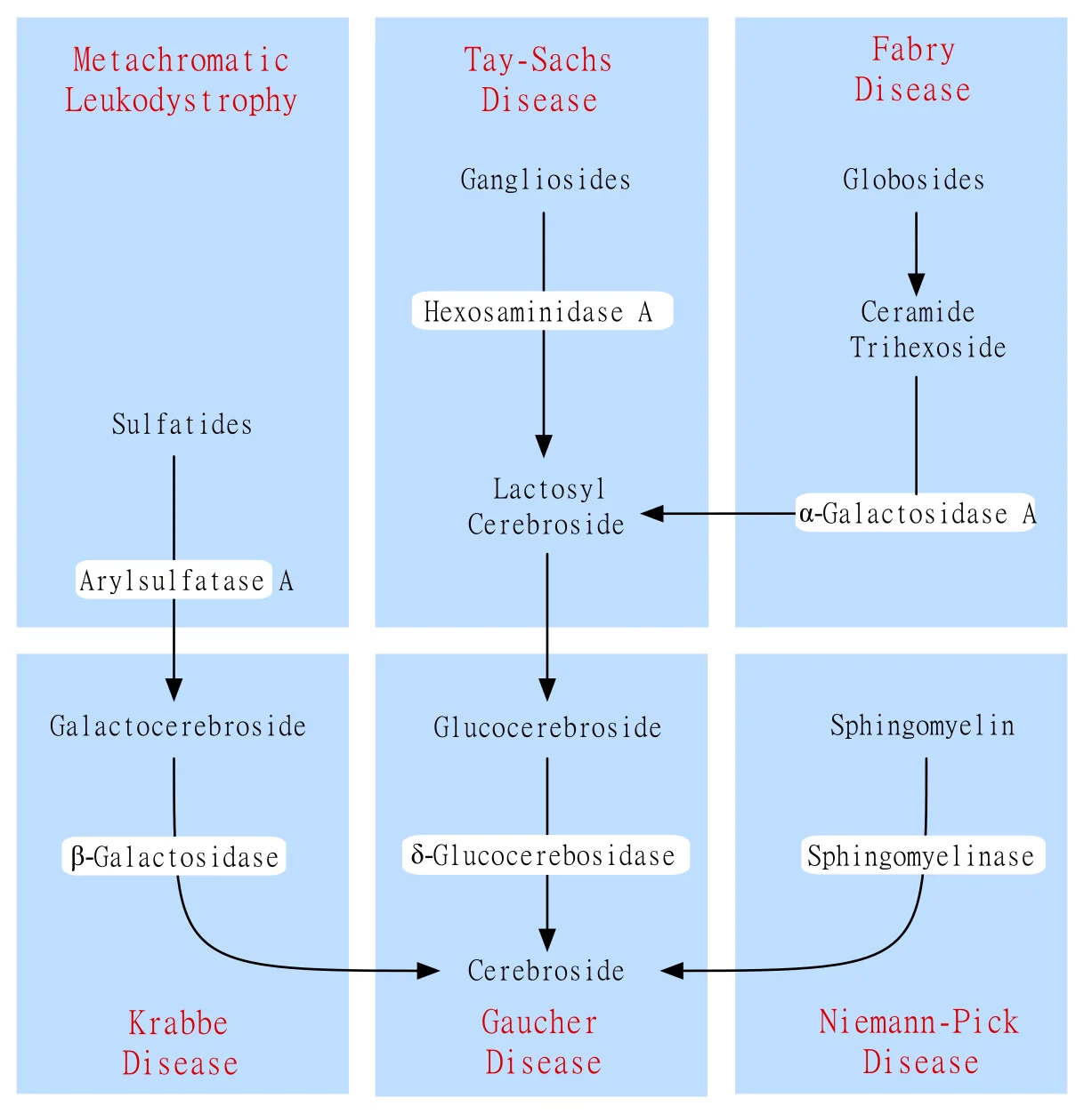
Long description
Basic diagram illustrating metabolic pathways involved in lysosomal storage diseases

Lysosomal storage diseases are a large, heterogeneous group of inherited metabolic disorders characterised by the abnormal intra-lysosomal accumulation of undegraded macromolecules. From a first principles perspective, normal lysosomal function relies on a coordinated multi-step cellular pathway. Messenger RNA coding for a specific lysosomal enzyme is directed to membrane-bound ribosomes of the rough endoplasmic reticulum, where the enzyme polypeptide is constructed. An amino-terminal signal peptide then directs the polypeptide into the lumen of the endoplasmic reticulum. To ensure correct sorting to the lysosome, mannose-6-phosphate is added to the N-linked oligosaccharide side chains of the polypeptide. Many lysosomal hydrolases undergo further proteolytic cleavage before becoming fully activated within the acidic environment of the organelle. [3]
When a single hydrolase is deficient, the cell experiences an inability to degrade the rest of the macromolecule, resulting in the formation of a lysosomal storage disease. In contrast to classic enzyme deficiencies that result in the storage of large, undegraded macromolecules, lysosomal transport deficiencies cause small molecules to accumulate within lysosomes. Although there are many lysosomal transporters, only five have been definitively associated with human disease, a phenomenon likely explained by the redundancy of small molecule carriers preventing pathological accumulation. These five distinct transport defects are cystinosis, Salla disease or free sialic acid storage disease, cobalamin F disease, cobalamin J disease, and mucolipidosis type IV. [3]
Another important category is drug-induced phospholipidosis, which consists of the exaggerated storage of phospholipids inside lysosomes, causing them to acquire a characteristic multilamellar appearance. More than fifty different pharmaceutical compounds can cause this condition, including antiarrhythmics, antimalarials, antipsychotics, and antibacterials. All of these offending compounds share the chemical property of being cationic amphiphilic drugs, possessing a hydrophobic ring and a hydrophilic side chain charged with a cationic amine group. Drug-induced phospholipidosis is considered an acquired form of lysosomal storage disorder because the drugs inhibit lysosomal phospholipases. Histopathologically, affected cells acquire a foamy appearance, and ultrastructurally there is storage of multi-lamellar whorled membranes, often referred to as myeloid bodies. [3]
DefinitionPathophysiological Hallmark
Lysosomal storage diseases arise primarily from single-enzyme defects causing macromolecular engorgement, or transporter defects leading to small molecule entrapment.
Patients with lysosomal storage disorders are generally normal at birth, with clinical symptoms typically developing over the course of the first year of life. However, careful pathological investigation reveals that microscopic findings can appear much earlier in the developing fetus. For instance, in a 9-week fetus with Tay-Sachs disease, lamellar and granular inclusions are already present within developing brain neurons. In fetuses ranging from 12 to 22 weeks of gestation, these lysosomal inclusions assume a more typical morphology of membranous cytoplasmic bodies and can be found in anterior horn cells of the spinal cord, ganglion cells of the retina, the enteric plexus, and spinal ganglia. [3]
Because safe and efficacious treatments exist for only a fraction of known lysosomal disorders—with others remaining under active investigation such as Niemann-Pick disease, Sly syndrome, and Farber disease—early detection before the onset of irreversible tissue changes is of paramount clinical importance. This applies to bone marrow transplantation in certain enzyme deficiencies and to cysteamine treatment for cystinosis [P348]. Consequently, newborn screening programs provide an eno [3]
Diagnostic Screening Pathways for Lysosomal Disorders
- Newborn Specimen CollectionDry blood spots obtained routinely.
- Question: Screening Methodology?
Enzyme Activity
- Direct MeasurementTandem mass spectrometry or digital microfluidics fluorometric assays.
Protein Abundance
- Immunological AssaysQuantifying the absolute level of specific lysosomal enzymes.
Metabolite Profiling
- Biomarker AssayingMeasuring accumulated substrates or pathological ratios.
- Confirmatory Testing & Genetic AnalysisReferral to metabolic center for molecular confirmation.
Various screening techniques have been proposed and implemented internationally:
- Direct measurement of enzyme activity, performed using either tandem mass spectrometry or digital microfluidics fluorometric assays [3]
- Measuring the abundance of specific lysosomal enzymes through immunological assays [3]
- Assaying characteristic metabolic biomarkers [3]
Several states and countries have published extensive epidemiological data regarding newborn screening for these conditions. For example, after screening 110,000 newborns in Washington state, investigators estimated a prevalence of 1 in 7,800 for Fabry disease males, 1 in 27,800 for Pompe disease, and 1 in 35,500 for Hurler disease, which is two to four times greater than previous prevalence estimates derived purely on clinical grounds. Missouri reported its experience screening 43,701 specimens during the first six months of a pilot study, discovering a detection rate of 1 in 2,913 for Fabry disease, 1 in 5,463 for Pompe disease, 1 in 14,567 for Hurler disease, and 1 in 43,701 for Gaucher disease. [3]
International data mirrors these high detection rates. Pilot studies from Austria involving 34,736 newborns demonstrated an incidence of Fabry disease of 1 in 3,859 newborns, Pompe disease in 1 in 8,684 births, and Gaucher disease in 1 in 17,368 births. Newborn screening in Taiwan successfully identified the cardiac variant of Fabry disease in 1 in 1,600 to 2,450 male newborns. An Italian study screening 37,104 newborn males for Fabry disease found an overall incidence of 1 in 3,100 to 4,600, noting a ratio of later-onset to classic-onset disease of 7 to 11 to 1. This consistently observed increase in disease incidence when deploying newborn screening is directly related to an increased diagnostic pickup of later-onset forms of the condition, regardless of the specific lysosomal storage disorder being evaluated. [3]
The sphingolipidoses and mucopolysaccharidoses represent major subsets of lysosomal storage disorders where complex lipids or carbohydrate polymers accumulate due to single-enzyme blocks. A prime historical and biological paradigm within the sphingolipidoses is Tay-Sachs disease, alongside related conditions such as Gaucher disease and Niemann-Pick disease. Gaucher disease serves as the single most common lysosomal storage disease overall, operating as an autosomal recessive disorder caused by a deficiency of glucocerebrosidase, also known as acid beta-glucosidase. This vital enzyme normally converts glucocerebroside to ceramide and glucose. As a direct consequence of this enzymatic defect, glucocerebroside—an important intermediate in the catabolism of globoid and gangliosides—accumulates massively within the reticuloendothelial system. [3]
| Disorder | Deficient Enzyme / Protein | Accumulating Substrate | Key Clinical / Histopathological Features |
|---|---|---|---|
| Gaucher Disease | Glucocerebrosidase (Acid beta-glucosidase) | Glucocerebroside (GL1) | Reticuloendothelial storage, hepatosplenomegaly, bone involvement |
| Niemann-Pick Type A & B | Sphingomyelinase (implied) | Sphingomyelin, glucosylceramide, GM2 | Reticuloendothelial and hepatic storage, neuronal storage in Type A |
| Tay-Sachs Disease | Beta-hexosaminidase A (implied) | GM2 ganglioside | Fetal neuronal inclusions, progressive neurodegeneration, macular cherry-red spot |
| Fabry Disease | Alpha-galactosidase A (implied) | Globotriaosylceramide (GL3) | Later-onset and classic variants, cardiac and renal involvement |
To further illustrate the pathological spectrum, Niemann-Pick disease types A and B demonstrate extensive storage material within reticuloendothelial cells, hepatocytes, pulmonary macrophages, and syncytiotrophoblast and villus stromal cells. Detailed biochemical analyses, such as those documented by Greenbaum and colleagues, establish that total extracts of brain glycolipids in Niemann-Pick disease type A contain pathologically increased levels of glucosylceramide, dihexoside, trihexoside, and GM2 ganglioside. In type A disease, there is predominantly neuronal sphingomyelin storage with lesser vascular involvement, whereas neutral glycolipid dominates within the vascular wall. Furthermore, hepatic storage of glycogen has been described specifically in type B Niemann-Pick disease, a biochemical overlap that has historically led to its clinical misdiagnosis as a primary glycogen storage disorder. [3]
Clinical pearlBiochemical Mimicry
Hepatic glycogen storage in Niemann-Pick disease type B can mimic glycogen storage disorders, requiring careful enzymatic confirmation.
In Tay-Sachs disease, the fundamental defect impairs the degradation of GM2 ganglioside, leading to its accumulation inside the central nervous system. As noted earlier, pathological evidence of this storage is exceptionally early; in a 9-week human fetus, lamellar and granular inclusions are clearly visible within developing brain neurons. By 12 to 22 weeks of gestation, these findings mature into distinct membranous cytoplasmic bodies located prominently within the anterior horn cells of the spinal cord, ganglion cells of the retina, the enteric plexus, and spinal ganglia. This early neural deposition explains the profound neurodegenerative course observed postnatally.
Fabry disease is an X-linked glycosphingolipid storage disorder caused by the deficiency of the lysosomal enzyme alpha-galactosidase A, resulting in the systemic accumulation of globotriaosylceramide within various tissues, including the vascular endothelium, kidneys, heart, and nervous system. With the advent of expanded newborn screening programs globally, our understanding of the true epidemiological prevalence and phenotypic spectrum of Fabry disease has evolved dramatically.
Population-based newborn screening data underscores that Fabry disease is significantly more common than previously appreciated through traditional clinical identification methods. For example, in Washington state, screening of 110,000 newborns revealed an estimated prevalence for Fabry disease males of 1 in 7,800, which is two to four times greater than estimates derived from historical clinical presentations. Similarly, a pilot study from Missouri encompassing 43,701 specimens demonstrated a striking detection rate of 1 in 2,913 for Fabry disease.
European pilot studies corroborate these high frequencies. In Austria, screening of 34,736 newborns demonstrated a population incidence for Fabry disease of 1 in 3,859 newborns. In Taiwan, newborn screening initiatives led to the specific identification of the cardiac variant of Fabry disease in 1 in 1,600 to 2,450 male newborns. Furthermore, an Italian screening study evaluating 37,104 newborn males found an overall incidence ranging from 1 in 3,100 to 4,600, establishing a specific ratio of later-onset to classic-onset disease of 7 to 11 to 1. [3]
Exam trapPhenotypic Skewing
Newborn screening detects high rates of later-onset Fabry variants, altering classic clinical paradigms and raising complex counselling challenges.
The observation of an increased incidence of disease when utilizing newborn screening is consistently related to an increased diagnosis of later-onset forms of the condition, a phenomenon that applies across nearly all lysosomal storage disorders subjected to population screening. These later-onset variants often present with isolated cardiac hypertrophy, renal failure, or cerebrovascular events in adulthood, contrasting sharply with the classic multi-organ phenotype of childhood onset. Early detection via screening remains critical before the development of irreversible fibrosis and organ failure, enabling timely therapeutic interventions. [3]
Lysosomal storage disorders frequently belong to the glycosphingolipidoses, a group of metabolic conditions where complex lipids accumulate due to specific enzyme deficiencies. Sphingolipids consist of a sphingosine long-chain amino alcohol combined with a fatty acid to form ceramides. Adding a single sugar produces a cerebroside, while attaching a sulfate group yields a sulfatide. When a polysaccharide is added, the resulting molecule is a globoside, and incorporating one or more N-acetylneuraminic acid molecules creates a ganglioside, where sialic acid always acts as a terminal sugar. The range of stored materials across these disorders includes ceramides, cerebroside, other ceramide hexosides, sulfatides, sphingomyelins, gangliosides, and lipofuscins. [3]
Gaucher disease specifically arises from impaired glucocerebrosidase function. This enzyme also contributes to normal skin barrier function. In type 2 Gaucher disease, rare individuals present with congenital ichthyosis. While light microscopy of skin samples from patients with type 1 and type 3 disease remains normal, skin biopsies from type 2 cases demonstrate dense hyperkeratosis, epidermal hyperplasia, and inflammation. Ultrastructural analysis of the epidermis in type 2 patients reveals abnormal arrays of loosely packed lamellar bodies within the stratum corneum, regardless of clinical ichthyosis. [3]
Pathology in type 2 Gaucher disease can initiate during the fetal and placental stages. The placenta may appear edematous, harboring storage material inside Hofbauer placental histiocytes and intravillus vessel cells. Fetuses examined at 17 weeks and 20 weeks of gestation show storage cells concentrated within the liver and spleen, though without overt neuropathological changes. Conversely, a fetus at 26 weeks of gestation exhibits extensive neuronal loss and gliosis within regions containing the most mature neurons, specifically the spinal cord, brainstem, thalamus, and basal ganglia. Fetal Gaucher cells might lack the well-delineated striations seen in mature patients, appearing foamy instead, yet tubular cytosomes remain identifiable via electron microscopy. Furthermore, the coexistence of Gaucher disease and hydrops fetalis is documented in multiple cases. [3]
DefinitionGaucher cell morphology
The cytoplasm of a mature Gaucher cell is packed with rod-shaped inclusions bound by a single membrane, creating a classic crumpled silk appearance. [3]
The characteristic appearance of the Gaucher cell serves as a diagnostic indicator, though bone marrow biopsy should not be utilised as the primary method for diagnosis. Macrophages in other sphingolipidoses, such as Niemann-Pick disease, contain multiple intracytoplasmic droplets of uniform size, yielding a foamy morphology rather than crumpled silk. Cells resembling Gaucher cells can occasionally appear in multiple myeloma, leukemias, thalassemia, and congenital dyserythropoietic anemia. However, true Gaucher cells are differentiated from these pseudo-Gaucher cells by much stronger expression of HLA-DR on immunohistochemistry. Additionally, erythrophagocytosis by Gaucher cells is frequent, alongside abundant intracellular hemosiderin. [3]
Biomarkers play a major role in detection. The activity of chitotriosidase, a human chitinase secreted by macrophages, is strikingly elevated in Gaucher disease and raised to varying extents in many other lysosomal storage disorders and macrophage-activation states. Because the assay requires only simple equipment and remains widely available, many laboratories propose it as an initial screening test when a lysosomal storage disorder is suspected, despite a lack of absolute specificity and the presence of a common 24-base-pair duplication in exon 10 of the CHIT1 gene that causes partial or complete enzyme deficiency in some individuals. [4]
Pharmacological chaperone therapy represents an evolving therapeutic avenue, though its applicability varies by mutation type. Chaperone-responsive mutations are relatively frequent in Gaucher disease, highlighted by the N370S mutation which accounts for 34 percent of Gaucher alleles in non-Ashkenazi patients and 71 percent of alleles in Ashkenazi patients. To evaluate enzyme replacement therapy efficacy in individual patients and manage potential infusion-related reactions, treatment must be overseen by experienced physicians working in specialized centres with expertise in managing lysosomal storage diseases. [1] [4]
| Feature | Finding |
|---|---|
| Ultrastructure | Rod-shaped inclusions bound by a single membrane |
| Immunohistochemistry | Strong expression of HLA-DR |
| Screening biomarker | Markedly elevated chitotriosidase activity |
| Common allele (Ashkenazi) | N370S accounting for 71 percent |
Niemann-Pick diseases share overlapping clinical features with other glycosphingolipidoses and macrophage-storage disorders, though distinct biochemical and cellular markers separate them. Acid sphingomyelinase deficiency (ASMD) presents distinct biomarker profiles compared to Gaucher disease. In ASMD, elevation of chitotriosidase activity is generally modest and inconsistent, and this elevation fails to differentiate ASMD from Niemann-Pick type C or several other conditions featuring hepatosplenomegaly. [4]
Pathologically, macrophage morphology provides clues during differential diagnosis. Unlike the crumpled silk appearance of Gaucher macrophages, the macrophages observed in Niemann-Pick disease possess multiple intracytoplasmic droplets of uniform size, which grants them a distinctly foamy appearance. Marked lipid storage can manifest remarkably early, identifiable as early as in a 20-week fetus. Furthermore, partial sphingomyelinase deficiency observed exclusively in cultured cells represents a variable, secondary consequence of lysosomal cholesterol esterification. [3]
Clinical pearlMacrophage comparison
Niemann-Pick macrophages display uniform intracytoplasmic droplets resulting in a foamy morphology, distinguishing them from the tubular, crumpled-silk Gaucher cells.
Cellular pathology in related lipid storage disorders highlights how defects in organelle fusion disrupt degradation pathways. For example, inactivation of mucolipin-1 causes abnormal accumulation of lipids and other compounds inside late endosomes because their contents fail to degrade within lysosomes due to impaired fusion with those organelles. Simultaneously, the resulting lysosomal calcium efflux through mucolipin-1 activates calcineurin, which promotes nuclear translocation of TFEB, a master transcriptional regulator responsible for controlling lysosomal biogenesis and autophagy. [3]
Diagnostic and evaluation pathway for lysosomal storage disorders
- Clinical suspicion of lysosomal storage disorderPresence of hepatosplenomegaly or cytopenia.
- Question: Perform initial screening?
Yes
- Assay chitotriosidase activitySimple equipment required; check for CHIT1 exon 10 duplication.
- Question: Marked elevation?
Gaucher suspected
- Evaluate genotype and HLA-DR expressionTest for common alleles like N370S.
Modest or non-specific elevation
- Investigate ASMD or Niemann-Pick type CDifferentiate using specialized enzyme and lipid assays.
No
- Direct specialist referralEnsure management occurs in an experienced centre.
Krabbe disease is a severe inherited leukodystrophy caused by the deficiency of the lysosomal hydrolase beta-galactocerebrosidase [P418]. The condition was first described by the Danish neurologist Knud Haraldsen Krabbe in 1916, and the underlying enzymatic defect was subsequently identified in 1970. From a genetic perspective, Krabbe disease is an autosomal recessive disorder caused by mutations leading to a deficiency of galactocerebrosidase. This specific lysosomal enzyme is normally responsible for [3][4]
To understand the pathophysiology from first principles, one must examine the normal distribution of the substrate. Galactocerebroside, also known as galactosylceramide, is a sphingoglycolipid consisting of sphingosine, a fatty acid, and galactose. Under normal physiological conditions, it is present almost exclusively in the myelin sheath. Because of the enzyme block in Krabbe disease, galactosylceramide accumulates within both the peripheral and central nervous systems. Interestingly, rather than accumulating inertly in the white matter, galactocerebroside is catabolized through an alternative pathway that yields psychosine, or galactosphingosine. Psychosine is highly toxic to oligodendrocytes, the specialized glial cells responsible for myelin production in the central nervous system, driving the rapid demyelination characteristic of the disease. [3]
DefinitionLeukodystrophy Mechanism
Krabbe disease is a fatal neurodegenerative leukodystrophy where alternative catabolism of unbranched galactocerebroside generates psychosine, causing oligodendrocyte destruction.
Clinically, late-onset variants of Krabbe disease can present diagnostic challenges because they mimic other neurological conditions. Late-onset Krabbe disease has been misdiagnosed as motor neuron disease, hereditary spastic paraplegia, multiple sclerosis, and Charcot-Marie-Tooth disease. When evaluating suspected cases, clinicians utilize imaging modalities. Aside from the characteristic leukodystrophy seen on magnetic resonance imaging, patients with Krabbe disease can also present with intracranial calcifications, which are much better assessed by computed tomography. [3]
| Feature | Description |
|---|---|
| Inheritance | Autosomal recessive |
| Deficient Enzyme | Galactocerebrosidase |
| Toxic Intermediate | Psychosine (galactosphingosine) |
| Imaging Finding | Intracranial calcifications on computed tomography |
Krabbe Disease Pathophysiology
- Enzyme DefectGalactocerebrosidase deficiency prevents normal degradation of galactocerebroside.
- Question: Alternative Pathway Active?
Yes
- Psychosine ProductionSubstrate is shunted to yield galactosphingosine (psychosine).
- Oligodendrocyte ToxicityPsychosine accumulation destroys oligodendrocytes, causing fatal neurodegeneration.
No
- StorageDirect accumulation occurs in central and peripheral nervous systems.
Metachromatic leukodystrophy is an inherited lysosomal storage disease with an estimated frequency of 1 in 40,000. The condition was first described by Scholz in 1925, although it was not until decades later that Scholz's preserved brain samples were shown to stain metachromatically. Similar clinical cases were subsequently reported by Bielschowsky and Henneberg in 1928, and by Greenfield in 1933, which accounted for the historical nomenclature of Scholz-Bielschowsky-Henneberg syndrome, or Greenfield disease. The specific enzymatic defect responsible for the disorder was identified in 1963. [3]
Metachromatic leukodystrophy is an autosomal recessive lysosomal storage disease caused by a deficiency of arylsulfatase A, designated as enzyme commission number 3.1.6.8, due to mutations in the ARSA gene. Arylsulfatase A normally degrades sulfated glycolipids. One of its major physiological substrates is cerebroside 3-sulfate, also known as galactosyl sulfatide or cerebroside sulfate, which is a lipid found mainly in myelin membranes where it accounts for 3% to 4% of total membrane lipids. The enzyme performs its catalytic function by removing the sulfate group from the galactose residue of cerebroside 3-sulfate, a reaction that requires the assistance of an activator protein called saposin B. Deficiencies of this activator protein cause a very rare clinical disease that is clinically indistinguishable from metachromatic leukodystrophy. [2] [3]
Clinical pearlSubstrate Accumulation
In metachromatic leukodystrophy, galactosyl sulfatide accumulates in the white matter of the central nervous system and within peripheral nerves. [3]
When arylsulfatase A is deficient, galactosyl sulfatide and, to a lesser extent, lactosyl sulfatide accumulate within the central nervous system white matter, peripheral nerves, and certain visceral organs including the kidney and gallbladder. Because these lipids are stored in excessive amounts, they are also excreted in large quantities in the urine. In histological preparations, these accumulated sulfatides form spherical granular masses that characteristically stain metachromatically. [3]
The systemic and neurological consequences of this storage process are profound. Reduced arylsulfatase A activity results in the accumulation of sulfatides in both the central nervous system and the peripheral nervous system, which triggers progressive dysmyelination, neuroinflammation, and neurodegeneration. Clinically, these cellular events translate into progressive motor and cognitive deterioration, marked by the steady loss of motor and neurocognitive functions, and ultimately resulting in death. In addition to nervous system pathology, sulfatides also accumulate in visceral organs such as the gallbladder and kidneys. [2]
| Parameter | Characteristic |
|---|---|
| Frequency | 1 in 40,000 |
| Genetic Defect | Mutations in the ARSA gene |
| Deficient Enzyme | Arylsulfatase A |
| Major Substrate | Cerebroside 3-sulfate |
Metachromatic Leukodystrophy Pathway
- ARSA MutationMutations in the ARSA gene cause arylsulfatase A deficiency.
- Question: Enzyme Functional?
No
- Sulfatide AccumulationCerebroside 3-sulfate accumulates in CNS, peripheral nerves, and viscera.
- NeurodegenerationProgressive dysmyelination, neuroinflammation, and motor-cognitive decline.
Yes
- Normal DegradationSulfatides are successfully degraded with saposin B assistance.
Mucopolysaccharidosis type II, also known as Hunter syndrome, was first described by Major Charles Hunter in the year 1917. It is classified as an X-linked recessive disease that arises from a deficiency of the lysosomal enzyme iduronate-2-sulphatase, which is abbreviated as I2S. The official OMIM designation for this genetic condition is OMIM +309900. [1]
From a biochemical perspective, the normal function of the iduronate-2-sulphatase enzyme is to cleave O-linked sulphate moieties from specific glycosaminoglycans, namely dermatan sulphate and heparan sulphate. This cleavage reaction represents the initial step in the normal degradative pathway of these macromolecules. When the enzyme is deficient, it leads to a progressive pathological lysosomal storage of glycosaminoglycans across nearly all cell types, tissues, and organs in the body. [1]
Hunter syndrome is clinically characterized as a severe, progressive, multisystemic disorder. Because it has the potential to cause pathology and disease manifestations in most body systems, it is typically fatal in either the second or the third decade of life. [1]
DefinitionHunter syndrome genetics
Hunter syndrome is an X-linked recessive lysosomal storage disease caused by iduronate-2-sulphatase deficiency, leading to dermatan and heparan sulphate accumulation.
Management of this multisystemic condition requires a comprehensive and holistic approach. Ideally, patient care should be centralized in major medical centers that possess ready access to all relevant medical specialties. A multidisciplinary team is essential, particularly for individuals who suffer from severe neurological involvement. Although modern medical interventions have advanced, central nervous system disease remains a major challenge, and innovative therapeutic approaches will be required to address it fully. [1]
Enzyme replacement therapy represents an important new treatment modality that holds the potential to help many patients, provided that it is initiated early in the course of the disease. The fundamental goal of enzyme replacement therapy is to address the underlying metabolic defect by supplying functional exogenous enzymes to substitute for deficient or defective endogenous ones. [1]
Clinical pearlTiming of ERT
Enzyme replacement therapy is most effective when administered early in the disease course before irreversible tissue and organ damage occurs.
In the broader context of lysosomal storage diseases, enzyme replacement strategies have achieved notable clinical success for systemic manifestations. For example, recombinant human enzymes are often modified for enhanced cellular uptake, such as targeting via mannose receptors on macrophages. These therapeutic preparations are typically administered via periodic intravenous infusions according to established dosing schedules. While generally well tolerated, these infusions carry a significant financial burden, costing several hundred thousand dollars per patient per year. [3]
Adverse events can occur during administration. Approximately 15% of patients may develop antibodies against the infused enzyme, depending heavily on the specific preparation used, though most of these antibodies are not neutralizing. Furthermore, some individuals can experience hypersensitivity reactions, including urticaria and pruritus linked to the infusions, which are generally manageable with standard antihistamines. [3]
For other conditions sharing similar pathophysiological mechanisms, such as acid sphingomyelinase deficiency, enzyme replacement utilizing human recombinant enzymes acts as a disease-modifying treatment specifically indicated for non-central nervous system manifestations. Regulatory approvals for such specialized enzyme therapies have been secured across multiple global jurisdictions including Brazil, Japan, Europe, and the United States of America. [4]
When managing lysosomal storage diseases and administering complex biologic therapies like enzyme replacement, clinicians must navigate several recurring clinical and biochemical pitfalls.
A primary limitation in treating these conditions is the impermeable nature of the blood-brain barrier. While systemic manifestations such as hepatosplenomegaly, skeletal changes, and somatic storage can be significantly ameliorated by intravenous enzyme infusions, central nervous system disease remains notoriously difficult to treat. Standard intravenous enzyme replacement therapies do not effectively cross into the central nervous system in sufficient quantities to reverse established neurological degeneration. Consequently, patients with severe neurological involvement continue to experience progressive neurodegeneration even while their systemic symptoms are being actively managed.
Another significant pitfall involves the timing of therapeutic initiation. Enzyme replacement therapy cannot reverse dense, established architectural fibrosis, skeletal deformities, or severe organ scarring. Therefore, delaying treatment until late in the disease course drastically curtails the clinical benefit.
Exam trapThe CNS barrier trap
Expecting standard intravenous enzyme replacement therapy to halt neurodegenerative decline ignores its poor penetration across the blood-brain barrier.
Immunological complications also present clinical hurdles. Although the majority of treatment-emergent antibodies are non-neutralizing, antibody formation can occasionally alter the pharmacokinetic profile or efficacy of the therapeutic enzyme. Clinicians must remain vigilant for acute infusion-associated reactions, such as urticaria and pruritus, ensuring that appropriate premedications or antihistamines are readily available. Finally, managing systemic complications requires careful multidisciplinary coordination; for instance, overlapping symptoms such as a bleeding tendency require direct haematological consultation when severe thrombocytopenia is present.
In clinical practice, the management of severe multisystemic lysosomal storage disorders requires a highly organized infrastructure. Because these conditions impact nearly every organ system and carry a high mortality rate in the second or third decade of life, care cannot be handled by a single clinician.
Multidisciplinary Management Pathway for Lysosomal Storage Diseases
- Initial PresentationMultisystemic symptoms, developmental or somatic signs.
- Question: Specialist Centre Access?
Yes
- Centralise care in a major medical centre with all specialties
- Question: Severe Neurological Involvement?
Yes
- Implement holistic multidisciplinary care and palliative support
No
- Evaluate for early enzyme replacement therapy
No
- Establish urgent link with regional specialist and multidisciplinary teams
Practical care is centered within major specialized medical hubs that maintain immediate access to multiple medical specialties. When a patient is diagnosed with a condition such as Hunter syndrome or another severe metabolic storage disorder, the specialist team coordinates closely with community care providers to ensure continuous monitoring.
The day-to-day application of therapy focuses on addressing existing and impending complications, comprehensive symptom management, and administering disease-modifying agents where available. For conditions possessing approved biologic treatments, infusions are scheduled regularly in outpatient infusion suites or specialized daycare units. Monitoring involves tracking somatic growth, joint mobility, respiratory function, and cardiac status. When hematologic abnormalities arise, such as a variable bleeding tendency linked to underlying platelet or coagulation defects, prompt consultation with a haematologist is integrated into the routine care plan to evaluate and manage severe thrombocytopenia safely. [4]
- Hunter syndrome is an X-linked recessive lysosomal storage disease caused by iduronate-2-sulphatase deficiency.
- The deficient enzyme normally cleaves sulphate moieties from dermatan sulphate and heparan sulphate.
- Progressive intracellular glycosaminoglycan storage leads to a severe, multisystemic disorder usually fatal in the second or third decade.
- Ideal management requires a centralized, multidisciplinary approach in major medical centers.
- Enzyme replacement therapy can help many patients if started early, though central nervous system disease remains a major challenge.
- Infusion-associated reactions like urticaria and pruritus can occur and are treatable with antihistamines.
Answer each one in your head first, then open the answer.
What is the inheritance pattern of Hunter syndrome?
Show answerHide answer
X-linked recessiveWhich specific enzyme is deficient in Hunter syndrome?
Show answerHide answer
Iduronate-2-sulphataseWhat are the two major glycosaminoglycans that fail to degrade properly in Hunter syndrome?
Show answerHide answer
Dermatan sulphate and heparan sulphateWhat is the typical age range of mortality for severe untreated Hunter syndrome?
Show answerHide answer
Second or third decade of lifeHow are mild infusion-related urticaria and pruritus typically treated during enzyme replacement therapy?
Show answerHide answer
Antihistamines
- Wraith JE, Scarpa M, Beck M, et al. Mucopolysaccharidosis type II (Hunter syndrome): a clinical review and recommendations for treatment in the era of enzyme replacement therapy. European journal of pediatrics 2008;167(3):267-277. doi:10.1007/s00431-007-0635-4. Accessed .
- Fumagalli F, Calbi V, Natali Sora MG, et al. Lentiviral haematopoietic stem-cell gene therapy for early-onset metachromatic leukodystrophy: long-term results from a non-randomised, open-label, phase 1/2 trial and expanded access. Lancet (London, England) 2022;399(10322):372-383. doi:10.1016/s0140-6736(21)02017-1. Accessed .
- Ferreira CR, Gahl WA. Lysosomal storage diseases. Translational science of rare diseases 2017;2(1-2):1-71. doi:10.3233/trd-160005. Accessed .
- Geberhiwot T, Wasserstein M, Wanninayake S, et al. Consensus clinical management guidelines for acid sphingomyelinase deficiency (Niemann-Pick disease types A, B and A/B). Orphanet journal of rare diseases 2023;18(1):85. doi:10.1186/s13023-023-02686-6. Accessed .
Figure credits
Educational content for medical students and doctors. It is not a substitute for clinical judgement or local guidelines.